www.biogeosciences.net/8/2547/2011/ doi:10.5194/bg-8-2547-2011
© Author(s) 2011. CC Attribution 3.0 License.
Biogeosciences
A laboratory experiment of intact polar lipid degradation
in sandy sediments
J. Logemann∗, J. Graue∗, J. K¨oster, B. Engelen, J. Rullk¨otter, and H. Cypionka
Institute for Chemistry and Biology of the Marine Environment (ICBM), Carl von Ossietzky University of Oldenburg, P.O. Box 2503, 26111 Oldenburg, Germany
∗These authors contributed equally to this work.
Received: 7 March 2011 – Published in Biogeosciences Discuss.: 24 March 2011
Revised: 26 August 2011 – Accepted: 3 September 2011 – Published: 13 September 2011
Abstract.Intact polar lipids (IPLs) are considered biomark-ers for living biomass. Their degradation in marine sedi-ments, however, is poorly understood and complicates inter-pretation of their occurrence in geological samples. To in-vestigate the turnover of IPLs, a degradation experiment with anoxic sandy sediments from the North Sea was conducted. Intact cells of two organisms that do not naturally occur in North Sea sediments were chosen as IPL sources: (i) Saccha-romyces cerevisiae, representative for ester-bound acyl lipids that also occur inBacteria, and (ii) the archaeonHaloferax volcanii, representative for ether-bound isoprenoid lipids. Surprisingly, IPLs with phosphoester-bound head groups showed approximately the same degradation rate as IPLs with glycosidic head groups. Furthermore, the results indi-cate a relatively fast degradation ofS. cerevisiaeIPLs with ester-bound moieties (analogs of bacterial membrane lipids) and no significant degradation of archaeal IPLs with ether-bound moieties. Pore water and 16S rRNA-based DGGE analysis showed only a minor influence of the IPL source on microbial metabolism and community profiles. Due to our results, the IPL-based quantification ofArchaeaand Bacte-riashould be interpreted with caution.
1 Introduction
Intact polar lipids (IPLs) have widely been used as biomark-ers for living organisms in sediments and water columns for several years (e.g. Zink et al., 2003; Sturt et al., 2004; Bid-dle et al., 2006; Ertefai et al., 2008; Rossel et al., 2008; Schubotz et al., 2009; Van Mooy et al., 2009).
Addition-Correspondence to:J. Logemann ([email protected])
ally, IPLs are applied as chemotaxonomic markers as some of these molecules are representative for specific microbial clades. The chemotaxonomic information of IPLs is based on the combination of various head groups with different side chains that are attached to a glycerol backbone by two different bonding types. In general, bacterial and eukaryal cytoplasma membranes contain intact polar lipids with non-isoprenoidal ester-bound fatty acid moieties. In contrast, ar-chaeal IPLs contain ether-bound isoprenoid moieties. This difference between bacterial and archaeal IPLs has been used to quantify Bacteria and Archaea in sediments and water samples (e.g. Rossel et al., 2008; Lipp et al., 2008; Schubotz et al., 2009; Van Mooy et al., 2009). Thus, IPL analy-sis is valuable as an alternative or complementary technique to standard microbiological methods. On a higher chemo-taxonomic level, ammonia-oxidizing bacteria can be iden-tified by the presence of ladderane lipids (Bouman et al., 2006; Jaeschke et al., 2009). Intact polar lipids with mixed ether/ester-bound moieties attached to the glycerol back-bone were found in some strains of sulfate-reducing bacteria (R¨utters et al., 2001). Separateδ13C analysis of polar head groups or non-polar core lipids of IPLs can be used to gain information on the metabolism of their producers (Boschker et al., 1998; Lin et al., 2010; Takano et al., 2010).
origin after sediment burial. Using different variables sig-nificantly influenced the results showing that modeling alone cannot resolve the preservation issue and that experimental data are needed.
The quantification ofBacteria andArchaea in the deep marine biosphere by IPL analysis and fluorescence in situ hy-bridization (FISH) in comparison to catalyzed reporter depo-sition fluorescence in situ hybridization (CARD-FISH) and quantitative polymerase chain reaction (q-PCR) analysis has been controversially discussed in several studies (e.g. Schip-pers et al., 2005; Biddle et al., 2006; Lipp et al., 2008). A reason for the contradictory abundances ofArchaea and Bacteriain these studies may be that ether-bound archaeal IPLs are more stable than their ester-bound bacterial coun-terparts, which in turn may lead to an overestimation of ar-chaeal cell numbers. On the other hand, q-PCR-based inves-tigations may underestimate the yields of archaeal 16S rRNA genes, as indicated by Lipp et al. (2008) and Teske and Sørensen (2008). However, the lack of a broad study on the degradation of intact polar lipids, which includes lipids with phosphoester and glycosidic head groups as well as ester- and ether-bound moieties occurring inBacteriaandArchaea, re-spectively, makes it necessary to revisit the degradation of IPLs to ensure the robustness of this proxy.
We designed a degradation experiment to answer three general questions: what are the degradation rates of IPLs? Are there differences between ester- and ether-bound intact polar lipids and what is the influence of the bonding type of the head group upon lipid degradation? The main degrada-tion experiment was accompanied by two controls: the first control was intended to assess any processes that are not me-diated by microorganisms but still lead to the degradation of the added IPLs. This control is subsequently named “abiotic control”. No cell material was added to the second control. It was used to investigate the development of the microbial community without any further substrate addition under lab-oratory conditions. This control is subsequently named “un-treated control”.
2 Material and methods
2.1 Experimental setup
The incubation vessels for the degradation experiment and the untreated control had a total volume of 2.5 l each and were filled with 3 kg wet sediment (water content 29 % wt, 2.13 kg dry wt). The sediment used in this ex-periment had been freshly collected in November 2009 on Janssand, a sandplate located approximately 3 km south of Spiekeroog Island, North Sea, Germany (53◦44.178′N and 07◦41.974′E). For sampling, the top centimeter of oxic sur-face sediment was removed until only black anoxic sedi-ment was visible. The underlying sedisedi-ment was transferred into plastic containers, which were sealed by a lid,
trans-ported to the laboratory and stored at 4◦C for one week prior to further use. The total organic carbon (TOC) con-tent was 0.23 %. It was calculated as the difference between total carbon (Vario EL Cube, Elementar Analysensysteme GmbH, Germany) and inorganic carbon (UIC CO2
coulome-ter). The pore water concentration of sulfate was equal to the sea water concentration (28 mM). A total of 2.5 g Saccha-romyces cerevisiaebiomass (elemental composition, of dry mass: 45.1 % C, 7.9 % H, 7.9 % N and 0.4 % S) as source for ester-bound IPLs and 1.25 g ofHaloferax volcanii(18.9 % C, 3.6 % H, 4.7 % N and 0.7 % S) as source for ether-bound IPLs were added to the sediment in the incubation vessels used to study IPL degradation.
As calculated from the equation of Adams et al. (1990), the dry weight of a single Saccharomyces cerevisiae cell is approximately 60 pg. With a total sediment volume of 1.5 l 2.8×10⁷cells ml−1 sediment were added. The cell
size of a singleHaloferax volcaniicell is 1–3×2–3 µm and
0.4–0.5 µm thick (Mullakhanbhai and Larsen, 1975). As-suming a water content of 80 %, this leads to a dry weight of 0.16–0.9 pg per single cell ofHaloferax volcanii. With 1.25 g of dry Haloferax biomass, approximately 9.3×10⁸–
5.2×10⁹cells ml−1 were added. The elemental
composi-tion of the dry biomass used in this experiment indicates a contamination with inorganic material. Thus, the calculated cell number may be too high but still in the correct order of magnitude. After addition of cell material to the incubation vessel, the sediment was homogenized for 6 h on a mixing device by slow rotation (12 rpm). The experimental parame-ters for the untreated control were the same as for the degra-dation experiment but no inactive cell material was added. To prevent contamination with microorganisms due to sam-pling, several 100 ml bottles were used instead of a single 2.5 l incubation vessel for the abiotic control. The bottles contained 50 g of sediment, 50 mg dry inactive biomass of S. cerevisiaeand 25 mg dry inactiveH. volcaniibiomass and were closed with rubber stoppers. The incubation vessels of the abiotic control were autoclaved after addition of the intact polar lipid-containing cell material.
2.2 Source material for intact polar lipids
As sources for intact polar lipids, two different organisms were used which do not occur in North Sea sediment. Ether-derived IPLs (diphytanylglycerols=DPGs) were originated
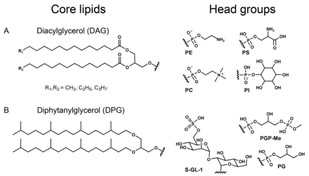
Fig. 1. Chemical structures of IPLs characteristic for the two organisms used in this study: (A): diacylglycerol ester-bound core lipids (DAGs) ofSaccharomyces cerevisiaebound to: phosphatidylethanolamine (PE), phosphatidylcholine (PC), phosphatidylinositol (PI) and phosphatidylserine (PS);(B): diphytanylglycerol ether-bound core lipids (DPGs) ofHaloferax volcaniibound to: sulfono diglyco dialkyl-glycerol S-GL-1 (nomenclature according to Sprott et al., 2003), phosphatidyldialkyl-glycerol (PG) and phosphatidyldialkyl-glycerol methylphosphate (PGP-Me).
as tracers to monitor the degradation of ether-bound IPLs. Commercially available Saccharomyces cerevisiae(baker’s yeast; Fala GmbH, Germany) was used as source for IPLs with ester-bound acylic moieties (diacylglycerols=DAGs)
and phosphoester head groups. S. cerevisiae cells harbor a broad variety of IPLs which were partly used as tracers because these IPLs are not synthetized by the natural sedi-ment microbial community. S. cerevisiaeand the harvested archaeal cells were freeze-dried and stored at−20◦C.
Be-fore use in the experiment,H. volcaniiandS. cerevisiaecells were pasteurized at 100◦C for 30 min. Thereafter, part of the cells was transferred to fresh medium (“Haloferax sul-furifontis medium” and yeast extract medium, respectively) to confirm that the cells were no longer alive and unable to grow.
Identification of S. cerevisiae lipids was achieved by HPLC-MS/MS experiments and comparison of mass spec-tral data to commercially available standards. The lipids of H. volcaniiwere identified by MS/MS experiments and com-parison with published results of lipid structures (Sprott et al., 2003). In this experiment we monitored the contents of 16 intact polar lipids, 13 of them were ester-bound and 3 ether-bound.
2.3 Incubation parameters and sampling
After starting the experiment, the incubation vessels were stored at room temperature in the dark. Before sampling,
the sediment was homogenized for a minimum of 20 min on a mixing device. Headspace gas samples were taken with a syringe directly before sampling the sediment. For sedi-ment and pore water sampling, the incubation vessels were transferred into a glove box with an oxygen-free nitrogen (99 %) and hydrogen (1 %) atmosphere. Samples were taken as triplicates, aliquots of 8–12 g sediment for IPL analysis and 3–5 g sediment for RNA extraction. Pore water (1–2 ml) was extracted with rhizones (Rhizon CSS 5 cm, Rhizosphere Research Products, Wageningen, The Netherlands) from the samples that were collected for IPL analysis. Our previous investigations had ensured the absence of IPLs in the pore water samples (data not shown). Until further processing, all samples were stored at−20◦C. The samples for RNA extrac-tion were kept at−80◦C. After sampling, the headspace of the incubation vessels was flushed with nitrogen to remove traces of hydrogen that was used in the glove box to sustain the oxygen-free atmosphere. The constantly increasing pH was adjusted at each sampling point to values between 7 and 7.5 by addition of hydrochloric acid. After day 21, sulfate concentrations were adjusted to approximately 28 mM by the addition of 1 M Na2SO4when concentrations dropped below
10 mM. After day 76, when hydrogen sulfide reached toxic concentrations, it was expelled by flushing the headspace with CO2. After each CO2flushing the headspace was
2.4 Lipid extraction
Wet sediment was extracted using the Bligh & Dyer method modified according to Sturt et al. (2004). In the first three extraction steps, a single-phase mixture of methanol, dichloromethane and phosphate buffer (2:1:0.8, v/v/v) was used. In the following three extraction steps, trichloroacetic acid replaced the phosphate buffer. The combined extracts were collected in a separatory funnel. Phase separation was achieved by addition of dichloromethane and water to a fi-nal mixture of 1:1:0.9 (v/v/v, methanol, dichloromethane, aqueous phase). The organic phase containing the IPLs was removed and the aqueous phase washed three times with dichloromethane. The dry extracts were stored in a freezer at−20◦C until further use.
2.5 HPLC-MS
Intact polar lipids were analyzed by HPLC (2695 sepa-ration module, Waters, Milfort, USA) coupled to a time-of-flight mass spectrometer equipped with an electrospray source (Micromass, Q-TOF micro, Waters, Milfort, USA). HPLC separation was achieved on a diol phase (Lichro-spher100 Diol 5 µm, CS – Chromatographie Service, Langer-wehe, Germany) using a 2×125 mm column. A flow rate of 0.35 ml min−1 was employed with the following solvent
gradient: 1 min 100 % A, increasing over 20 min to 35 % A, 65 % B using a concave curvature, followed by 40 min of re-conditioning (R¨utters et al., 2001). Eluent A was a mixture ofn-hexane,i-propanol, formic acid and a solution of 25 % ammonia in water (79:20:1.2:0.04 by volume), eluent B was
i-propanol, water, formic acid and a solution of 25 % ammo-nia in water (88:10:1.2:0.04 by volume). After addition of an injection standard (O-PE, phosphatidyl ethanolamine diether with two C16 alkyl moieties) to every sample, the extracts
were dissolved in the starting eluent and directly analyzed. In this study, we exclusively report the change of those com-pounds that were added with the inactive cell material and did not occur in the natural sediment. Due to the lack of analytical standards for the archaeal glycolipids used, it was not possible to determine the absolute concentrations of these compounds. Instead, ratios of peak areas of the monitored compounds to the peak area of the injection standard for each sample were calculated. To compare the results of ester-bound and ether-ester-bound IPLs the same procedure was applied also toSaccharomyces-derived IPLs. Peak areas were deter-mined by integration of mass traces. Since all samples had the same matrix background, this procedure should give rea-sonable results without any influence of changing ionization. The analytical error varied between 0.5 % and 7 % depend-ing on the investigated IPL and was determined by repeated analysis of the same samples taken at three different times. The limit of detection in general depends on the ionization efficiency for every analyzed compound and typically lies
between 2 and 10 ng per injection and IPL for the mass spec-trometer used.
2.6 Chemical analyses
Sulfate concentrations were measured by an ion chro-matograph with an LCA A24 anion separation column (both Sykam, F¨urstenfeldbruck, Germany) at 60◦C fol-lowed by conductivity detection. The eluent consisted of 0.64 g sodium carbonate, 0.2 g sodium hydroxide, 150 ml ethanol and 2 ml modifier (0.1 g 4-hydroxybenzonitrile/10 ml methanol) filled up to 1 l with distilled water. The flow rate was set to 1.2 ml min−1. Prior to analysis the samples were
diluted 1:100 in eluent without modifier.
The concentrations of gaseous compounds were deter-mined by an 8610C gas chromatograph (Schambeck SFD GmbH, Honnef, Germany). Analysis was carried out with argon (1 ml min−1) as carrier gas and at a column oven
tem-perature of 40◦C. For analysis of molecular hydrogen and methane a molecular sieve 13X packed column was used, whereas carbon dioxide was separated by a HayeSep D packed column. A thermal conductivity detector (256◦C) and a flame ionization detector (380◦C) were connected in series for detection of the gases.
Sulfide concentrations were determined photometrically as described by Cord-Ruwisch (1985).
2.7 Calculation of degradation rates
Degradation rates were calculated with the following equa-tion described by Schouten et al. (2010):
Ct=Ci·e−k
′t
, (1)
withCt=concentration at timet,Ci=initial concentration
andk′being the kinetic degradation constant. This method allows calculating degradation rates from degradation curves without curve fitting. The degradation constantsk′were cal-culated for every time point separately using data of day zero and the respective time point. Values for the calcula-tion of degradacalcula-tion rates of phosphatidylethanolamine dia-cylglycerol (PE-DAG) and glycol diphytanylglycerol (GL-DPG) were taken from Figs. 2 and 3 (beach sediments) in Harvey et al. (1986). For calculating degradation rates of IPLs investigated in this study, we used mean values of and ether-bound IPLs. Due to increasing contents of ester-and ether-bound IPLs, no degradation rates could be calcu-lated for the first 5 and first 9 days, respectively.
2.8 Determination of total cell numbers
30 ml methanol, 2 ml of 25 % aqueous glutardialdehyde so-lution, 5 ml Tween 80) and incubated at room temperature overnight. For detaching cells from particles, the sediment slurries were incubated for 15 min at 35◦C in an ultrasonic bath (35 kHz, 2×320 W per period; Sonorex RK 103 H,
Bandelin, M¨orfelden-Walldorf, Germany). Homogenized aliquots of 20 µl were equally dispensed on a clean micro-scope slide in a square of 20×20 mm. The slide was dried on a heating plate at 40◦C. A drop of 12 µl staining solution (190 µl Moviol, 5 µl SYBR Green I, 5 µl 1 M ascorbic acid in TAE buffer) was placed in the center of a 20×20 mm cov-erslip, which was then placed on the sediment sample. After 10 min of incubation, 20 randomly selected fields or at least 400 cells were counted for each sediment sample by epifluo-rescence microscopy.
2.9 RNA extraction and quantification
Total RNA was extracted from 1 g sediment using the All-Prep DNA/RNA Mini Kit (Qiagen, Hilden, Germany) ac-cording to the manufacturer’s instructions. For cell disrup-tion, 1 g sediment and 1 ml RLT Buffer were added to 1 g glass beads (0.18 mm diameter, Sartorius, G¨ottingen, Ger-many). Samples were homogenized for 90 s using a Mini Beadbeater (Biospec Products, Bartlesville, USA).
For quantification, 100 µl of RiboGreen (Invitrogen, Eu-gene, USA) solution (diluted 1:200 in TE buffer; pH 7.5) were added to 100 µl of RNA extract (each sample diluted 1:100 in TE buffer; pH 7.5) and transferred to a microtiter plate. Serial dilutions (200 to 1 ng µl−1)ofE. coli16S and
23S ribosomal RNA (Roche, Grenzach-Wyhlen, Germany) were treated as described above and served as a calibration standard in each quantification assay. Fluorescence was mea-sured at an excitation of 485 nm and an emission of 520 nm. 2.10 Quantitative reverse transcription PCR
(qRT-PCR)
Bacterial and archaeal 16S rRNA gene copy numbers were determined by quantitative reverse-transcription PCR using the OneStep RT-PCR Kit (Qiagen, Hilden, Germany). The primer pairs 519f/907r and s D Arch 0025-a-S-17/s-D-Arch-0344-a-S-20 were used to quantify bacterial and archaeal RNA, respectively. Primer sequences of these two domains are given in Wilms et al. (2007). Each 25 µl PCR reaction contained 15.9 µl nuclease-free water, 5×RT-PCR Buffer
(Qiagen, Hilden, Germany), 0.4 mM dNTP Mix (Qiagen, Hilden, Germany), 0.2 µM of each primer, 0.1 µl of a 1:500 diluted SYBR Green I solution (Molecular Probes, Eugene, OR, USA), 1 µl One Step Enzyme Mix (Qiagen, Hilden, Ger-many) and 1 µl standard (109to 102gene copies per µl) or environmental target RNA. Thermal cycling comprised a re-verse transcription step for 30 min at 50◦C, followed by an initial denaturation step for 15 min at 95◦C, 40 cycles of amplification (10 s at 94◦C, 20 s at 54◦C for bacterial RNA
quantification or 48◦C for archaeal RNA quantification, 30 s at 72◦C and 20 s at 82◦C) and a terminal step (2 min at 50◦C). After each run, a melting curve was recorded between 50◦C and 99◦C to ensure that only specific amplification had occurred.
As standards for bacterial gene targets, 16S and 23S ribo-somal RNA ofE. coli(Roche Diagnostics GmbH, Grenzach-Wyhlen, Germany) were used. A PCR product was used as standard for quantification ofArchaea. Archaeal primer se-quences and PCR conditions are given in Wilms et al. (2007). For each amplification the OneStep RT-PCR Kit was used according to the manufacturer’s instructions. All PCRs con-tained a reverse transcription step (30 min, 50◦C) prior to amplification.
3 Results
The monitored microbial processes demonstrated similar trends in the degradation experiment and the untreated con-trol. In contrast, the abiotic control showed no sign of sulfate reduction, methanogenesis, IPL degradation, fermentation or any other microbial activity as demonstrated by the stability of all measured parameters (data not shown).
3.1 Sulfate and methane data
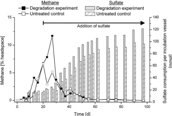
Sulfate reduction and methanogenesis are terminal anaero-bic mineralization processes. The concentrations of sulfate and methane were monitored to assess the current metabolic status of the experiment. Sulfate was completely consumed within the first 9 days (Fig. 2). Between day 9 and day 20, the sulfate concentration remained below the detection limit, un-til sulfate was refilled. Sulfate was replenished to mimic the natural environment. At the end of the experiment, sulfate was consumed more slowly than at the beginning, indicating the depletion of electron donors. The sulfate concentration decreased to 9 mM on day 97. Large amounts of methane were only detected in the absence of sulfate. The slightly decreasing values for sulfate consumption were in the range of the analytical error. The concentration of dissolved sul-fide in the pore water remained relatively low (6 mM) until day 27. The maximum concentration of 38 mM was reached on day 76. Oxygen was never detected in any incubation vessel.
3.2 Degradation of intact polar lipids
Fig. 2.Methane production and cumulative sulfate consumption in the degradation experiment and the untreated control. Sulfate consumption for each time point was calculated by addition of measured day-to-day losses. Methane concentrations are given in percentage of incubation vessel headspace. After day 20 sulfate was refilled when the concentration decreased below 10 mM as indicated by the arrow.
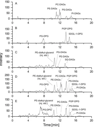
degradation experiment (Fig. 3d). After an incubation pe-riod of 97 day substantial amounts of ester-bound IPLs of S. cerevisiaewere degraded, whereas ether-bound IPLs of H. volcaniiwere still present (Fig. 3e).
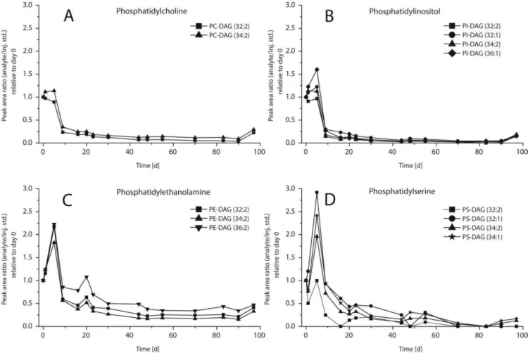
The amounts of ester-bound IPLs decreased in the course of the experiment whereas those of ether-bound IPLs re-mained stable (Figs. 4–6). The head groups had no visible influence on the observed degradation pattern. The amounts of ester-bound IPLs with PC and PI head groups did not show any significant change in the first days of the degrada-tion experiment. Beginning at day 5, they decreased rapidly over 5 days followed by a phase of moderate loss until day 90. In case of phosphatidylethanolamine-diacylglycerol (PE-DAG) and phosphatidylserine-diacylglycerol (PS-DAG) highest amounts were found on day 5 (Fig. 4c and d). Subse-quently, the signal decreased over the rest of the experiment. In contrast to this, the amounts of all ether-bound IPLs (Fig. 5) scattered but did not decrease significantly until the end of the degradation experiment. Ester- and ether-bound IPLs in the abiotic control showed behavior similar to each other with slightly decreasing values in the course of the ex-periment (Fig. 6). The amounts of IPLs in the degradation experiment increased within the first days while the abiotic control did not show this effect. The archaeal ether lipids in the degradation experiment remained elevated by about 50 % relative to the abiotic control throughout the entire experi-ment.
Samples from the untreated control were taken in the same sampling intervals as in the degradation experiment. Most of the IPLs added to monitor the degradation were not detected in the untreated control. Exceptions were the IPLs PE-DAG 34:2 and PE-DAG 36:2, but compared to the corresponding PE-DAGs in the added biomass their total amount was low (less than 3 %). However, other IPLs such as sulfoquinovosyl diacylglycerol (SQ-DAG) and phosphatidylglycerol-diacylglcerol (PG-DAG) were identi-fied in the untreated control. Additionally, PE-DAGs with side chains different from those in the degradation experi-ment were found (31:0, 31:1, 33:1, 33:2, 35:2). SQ-DAG was the most prominent IPL in the untreated control. It showed increasing abundance from day 0 to day 23 and then a de-crease to the starting value at a moderate rate after a ma-jor drop between days 23 and 27. PE-DAG and PG-DAG showed an increase between day 0 and day 5 and returned to the starting values in the course of the experiment.
3.3 IPL degradation rates
Fig. 3. HPLC-ESI-MS base-peak intensity (BPI) chromatograms in negative ion-mode of(A): total extract ofSaccharomyces cere-visiae;(B): total extract ofHaloferax volcanii;(C): total extract of the untreated control at day 0;(D): total extract of the degradation experiment, sample taken at day 0;(E): total extract of the degra-dation experiment, sample taken at day 97. For abbreviations see Fig. 1. Please note that the observed peak intensities cannot be di-rectly transferred into concentrations.
ether-bound IPLs (Fig. 6), the relation for their kinetic degra-dation constants was less linear (Fig. 7).
3.4 Succession in microbial abundance, diversity and activity
The development of the microbial communities was moni-tored to obtain background information for the degradation of IPLs. The total cell counts of the degradation exper-iment and the untreated sedexper-iment showed the same trend and decreased only slightly during the experiment (Fig. 8). The total cell numbers of the degradation experiment were marginally higher than those of the untreated control. The numbers of bacterial 16S rRNA targets were one to two orders of magnitude higher than those of Archaea. Be-tween days 7 and 16, the bacterial 16S rRNA copy num-bers dropped significantly before returning to their previous
values. The number of archaeal 16S rRNA copies showed a generally increasing trend. After day 20, the numbers of both bacterial and archaeal 16S rRNA targets remained rel-atively constant. The RNA content of the sediment ranged from 80 to 4800 ng cm−3. Ravenschlag et al. (2000)
de-termined an rRNA content of 0.9 to 1.4 fg rRNA per cell for two sulfate-reducing bacteria from surface sediments. These values were used to assess the total cell numbers of our study. The calculated values range between 8.9×10⁷ and 3.4×10⁹cells cm−3, which fits nicely to our total cell
counts. The analysis of fermentation products showed no sig-nificant difference between the degradation experiment and the untreated control (Fig. S1). The bacterial community profiles of the degradation experiment and the untreated sed-iment looked similar to each other (Fig. S2). Initially, the community structure was highly diverse. This diversity de-creased in the course of the experiment probably due to a diminishing substrate spectrum as indicated in Fig. S1.
The archaeal community pattern showed minor differ-ences between the degradation experiment and the untreated control (Fig. S3). H. volcaniiwas only detected at the very first sampling point in the degradation experiment. In the beginning of the experiment, when sulfate was still present, no methanogenic archaea were detected (Fig. S3). Only af-ter depletion of sulfate the rRNA of methanogens was found. The presence and activity of these organisms were supported by methane production observed in the absence of sulfate. In all samples, the content of eukaryotic RNA was too low to obtain sufficient PCR products to prepare a DGGE with eukaryotic primers.
3.5 Total organic carbon
The carbon content was analyzed at five time points of the degradation experiment and the untreated control. The differ-ence between the degradation experiment and the untreated control reflected the amount of organic matter that was added to the degradation experiment with the inactive cell material of S. cerevisiaeandH. volcanii. We added 1.36 g of cell-derived organic carbon which increased the TOC content of the natural sediment from 0.23 % Corg(4.9 g) to 0.29 % Corg
(6.2 g). No pronounced difference was visible between the degradation experiment and the untreated control for all other parameters.
3.6 Effects of sediment-derived organic matter on microbial processes
Fig. 4.Relative concentrations of ester-bound IPLs with different head groups in the degradation experiment vs. time in days (normalized to day 0). Core lipid structures are given as sum of fatty acids (e.g. 32:2) where 32 represents the number of carbon atoms and 2 represents the number of double bonds in the core lipid structure. See Fig. 1 for abbreviations.
consumption, methane production and fermentation (Figs. 2 and S1). Roughly 10 % of the cellular dry weight consists of lipids (Stouthamer, 1979) and the IPL content is even lower. Therefore, the absolute amount of IPLs that were added with the biomass ofH. volcaniiandS. cerevisiaeshould have been low compared to other organic compounds present in the sed-iment itself. Accordingly, the fermentation products do not only reflect IPL degradation but mainly degradation of the organic matter inherited from the natural sediment (Fig. S1).
4 Discussion
In this experiment, ester-bound bacteria-like IPLs were de-graded faster than ether-bound archaeal IPLs while an influ-ence of the structure and the bonding type of the head group was not detected.
4.1 Quality assessment of experimental design and data
Haloferax volcaniiandSaccharomyces cerevisiaedo not oc-cur naturally in tidal-flat sediments. Thus, it might be sus-pected that the observed degradation rates of IPLs turn out to be different from those of IPLs in the natural microbial community. However, the source of ether- and ester-bound IPLs should have no influence on the degradation rate since the chemical structures and the bonding types of the added material also occur naturally in IPLs found in Wadden Sea sediments (R¨utters et al., 2001). Nevertheless, it is necessary to use cell material that is not indigenous to the sediment matrix to monitor the degradation of individual IPLs without confusing them with the inherited IPL inventory.
Fig. 5. Relative concentrations of ether-bound IPLs with different head groups in the degradation experiment vs. time in days (nor-malized to day 0). Abbreviations: sulfono diglyco dialkylglycerol (S-GL-1-DPG, according to Sprott et al., 2003), diphytanylglyc-erol (PG-DPG) and diphytanylglycdiphytanylglyc-erol methylphosphate (PGP-Me-DPG).
glycoprotein (Sumper et al., 1990), whereas the cell wall of Saccharomyces cerevisiae consists mainly of glucan poly-mers, chitin and glycoproteins (Levin, 2005). Thus, the cell walls are likely to differ in stability. As archaeal S-layers are exceptionally stable, one might assume that the cells were still largely intact during the experiment and thus, the ether-bound lipids were not readily available for degradation. There are several arguments that this problem did not occur in our experimental set-up:
Although approximately 1×10⁹ H. volcaniicells ml−1
were added, no difference in total cell counts was observed between the degradation experiment and the untreated con-trol at the beginning of the experiment.
The amount of archaeal 16S rRNA of the untreated con-trol was even lower than in the degradation experiment at the beginning of degradation.
The RNA of H. volcanii was only detected at the very first sampling point of the experiment. The rapid degrada-tion of the RNA indicates disintegradegrada-tion of theH. volcanii cells. RNA-based community analyses are often used to de-termine the active part of a community since the RNA con-tent of cells can be correlated with cellular activity (Lee and Kemp, 1994; Wagner, 1994). However, stable isotope prob-ing (SIP) experiments have shown that the RNA is detectable for days even when the microbes are not active (Graue et al., 2011). In our experiment we found RNA of sulfate-reducing bacteria in the absence of sulfate, supporting the finding that RNA is not degraded immediately when these organisms are inactive (Fig. S2).
Most importantly, however, the glycoprotein cell wall of halophilic archaea of the order Halobacteriales requires a high NaCl concentration for stability and cells almost instan-taneously lyse in the absence of salt as described by Mohr and Larson (1963) and Kushner (1964). H. volcanii
addi-Fig. 6.Sum of ester- and ether-bound IPLs in the degradation exper-iment and the abiotic control given as relative concentrations nor-malized to day 0. Closed symbols: degradation experiment; open symbols: abiotic control.
tionally requires high concentrations of magnesium (Cohen et al., 1983). As theH. volcanii cells were pasteurized in distilled water, it is unlikely that the cells remained intact.
Finally, cultivation attempts with pasteurized cells showed no growth which indicates that the H. volcanii cells were killed during the experiment. Thus, according to our line of argument it is very unlikely that the added biomass contained intact cells protecting IPLs from degradation.
If the degradative capacity of the microbial community is exceeded due to the high load of organic matter this would af-fect the degradation rates. However, high organic matter con-centrations in Wadden Sea sediments are not unusual. They are caused e.g. by burial of algal blooms during storm events. In a study of Neira and Rackemann (1996) the degradation of algae biomass was investigated in situ. The amount of intro-duced total organic matter was 300 times higher than in our experiment. Nevertheless, the organic matter was degraded within two month. Even if the conditions of this study are not completely the same, we can assume that the degradative capacity in our study is not a limiting factor.
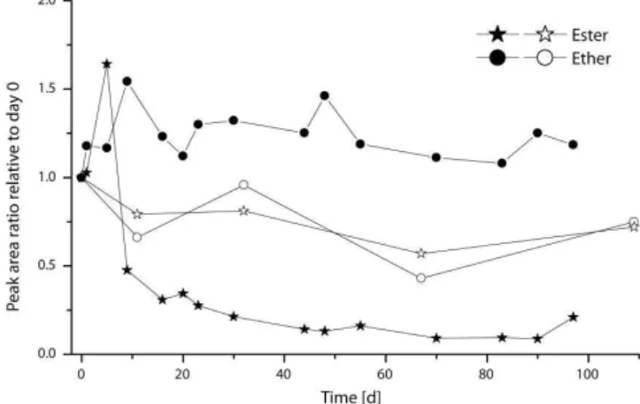
Fig. 7.Log-log plot of calculated kinetic degradation constants of ester-bound and ether-bound IPLs (dashed lines) with time in combination with three incubation experiments from Harvey et al. (1986, dotted lines). Closed symbols indicate aerobic degradation and open symbols anaerobic degradation. The plot was prepared as described by Schouten et al. (2010).
Fig. 8.Bacterial and archaeal 16S rRNA copies and total cell counts (TCC) in the course of the experiment. The number of bacterial and archaeal 16S rRNA targets are given in copies per ng of extracted RNA, whereas the total cell counts are given in cells per cm3 sed-iment. Closed symbols: degradation experiment; open symbols: untreated control.
explanation. We tried to minimize this effect by the design of the incubation vessel and intense mixing prior to every sam-pling. In addition to this, the sediment was resuspended by shaking directly before opening the incubation vessels in the anaerobic chamber. Other reasons for scattering IPL values may be varying extraction efficiencies or changing matrix ef-fects during ESI ionization.
There are several possible explanations for the elevated amounts of archaeal ether lipids in the degradation experi-ment relative to the abiotic control (Fig. 6). The higher val-ues for ether-bound IPLs may be caused by the presentation of data normalizing to day 0. If the sediment despite all cau-tion was not completely homogenized during the first sam-pling, this affects all data points. Furthermore, the sediment of the abiotic control was autoclaved. This thermal treat-ment may have changed the structure of organic matter lead-ing to a stronger adsorption. Another explanation may be that the microbial community in the degradation experiment has a positive effect on the release of adsorbed IPLs lead-ing to a high recovery of those molecules. Nevertheless, all mentioned difficulties do not have any impact on the general results of this study.
4.2 Influence of bonding types and structural moieties on IPL degradation
two structurally different bonding types of head groups were present – one glycosidic (S-GL-1) and two phosphoester (PG-DPG and PGP-Me-DPG; Fig. 1) head groups. However, the same degradation patterns were observed for both bind-ing types. In general, compared to glycosidic bonds, phos-phoester bonds are chemically more labile, since phospho-esters can be hydrolyzed by acids and bases whereas glyco-sidic bonds can usually only be hydrolyzed by acids (Beyer and Walter, 1991). Correspondingly, Harvey et al. (1986) found that glycosidic ether lipids were more stable than phos-pholipids with ester-bound moieties. However, they investi-gated the degradation of a glycosidic ether-bound lipid and a phosphoester ester-bound lipid. Thus, the study of Harvey et al. (1986) cannot be used to answer the question which part of an IPL-molecule (head group or moiety bonding type) is responsible for the observed differential stability of bacterial and archaeal IPLs.
According to our results, we assume that the differences in chemical stability of IPLs play only a minor role during the degradation of IPLs, at least in the investigated system. Since not only the head groups may have an influence on IPL degradation, also the structurally different core lipids have to be considered as possible factor for the observed degradation patterns. In general, complex molecules like lipids and pro-teins are hydrolyzed by exo- or ectoenzymes which are re-leased by prokaryotic cells (Cypionka, 2010). Therefore, we conclude that microbiological enzymatic processes are the driving force in IPL degradation as also suggested by White et al. (1979) and Harvey et al. (1986) rather than expected chemical stabilities alone.
4.3 IPL degradation rates
The kinetic degradation constants of ester-bound IPLs (Fig. 7) in the beginning of our experiment are in the same range as those at the end of aerobic degradation of PE-DAG in beach sediments observed by Harvey et al. (1986). Measured signal intensities in the degradation experiment on day 7 were as high as expected from the amount of added biomass. Therefore, significant degradation did not occur during the first 7 days. This means that the kinetic degradation constants of ester-bound IPLs in our experiment were significantly lower than those determined by Harvey et al. (1986).
In case of ether-bound IPLs the kinetic degradation con-stants determined in our study lie between those calculated for the ether-bound IPL with a glycosidic head group under oxic and anoxic conditions. In all cases this is in accordance to the common knowledge that the turnover of organic matter is aerobically faster than anaerobically.
4.4 Microbial activity
Originally, the experiment was also planned as an enrichment culture for lipid-degrading bacteria. It turned out, however,
that the added cell material had only little influence on the community structure and the metabolic activity. This had a positive side-effect, since the processes which originally oc-cur in tidal flat sediments were not disturbed and superposed. Therefore, the selected set-up is suitable to reflect the natural IPL degradation in such sediments. The increasing total cell counts in the beginning of the experiment were also visible in the untreated control and thus were probably caused by stimulation due to mixing and the higher temperature (4◦C storage temperature and 20◦C during the experiment).
Approximately 10 % of theArchaeain Wadden Sea sed-iments are known to be methanogens (Wilms et al., 2007). They have a narrow substrate spectrum limited to simple molecules and are not known to degrade lipids. Probably, the main lipid degraders areBacteriawhich are more abundant thanArchaeain intertidal surface sediments (e.g. Beck et al., 2011). Anyhow, the physiological roles of most sedimentary archaea are not known (e.g. Teske and Sørensen, 2008) and therefore it cannot be excluded that they are also involved in the degradation of lipids.
Some studies indicate that Archaea use other enzymes thanBacteriato synthesize and degrade IPLs (Choquet et al., 1994; Daiyasu et al., 2005). The IPL degradation experiment was conducted with anoxic surface sediment which is dom-inated by bacteria affiliated toProteobacteria, Actinobacte-ria, Bacteriodetes, Firmicutes, SpirochaetesandChloroflexi, and archaea affiliated to Methanosarcinales, Thermococ-cales, MethanomicrobialesandMethanobacteriales(Wilms et al., 2006). The community structure of the deep biosphere, however, consists mainly of bacteria related toChloroflexi, Gammaproteobacteria and JS1 candidate group and of ar-chaea affiliated to the Miscellaneous Crenarchaeotic Group (MCG), Marine Benthic Group (MBG) and South African Goldmine Euryarchaeotal Group (SAGMEG) (Inagaki et al., 2006; Webster et al., 2006; Teske and Sørensen, 2008). As our results strongly suggest a microbial degradation of IPLs by enzymes, the community structure probably has a ma-jor influence on the degradation pattern. Thus, the observed degradation rates might be different if the experiment is re-peated with sediment from the deep biosphere or in sedi-ments where sulfate-reducing bacteria are not stimulated.
The question whether ester-bound lipids are recycled and used for membrane synthesis by Bacteria cannot be an-swered by our experimental-setup. Enzymes are described forEukaryaandProkaryathat facilitate bidirectional ATP-independent flipping of polar lipids across cytoplasmic mem-branes (Sanyal and Menon, 2009). The incorporation of ex-trinsic cell building blocks is energetically useful for mi-croorganisms because it is more efficient to recycle exist-ing molecules than to break them down to smaller molecules to synthesize them “de-novo”. Takano et al. (2010) added
13C-labeled glucose to marine sediment and found that the 13C was incorporated into the glycerol backbone of archaeal
membrane building blocks. To answer the question if IPLs can be recycled as intact molecules, stable-isotope probing (SIP) or14C-labeling would be suitable tools to follow the degradation and incorporation of IPLs and their resulting products.
4.5 DNA, RNA and intact polar lipids as biomarkers for living cells
DNA and IPLs are commonly used as biomarkers for living cells (e.g. Biddle et al., 2006; Wilms et al., 2006). Addi-tionally, RNA is used to determine the active part of micro-bial communities (e.g. Griffith et al., 2000). However, the successful application of these methods may be hampered by several aspects like varying extraction efficiencies, matrix effect and adsorption/desorption processes. The presence of DNA does neither prove the activity nor even the presence of the corresponding cells (Lorenz and Wackernagel, 1987; Josephson et al., 1993). In contrast, RNA is labile and the RNA content of cells can be correlated with cellular activ-ity (Lee and Kemp, 1994; Wagner, 1994). However, when cells are inactive or have a very slow metabolism, the RNA content might not be sufficient for detection. The stability of ether-bound IPLs in our study suggests that IPLs also do not reflect exclusively the living community. This shows that none of the mentioned tools can be used without caution but a combination of these tools may help to get a more accurate picture.
5 Conclusions
The quantification ofBacteriaandArchaeain the deep ma-rine biosphere by IPL and FISH analysis led to the assump-tion that this habitat is dominated by Archaea (Biddle et al., 2006; Lipp et al., 2008). In contrast, catalyzed reporter deposition fluorescence in situ hybridization (CARD-FISH) and quantitative polymerase chain reaction (q-PCR) suggest a predominance of Bacteria (Schippers et al., 2005; Ina-gaki et al., 2006). Our experiment shows that these inter-pretations have to be considered with caution, since ether-bound archaeal IPLs were clearly more stable than ester-bound bacteria-like IPLs over a period of 100 days. In na-ture, ether-bound IPLs may even be preserved for longer time periods since the enhanced temperature in the laboratory ac-celerated the degradation processes.
To give better insights into the degradation of IPLs in other environments like the deep biosphere, it is desirable to con-duct degradation experiments reflecting the in situ conditions of those habitats. For the deep biosphere this would mean that the experiments must be conducted under high pressure and over a long period since microbial turnover of organic matter is low. Furthermore, the addition of high substrate concentration would lead to a distorted image of ongoing
processes as the in situ available organic matter is recalci-trant.
On the other hand, IPL-based quantification may lead to an overestimation of archaeal cell counts as indicated by our study. Regarding the discussion above, molecular biologi-cal methods based on DNA or RNA can also be inaccurate. Thus, the abundances ofArchaeaandBacteriaobtained with both methods have to be interpreted with caution.
Supplementary material related to this article is available online at:
http://www.biogeosciences.net/8/2547/2011/ bg-8-2547-2011-supplement.pdf.
Acknowledgements. The authors are grateful to B. Kopke, B.
Buch-mann, H. Haase and P. Neumann for their technical assistance. Many thanks to Michael Seidel for sharing his experience in IPL degradation experiments that were conducted prior to this work. We thank K.-U. Hinrichs, A. Schippers and two anonymous referees for their constructive comments and suggestions. This work was financially supported by Deutsche Forschungsgemeinschaft (DFG) within the Research Group on “BioGeoChemistry of Tidal Flats” (FOR 432).
Edited by: K. Suzuki
References
Adams, A. E. M., Johnson, D. I., Longnecker, R. M., Sloat, B. F., and Pringle, J. R.: CDC42 andCDC43, two additional genes involved in budding and establishment of cell polarity in the yeast
Saccharomyces cerevisiae, J. Cell Biol., 111, 131–142, 1990.
Beck, M., Riedel, T., Graue, J., K¨oster, J., Kowalski, N., Wu, C. S., Wegener, G., Lipsewers, Y., Freund, H., B¨ottcher, M. E., Brumsack, H.-J., Cypionka, H., Rullk¨otter, J., and Engelen, B.: Imprint of past and present environmental conditions on micro-biology and biogeochemistry of coastal Quaternary sediments, Biogeosciences, 8, 55–68, doi:10.5194/bg-8-55-2011, 2011. Beyer, H. and Walter, W.: Lehrbuch der Organischen Chemie, 22.
Auflage, S. Hirzel Verlag, Stuttgart, 1–1030, 1991.
Biddle, J. F., Lipp, J. S., Lever, M. A., Lloyd, K. G., Sørensen, K. B., Anderson, R., Fredricks, H. F., Elvert, M., Kelly, T. J., Schrag, D. P., Sogin, M. L., Brenchley, J. E., Teske, A., House, C. H., and Hinrichs, K. U.: Heterotrophic archaea dominate sed-imentary subsurface ecosystems off Peru, P. Natl. Acad. Sci. USA, 103, 3846–3851, 2006.
Boschker, H. T. S., Nold, S. C., Wellsbury, P., Bos, D., de Graaf, W., Pel, R., Parkes, R. J., and Cappenberg, T. E.: Direct linking of microbial populations to specific biogeochemical processes by
13C-labelling of biomarkers, Nature, 392, 801–805, 1998.
Choquet, C. G., Patel, G. B., Beveridge, T. J., and Sprott, G. D.: Sta-bility of pressure extruded liposomes made from archaebacterial ether lipids, Appl. Microbiol. Biot., 42, 375–384, 1994. Cohen, S., Oren, A., and Shilo, M.: The divalent cation requirement
of Dead Sea halobacteria, Arch. Microbiol., 136, 184–190, 1983. Cord-Ruwisch, R.: A quick method for the determination of dis-solved and precipitated sulfides in cultures of sulfate-reducing bacteria, J. Microbiol. Meth., 4, 33–36, 1985.
Cypionka, H.: Grundlagen der Mikrobiologie, Auflage 4, Springer, Heidelberg, 1–337, 2010.
Daiyasu, H., Kuma, K.-I., Yokoi, T., Morii, H., Koga, Y., and Toh, H.: A study of archaeal enzymes involved in polar lipid synthesis linking amino acid sequence information, genomic contexts and lipid composition, Archaea, 1, 399–410, 2005.
Ertefai, T. F., Fisher, M. C., Fredricks, H. F., Lipp, J. S., Pearson, A., Birgel, D., Udert, K. M., Cavanaugh, C. M., Gschwend, P. M., and Hinrichs, K. U.: Vertical distribution of microbial lipids and functional genes in chemically distinct layers of a highly polluted meromictic lake, Org. Geochem., 39, 1572–1588, 2008. Graue, J., Engelen, B., and Cypionka, H.: Degradation of
cyanobac-terial biomass in anoxic tidal-flat sediments: A microcosm study of metabolic processes and community changes, ISME J., doi:10.1038/ismej.2011.120, in press, 2011.
Griffiths, R. I., Whiteley, A. S., O’Donnell, A. G., and Bailey, M. J.: Rapid method for coextraction of DNA and RNA from natural environments for analysis of ribosomal DNA- and rRNA-based microbial community composition, Appl. Environ. Microbiol., 66, 5488–5491, 2000.
Harvey, H. R., Fallon, R. D., and Patton, J. S.: The effect of organic-matter and oxygen on the degradation of bacterial membrane lipids in marine sediments, Geochim. Cosmochim. Ac., 50, 795– 804, 1986.
Inagaki, F., Nunoura, T., Nakagawa, S., Teske, A., Lever, M., Lauer, A., Suzuki, M., Takai, K., Delwiche, M., Colwell, F. S., Nealson, K. H., Horikoshi, K., D’Hondt, S., and Jørgensen, B. B.: Biogeographical distribution and diversity of microbes in methane hydrate-bearing deep marine sediments on the Pa-cific Ocean Margin, P. Natl. Acad. Sci. USA, 103, 2815–2820, doi:10.1073/pnas.0511033103, 2006.
Jaeschke, A., Rooks, C., Trimmer, M., Nicholls, J. C., Hopmans, E. C., Schouten, S., and Damste, J. S. S.: Comparison of ladderane phospholipid and core lipids as indicators for anaerobic ammo-nium oxidation (anammox) in marine sediments, Geochim. Cos-mochim. Ac., 73, 2077–2088, doi:10.1016/j.gca.2009.01.013, 2009.
Josephson, K. L., Gerba, C. P., and Pepper, I. L.: Polymerase chain reaction detection of nonviable bacterial pathogens, Appl. Envi-ron. Microbiol., 59, 3513–3515, 1993.
Kushner, D. J.: Lysis and dissolution of cells and envelopes of an extremely halophilic bacterium, J. Bacteriol., 87, 1147–1156, 1964.
Lee, S. H. and Kemp, P. F.: Single-cell RNA-content of natural marine planktonic bacteria measured by hybridization with mul-tiple 16S ribosomal-RNA-targeted fluorescent-probes, Limnol. Oceanogr., 39, 869–879, 1994.
Levin, D. E.: Cell wall integrity signaling inSaccharomyces cere-visiae, Microbiol. Mol. Biol. R., 69, 262–291, 2005.
Lin, Y. S., Lipp, J. S., Yoshinaga, M. Y., Lin, S. H., Elvert, M., and Hinrichs, K. U.: Intramolecular stable carbon isotopic analysis
of archaeal glycosyl tetraether lipids, Rapid Commun. Mass Sp., 24, 2817–2826, doi:10.1002/rcm.4707, 2010.
Lipp, J. S. and Hinrichs, K. U.: Structural diversity and fate of intact polar lipids in marine sediments, Geochim. Cosmochim. Ac., 73, 6816–6833, 2009.
Lipp, J. S., Morono, Y., Inagaki, F., and Hinrichs, K.-U.: Signifi-cant contribution of Archaea to extant biomass in marine subsur-face sediments, Nature, 454, 991–994, doi:10.1038/nature07174, 2008.
Lorenz, M. G. and Wackernagel, W.: Adsorption of DNA to sand and variable degradation rates of adsorbed DNA, Appl. Environ. Microbiol., 53, 2948–2952, 1987.
Lunau, M., Lemke, A., Walther, K., Martens-Habbena, W., and Si-mon, M.: An improved method for counting bacteria from sed-iments and turbid environments by epifluorescence microscopy, Environ. Microbiol., 7, 961–968, 2005.
Matos, A. R. and Pham-Thi, A. T.: Lipid deacylating enzymes in plants: Old activities, new genes, Plant Physiol. Bioch., 47, 491– 503, doi:10.1016/j.plaphy.2009.02.011, 2009.
Mohr, V. and Larsen, H.: On the structural transformations and ly-sis ofHalobacterium salinariumin hypotonic and isotonic solu-tions, J. Gen. Microbiol., 31, 267–280, 1963.
Mullakhanbhai, M. F. and Larsen, H.:Halobacterium volcaniispec. nov., a Dead Sea halobacterium with a moderate salt require-ment, Arch. Microbiol., 104, 207–214, 1975.
Neira, C. and Rackemann, M.: Black spots produced by buried macroalgae in intertidal sandy sediments of the Wad-den Sea: Effects on the meiobenthos, J. Sea Res., 36, 153–170, doi:10.1016/s1385-1101(96)90786-8, 1996.
Ravenschlag, K., Sahm, K., Knoblauch, C., Jørgensen, B. B., and Amann, R.: Community structure, cellular rRNA con-tent, and activity of sulfate-reducing bacteria in marine arc-tic sediments, Appl. Environ. Microbiol., 66, 3592–3602, doi:10.1128/aem.66.8.3592-3602.2000, 2000.
Rossel, P. E., Lipp, J. S., Fredricks, H. F., Arnds, J., Boetius, A., Elvert, M., and Hinrichs, K. U.: Intact polar lipids of anaerobic methanotrophic archaea and associated bacteria, Org. Geochem., 39, 992–999, doi:10.1016/j.orggeochem.2008.02.021, 2008. R¨utters, H., Sass, H., Cypionka, H., and Rullk¨otter, J.:
Monoalkylether phosphoslipids in the sulfate-reducing bacteria
Desufosarcina variablis and Desulforhabus amnigenus, Arch.
Microbiol., 176, 435–442, 2001.
Sanyal, S. and Menon, A. K.: Flipping Lipids: Why an’ What’s the Reason for?, ACS Chem. Biol., 4, 895–909, doi:10.1021/cb900163d, 2009.
Schippers, A., Neretin, L. N., Kallmeyer, J., Ferdelman, T. G., Cragg, B. A., Parkes, R. J., and Jørgensen, B. B.: Prokaryotic cells of the deep sub-seafloor biosphere identified as living bac-teria, Nature, 433, 861–864, 2005.
Schouten, S., Middelburg, J. J., Hopmans, E. C., and Sinninghe Damst´e, J. S.: Fossilization and degradation of intact polar lipids in deep subsurface sediments: A theoretical approach, Geochim. Cosmochim. Ac., 74, 3806–3814, 2010.
Schubotz, F., Wakeham, S. G., Lipp, J. S., Fredricks, H. F., and Hinrichs, K.-U.: Detection of microbial biomass by intact polar membrane lipid analysis in the water column and surface sed-iments of the Black Sea, Environ. Microbiol., 11, 2720–2734, 2009.
M., and Brisson, J. R.: Novel polar lipids of halophilic eubac-teriumPlanococcusH8 and archaeonHaloferax volcanii, BBA-Mol. Cell Biol. L., 1633, 179–188, 2003.
Stouthamer, A. H.: The search for correlation between theoretical and experimental growth yields, Int. Rev. Biochem, 21, 1–15, 1979.
Sturt, H. F., Summons, R. E., Smith, K., Elvert, M., and Hin-richs, K. U.: Intact polar membrane lipids in prokaryotes and sediments deciphered by high-performance liquid chromatogra-phy/electrospray ionization multistage mass spectrometry – new biomarkers for biogeochemistry and microbial ecology, Rapid Commun. Mass Sp., 18, 617–628, 2004.
Sumper, M., Berg, E., Mengele, R., and Strobel, I.: Primary struc-ture and glycosylation of the S-layer protein ofHaloferax vol-canii, J. Bacteriol., 172, 7111–7118, 1990.
Takano, Y., Chikaraishi, Y., Ogawa, N. O., Nomaki, H., Morono, Y., Inagaki, F., Kitazato, H., Hinrichs, K. U., and Ohkouchi, N.: Sedimentary membrane lipids recycled by deep-sea benthic ar-chaea, Nat. Geosci., 3, 858–861, doi:10.1038/ngeo983, 2010. Teske, A. and Sørensen, K. B.: Uncultured archaea in deep marine
subsurface sediments: have we caught them all?, ISME J., 2, 3– 18, 2008.
Van Mooy, B. A. S., Fredricks, H. F., Pedler, B. E., Dyhrman, S. T., Karl, D. M., Koblizek, M., Lomas, M. W., Mincer, T. J., Moore, L. R., Moutin, T., Rappe, M. S., and Webb, E. A.: Phytoplankton in the ocean use non-phosphorus lipids in response to phosphorus scarcity, Nature, 458, 69–72, 2009.
Wagner, R.: The regulation of ribosomal-RNA synthesis and bacterial-cell growth, Arch. Microbiol., 161, 100–109, 1994. Webster, G., Parkes, R. J., Cragg, B. A., Newberry, C. J.,
Weight-man, A. J., and Fry, J. C.: Prokaryotic community compo-sition and biogeochemical processes in deep subseafloor sedi-ments from the Peru Margin, FEMS Microbiol. Ecol., 58, 65–85, doi:10.1111/j.1574-6941.2006.00147.x, 2006.
White, D. C., Davis, W. M., Nickels, J. S., King, J. D., and Bobbie, R. J.: Determination of the sedimentary microbial biomass by extractable lipid phosphate, Oecologia, 40, 51–62, 1979. Wilms, R., Sass, H., Kopke, B., Koster, J., Cypionka, H., and
En-gelen, B.: Specific Bacterial, Archaeal, and Eukaryotic Com-munities in Tidal-Flat Sediments along a Vertical Profile of Several Meters, Appl. Environ. Microbiol., 72, 2756–2764, doi:10.1128/aem.72.4.2756-2764.2006, 2006.
Wilms, R., Sass, H., K¨opke, B., Cypionka, H., and Engelen, B.: Methane and sulfate profiles within the subsurface of a tidal flat are reflected by the distribution of sulfate-reducing bacteria and methanogenic archaea, FEMS Microbiol. Ecol., 59, 611–621, 2007.