Acta bot. bras. 24(3): 765-779. 2010.
Introduction
Bromeliaceae currently covers 58 genera and 3172 species (Luther 2008). The family is included in the order Poales (APG II 2003) and has a typically Neotropical geographic distribution (Smith & Downs 1974). Its representatives are epiphytic, rupicolous or terricolous herbs, whose fruits are classifi ed as capsules or berries (Benzing 2000).
Although the monophyletic origin of this family has been confi rmed (Ranker et al. 1990; Horres et al. 2000; Barfuss et al. 2005), the origins, phylogenetic relations and circumscriptions of the subfamilies, genera and species are as yet little known, and, above all, there is much diffi culty in delimiting genera and species. Therefore, the infrafamilial relations of Bromeliaceae have been the target of several investigations, but the phylogeny of the group is still being intensely discussed, with signifi cant differences among the authors.
Traditionally, Bromeliaceae is divided into three subfamilies: Pitcairnioideae, Tillandsioideae and Bromelioideae, distinguished especially by the morphology of fl owers, fruits and seeds (Smith & Downs 1974). However, Givnish et al. (2007) proposed a new arrangement for the family based on molecular data, increasing the number of subfamilies to eight (Brocchinioideae, Lindmanioideae, Tillandsioideae, Hechtioideae, Navioideae, Pitcairnioideae, Puyoideae and Bromelioideae). This new classifi cation
became more natural, so that the recognized subfamilies are monophyletic.
As for fruit morphology and anatomy, several studies have shown that they are used to delimit or resolve taxonomic problems among species, genera, tribes and even monotypical families, such as Tobe et al. (1992), Smitt et al. (1995), Doweld (1998), Decraene et al. (2000), Doweld (2001) and Moon & Hong (2006). Moreover, this type of investigation is essential to help understand the true origin of the fruits of angiosperms and to contribute to making their classifi cation more uniform. As the reproductive organ responsible for seed dispersal, through morphoanatomical studies, the fruit helps us to understand how the species are dispersed and these are important data for proposals to conserve biodiversity.
Despite what has been presented about the importance of structural studies in fruit, these are few in comparison to those on other plant organs, and very few angiosperm families have a representative number of publications in this fi eld, as in the case of Fabaceae and Anacardiaceae. In contrast to these, only two anatomical studies of fruit were recorded for Bromeliaceae, in Ananas comosus (Okimoto 1948) and Tillandsia tricholepis (Morra et al. 2002).
Therefore, the purpose of this study is to characterize and compare the morphoanatomy and ontogeny of the fruit of six species of Bromeliaceae – Aechmea calyculata and
1 Part of the Master’s dissertation of the fi rst Author
2 Universidade Federal do Rio Grande do Sul, Instituto de Biociências, Departamento de Botânica, Laboratório de Anatomia Vegetal, Porto Alegre, RS, Brasil
3 Author for correspondence: natiffagundes@yahoo.com.br
Morphoanatomy and ontogeny of fruit in Bromeliaceae species
1Natividad Ferreira Fagundes2,3 and Jorge Ernesto de Araujo Mariath2
Recebido em 26/04/2009. Aceito em 18/06/2010
RESUMO –(Morfoanatomia e ontogenia de fruto em espécies de Bromeliaceae). O presente estudo objetivou apresentar um panorama acerca da estrutura de frutos em Bromeliaceae, devido à extrema escassez de estudos nessa área; revelando a morfologia, anatomia e ontogenia dos frutos de seis espécies pertencentes a seis gêneros distintos, representativos de três subfamílias. Todas as espécies estudadas apresentam ovário tricarpelar e trilocular, com ob-turador na região da placenta. Dyckia maritima, Pitcairnia fl ammea, Tillandsia aeranthos e Vriesea carinata possuem frutos do tipo cápsula (septicida ou biscida), caracterizados pela presença de macroesclereídes no exocarpo ou endocarpo, fi bras na região ventral dos carpelos, seis linhas de deiscência e três feixes vasculares por carpelo. Aechmea calyculata e Billbergia nutans possuem frutos do tipo baga, com exocarpo e hipoderme como camadas mecânicas e muitos feixes vasculares por carpelo. Diversos caracteres úteis para a taxonomia de Bromeliaceae foram destacados, sendo os frutos ótimas ferramentas para a delimitação, principalmente, de gêneros e subfamílias. Em relação às cápsulas de Bromeliaceae, as macroesclereídes são fi rmadas como estruturas marcantes do pericarpo e a anatomia do fruto está intimamente relacionada ao tipo de deiscência. Os espessamentos de parede celular em “U” demonstram um caráter comum na família e passam a constar como estruturas ocorrentes também em frutos de monocotiledôneas.
Palavras-chave: anatomia, Bromeliaceae, desenvolvimento, ovário, pericarpo
ABSTRACT –(Morphoanatomy and ontogeny of fruit in Bromeliaceae species). This study aims to give an overall view of fruit structure in Bromeliaceae, since these studies are extremely scarce in this family, showing the morphology, anatomy and ontogeny of fruits of six species belonging to six different genera, representing three subfamilies. All species studied have a tricarpellary and trilocular ovary, with an obturator covering the placental region. Dyckia maritima, Pitcairnia fl ammea, Tillandsia aeranthos and Vriesea carinata have capsular fruits (septicidal or biscidal), characterized mainly by the presence of macrosclereids in the exocarp or endocarp, fi bers in the ventral region of the carpels, six dehiscence lines and three vascular bundles in each carpel. Aechmea calyculata and Billbergia nutans have fruits classifi ed as berries, presenting exocarp and hypodermis as mechanical layers and many vascular bundles in each carpel. Many useful characters for taxonomy of Bromeliaceae were revealed, so fruits are excellent tools for delimitation of genera and subfamilies. In relation to Bromeliaceae capsules, macrosclereids are conspicuous structures of the pericarp and fruit anatomy is greatly related to dehiscence type. The U-shaped cell wall thickenings are a very common character in this family and can be identifi ed as structures also occurring in monocot fruits.
Billbergia nutans (Bromelioideae), Dyckia maritima and
Pitcairnia fl ammea (Pitcairnioideae), Tillandsia aeranthos
and Vriesea carinata (Tillandsioideae) – corresponding to the three subfamilies that occur in the state of Rio Grande do Sul, presenting an overview of the structure of fruits in Bromeliaceae and correlating the data to the current phylogenies.
Materials and methods
The botanical material was obtained partly from the Bromeliaceae Collection in the Botanical Garden of Porto Alegre, under the following numbers: Aechmea calyculata, BROM 00022, BROM 00252, BROM 00393; Billbergia nutans, BROM 00106, BROM 00189, BROM 00739; Pitcairnia fl ammea, BROM 00140, BROM 00324, BROM 00335. The other species were collected in the counties of Porto Alegre, Viamão, Dom Pedro de Alcântara and Caraá, in Rio Grande do Sul, and the exsiccatae deposited in the ICN Herbarium of the Federal University of Rio Grande do Sul under numbers: Dyckia maritima, ICN 188806; Tillandsia aeranthos, ICN 188807; Vriesea carinata, ICN 144794.
The collection involved flowers and fruits at different stages of development, from three or more individuals. It should be pointed out that the fl owers in pre-anthesis are different from fl oral buds because they have fully developed fl oral verticilles. The material was dissected, and only the ovaries and fruits were preserved and fi xed in glutaraldehyde 1% and formaldehyde 4% in sodium phosphate buffer 0.1M, pH 7.2 (McDowell & Trump 1976). In order to prepare transverse, longitudinal and paradermal histological sections, the samples were passed through a sodium phosphate buffer 0.1M, pH 7.2 (Gabriel 1982), and later dehydrated in an ethylic series until they were included in hydroxyethylmethacrylate (Gerrits & Smid 1983). After this, the sections were made, 4 μm thick, in a Zeiss rotation microtome HM 340 E, and stained with Toluidine Blue O 0.05%, pH 4.4 (O’Brien & McCully 1981). A few histochemical tests were performed, such as Lugol for starch (Johansen 1940), Hydrochloric Acid 10% for calcium oxalate (Chamberlain 1932), Phenol for silica (Johansen 1940) and Ruthenium Red for pectins (Johansen 1940). The analyses and photomicrographs of the histological material were done under bright fi eld optical microscopy, using a Leica DM R microscope.
For scanning electron microscopy (SEM), the material was dehydrated in an ascending acetone series and then submitted to drying by the critical point method (Gersterberger & Leins 1978) using BAL-TEC CPD 030 equipment. After this, the samples were mounted on aluminum stubs and sputter coated with platinum in the BAL-TEC SCD 050 metalizing apparatus. The observations and electromicrographic record were performed in a JEOL 6060 microscope, under 10kV.
The terminology used is according to Roth (1977), for the defi nition of layers of pericarp, Spjut (1994) and Barroso et al. (1999), for the classifi cation of the type of fruit and type of dehiscence.
The following developmental stages were established to describe most of the species: I – undifferentiated ovary of fl oral buds; II – ovary and young fruit from fl owers in pre-anthesis, anthesis and post-anthesis; III – mature fruit and, when appropriate, dry fruit. The description of Vriesea carinata follows the stages: I – undifferentiated ovary of fl oral buds; II – ovary from fl owers in pre-anthesis; III – ovary and young fruit from fl owers in anthesis and post-anthesis; IV- mature, dry fruit. In species in which the ovary wall is fused to non-carpellary tissues, the characterization of the ovary and fruit results from the topographic analysis of the tissues.
Results
1. Bromelioideae species
Stage I – The ovary is tricarpellary and trilocular, with the presence of an obturator in the placental region, continuing the inner epidermis, and septal nectaries (not discussed in this study). These characters are common to the other
species studied. The ovary is inferior with the carpels completely fused, which present ramifi cations of the septal nectary in their lateral regions. The ovary wall is fused to non-carpellary tissues, so that they cannot be distinguished. The epidermises are unistratifi ed with rectangular cells in section, while the mesophyll is parenchymatous, pluristratifi ed and composed predominantly of isodiametric cells (Fig. 1); this can also be seen in the other species analyzed. The epidermises have anticlinally elongated cells (Fig. 1-2). In Aechmea calyculata, the outer epidermis has cells with dense cytoplasmic content and spherical silica bodies, as well as peltate scales distributed irregularly on the epidermal surface (Fig. 2). The mesophyll is composed of 18-22 cell layers, following a gradient with a decreasing cell volume from the central region to the one close to the peripheries (Fig. 1). Idioblasts containing calcium oxalate raphides frequently occur in the mesophyll. They occur in all carpel regions in B. nutans and in the dorsal and lateral regions of the carpels in A. calyculata (Fig. 1). The fusion of adjacent carpels along their entire laterals characterizes syncarpy as total, and the complete fusion of tissues, without distinguishing the limit between the carpels, characterizes it as true (Fig. 3-4). Each carpel presents many vascular bundles (Fig. 3-4) – about 15 in A. calyculata and 25 in B. nutans. In Billbergia nutans, the ovary has an outer surface with ribs, always nine of them (Fig. 4). In the central region of the ovary, there are six distinct lines: three in the septal region, extending only to the nectariferous region, and three ventral sutures of the carpels (Fig. 5). The obturator has elongated cells with dome-shaped outer periclinal faces. Starch grains are present in the ovary wall and in the non-carpellary tissues, a trait common to the other species studied. In this stage, anticlinal cell divisions are observed in the mesophyll, in a longitudinal plane.
Stage II – The ovary, and then the fruit (Fig. 3-4), undergo changes referring to cell enlargement and cell differentiation. In the fruit wall as a whole, there is an accumulation of pectins inside the cells (Fig. 6-8) and cell enlargement in a tangential direction (Fig. 6). The exocarp and adjacent cell layers (4-5 layers in A. calyculata and 2-3 layers in B. nutans) have little thickened cell walls, just as the outer periclinal wall of the endocarp (Fig. 7-8). In the exocarp a differentiation of stomata occurs with equal cell wall thickenings in the periclinal walls of the guard cells, which are elevated compared to the other exocarp cells in A. calyculata, and at the same level of the other epidermal cells in B. nutans. In A. calyculata, fi bers are differentiated around the vascular bundles, forming a bundle sheath (Fig. 9). The obturator cells differentiate forming labyrinths of wall ingrowths (Fig. 10).
Acta bot. bras. 24(3): 765-779. 2010.
767
Figures 1-10. Ovary and developing fruit of Bromelioideae, mostly in transverse section. 1. Ovary wall with idioblasts in the mesophyll (arrow) of A. calyculata. 2. Outer epidermis with peltate scales (longitudinal section) of A. calyculata. 3-4. General view of developing fruit of A. calyculata (asterisk: ramifi cation of the septal nectary) and B. nutans (arrows: ribs), respectively. 5. Six different rows of cells in the central region of the fruit of A. calyculata, with three ventral sutures (arrow) and three lines in the septal region (broad arrow). 6. Fruit wall of B. nutans. 7. Exocarp and hypodermal cell layers of B. nutans, showing cell wall thickening (arrow) and the pectic content of the cells (broad arrow). 8. Endocarp of B. nutans, with pectic cell content (arrow). 9. Vascular bundle wrapped in metabolically active bundle sheath fi bers (arrow: nucleus) of A. calyculata. 10. Obturator in the placental region of A. calyculata, with detail (arrow: cell wall labyrinth). en: endocarp, ex: exocarp, ie: inner epidermis, mc: mesocarp, me: mesophyll, ob: obturator, oe: outer epidermis, pl: placenta, ps: peltate scale, sf: bundle sheath fi bers, vb: vascular bundle. Bars= 50 μm (Fig. 1-2, 5-10); 200 μm (Fig. 3-4).
In A. calyculata, the fruit is formed also by the calyx (Fig. 11-13) and is identifi ed as an ellipsoid armed berry, hairy, about 0.9 cm long and dark-purple color, with persistent bract, corolla, gynoecium and androecium. The sepals are fused over about 1/3 of their length, and the apexes of the free portions form caudate, rigid projections.
The exocarp is represented by sclereids with U-shaped cell wall thickenings, with thickened anticlinal and inner periclinal walls; on the other hand, the sclereids of the
walls of the inner epidermis become thicker only in this region. In the sepal cohesion zone, there is a colenchymatous tissue with pectic cell content, close to the inner epidermis (Fig. 13). Throughout the fruit, the peltate scales are formed by an asymmetrical shield, non-organized into distinct disc and wing, with rather elongated extremities (Fig. 19-20), by 3-4 stalk cells and two basal cells, with the nearer cells from the exocarp and hypodermis disposed radially, forming a projection. Compounds are deposited inside the exocarp, hypodermis and endocarp cells, and they make the fruit dark-purple, almost black (Fig. 16-18).
In B. nutans, the fruit (Fig. 21) is an ovaloid, greenish berry, about 1.5 cm long, with a persistent calyx, corolla, androecium and gynoecium. Bundle sheath fibers are differentiated in the mesocarp (Fig. 22). Both the exocarp and the endocarp have longitudinally elongated cells with straight cell walls. The endocarp has an irregular pattern, in cell shape and in cell distribution, which follows only a tendency to longitudinal disposition (Fig. 23-26).
In both species, the mesocarp maintains the same number of cell layers and the same pattern of cell volume gradient and the 2-3 cell layers adjacent to the exocarp acquire thicker walls (Fig. 16, 23), which constitute a hypodermis.
The sclerenchymatous tissues are metabolically active due to the presence of cellular organelles, including the nucleus, inside the cells (Fig. 9). The stomata are located in exocarp depressions with the guard cells superposed on the under-arching subsidiary cells, and distributed longitudinally in relation to the fruit axis. The number of starch grains is reduced during development, until they disappear completely at fruit maturity. The ontogenesis of the fruit occurs from the apex towards the base and results in their growth in length and diameter, and this is mainly a consequence of the increased fruit perimeter, and not so much of pericarp thickness. The characteristics mentioned can also be seen in the other species studied.
2. Pitcairnioideae and Tillandsioideae species
Stage I – The ovary is superior, except for Pitcairnia fl ammea, which has a semi-inferior ovary, with the inferior region fused to non-carpellary tissues. Tissue fusion between the carpels is not complete, since it is possible to distinguish the united epidermises in the lateral region of the carpels, which characterizes syncarpy of the species as being false (Fig. 27-28). In Dyckia maritima and Pitcairnia fl ammea, the carpels are fused to each other only in the ventral regions (Fig. 27), while in Tillandsia aeranthos and Vriesea carinata, the carpels are fused over the length of their lateral regions (Fig. 28). In this way, syncarpy is characterized as partial in
D. maritima and P. fl ammea, because it is restricted to the ventral regions, and total in T. aeranthos and V. carinata. The ovary wall consists of two epidermises with elongated cells with dense content, the outer epidermis is in the anticlinal direction and the inner one in the periclinal
direction, in transverse section (Fig. 29). In P. fl ammea, the cell content is denser in the inner epidermis. In T. aeranthos, the cell content is quite dense compared to the other species. The mesophyll is composed of 6-8 cell layers in D. maritima
and T. aeranthos (Fig. 29), of 8-10 cell layers in P. fl ammea
and of 12-14 cell layers in V carinata. Six dehiscence lines are clearly seen, each formed by two rows of cells that are minuscule compared to the other carpellary tissues. Of these, three are located in the lateral region of the carpels (septa) and are characterized by the continuation of the outer epidermises of adjacent carpels (Fig. 30-31) and the other three are located in the ventral region of the carpels (placentation zone) constituting the ventral sutures (Fig. 30). In the mesophyll there are idioblasts containing calcium oxalate raphides, except for V. carinata. Their location varies in the carpel according to species – occurring in the lateral and ventral regions in D. maritima, in the lateral and dorsal regions in P. fl ammea and in the ventral region in T. aeranthos. Three vascular bundles are identifi ed per carpel, a main one in the dorsal region and two of a smaller caliber in the ventral region (Fig. 29-30). Cell divisions occur in the mesophyll of D. maritima, P. fl ammea and T. aeranthos, mostly in the anticlinal direction. The obturator is formed by elongated cells with dome-shaped outer periclinal faces in D. maritima and P. fl ammea. On the other hand, in T. aeranthos, the obturator cells undergo periclinal divisions, becoming very elongated and forming one to two strata (Fig. 32), while in V. carinata the obturator is represented by elongated cells.
Stage II – This stage only occurs in V. carinata. Cell divisions are found in the mesophyll, mainly in the anticlinal direction, and there is a clear, more intense proliferation in the inner region of this tissue (Fig. 33).
Acta bot. bras. 24(3): 765-779. 2010.
769
obturator cells divide, becoming rather elongated and forming uni- to bistratifi ed tissue. The outer surface of the fruit presents a smooth cuticular pattern except for P. fl ammea, which has a striate pattern (Fig. 42-43).
Stages IV (V. carinata) and III (other species) – The pericarp reaches maturity by differentiation of the sclerenchymatous tissues (Fig. 44-46, 51-53, 58-60, 66-67).
In D. maritima, the mature fruit (Fig. 44) has an ovaloid shape, about 0.5 cm long, brown color and with a persistent calyx and corolla. In this stage, the mucilage deposited in the cells close to the stomata disappears. In the dorsal vascular bundle, close to the phloem, and between the fi bers, some radially aligned cells remain parenchymatous, forming a dehiscence zone (Fig. 45). The exocarp cells acquire very thick walls and their shape is radially elongated in the transversal direction, and are characterized as macrosclereids (Fig. 45, 47-48). This tissue is unistratifi ed over most of its length, and is pluristratifi ed in restricted regions. The endocarp is constituted by sclereids with not very thick cell walls, which are tangentially elongated in the transverse direction (Fig. 45, 49-50).
In P. fl ammea, the fruit (Fig. 51) is characterized by its elongated, conical shape, length of about 0.9 cm and dark-green color, with persistent calyx, corolla, gynoecium and androecium. The exocarp cells are characterized by the softly elongated shape in the longitudinal direction, with a subtle deposition of cell wall on their outer periclinal faces (Fig. 52, 54-55). As in some mesocarp cells, the exocarp has a pectic cell content (Fig. 54-55). The endocarp cells acquire very thick and conspicuous U-shaped cell walls, and they have a radially and tangentially elongated shape in the transverse direction (Fig. 52, 56-57), being classifi ed as macrosclereids.
In T. aeranthos and V. carinata, the mature fruit (Fig. 58, 66) is elongated, dark-green, with persistent bract and calyx. In the former species, the fruit has a cylindrical shape and is about 2 cm long, while in the latter the fruit is obovoid and about 3 cm long. Both the exocarp and endocarp cells undergo deposition of U-shaped cell wall (Fig. 59, 61, 63, 67-68, 70). The conspicuous endocarp cells are characterized as macrosclereids, which show the lamellation of their walls as a refl ex of deposition (Fig. 65). As to shape, the exocarp sclereids are longitudinally elongated (Fig. 61-62, 68-69) while those of the endocarp are radially and tangentially elongated in the transverse direction (Fig. 63-64, 70-71).
The mesocarp is homogeneous in D. maritima and
T. aeranthos (Fig. 45, 59), and it is divided into two parenchymatous regions in P. fl ammea, the outer one with larger cells and the inner one with smaller cells (Fig. 52), and following a gradient of cell volume in V. carinata, which diminishes from the center towards the exocarp and endocarp (Fig. 67). There are 8-10 mesocarp cell layers in
D. maritima and T. aeranthos, 9-11 in P. fl ammea and 14-16 in V. carinata. In the mesocarp, a subtle thickening occurs in the outer periclinal walls of the cell layer adjacent to the exocarp, characterized as hypodermis (Fig. 45, 61, 68),
except for P. fl ammea. Around the dorsal vascular bundle, fi bers which form the bundle sheath are differentiated. These fi bers also form bundle sheath extensions in D. maritima
(Fig. 45) and a bundle sheath extension towards the endocarp in P. fl ammea (Fig. 53). In the ventral region of the carpels, a great amount of fi bers appear among the ventral vascular bundles and the dehiscence lines (Fig. 46). In T. aeranthos, these fi bers are inconspicuous due to their scarcity and to the thin layer of cell wall deposited (Fig. 60). In this stage, the quantity of idioblasts containing raphides in the mesocarp diminishes in D. maritima or even disappears in P. fl ammea
and T. aeranthos. Furthermore, a cell compression is found in the outer mesocarp, comprising around 1-2 cell layers in T. aeranthos (Fig. 59) and three layers in V. carinata (Fig. 67).
The dry fruit basically maintains the same structure as the fruit when it was fl eshy, and only a marked compression of some cell layers of the mesocarp is outstanding. After dehydration, dehiscence of the fruit takes place from the apex to the base, as a result of the concomitant rupture of the six dehiscence lines in the central region of the fruit, and besides this, of the dorsal vascular bundle in its middle region in D. maritima, where the parenchymatous cells are located in a radial row. In D. maritima, the capsule opens into six erect valves, each one corresponding to half a carpel, and the septal openings occur until the base and the openings of the locules occur over 2/3 of the fruit length. Therefore, the capsule is classifi ed as biscidal – because of the combination of septicidal dehiscence with loculicidal. In P. fl ammea, the capsule is of the septicidal type, with openings occurring over 2/3 of its length, showing three erect valves originating in each of the carpels, so that the valves remain joined at their apexes due to the persistent style. In T. aeranthos and
V. carinata, the capsule opens until its base in three valves which are completely folded back, and it is classifi ed as septicidal.
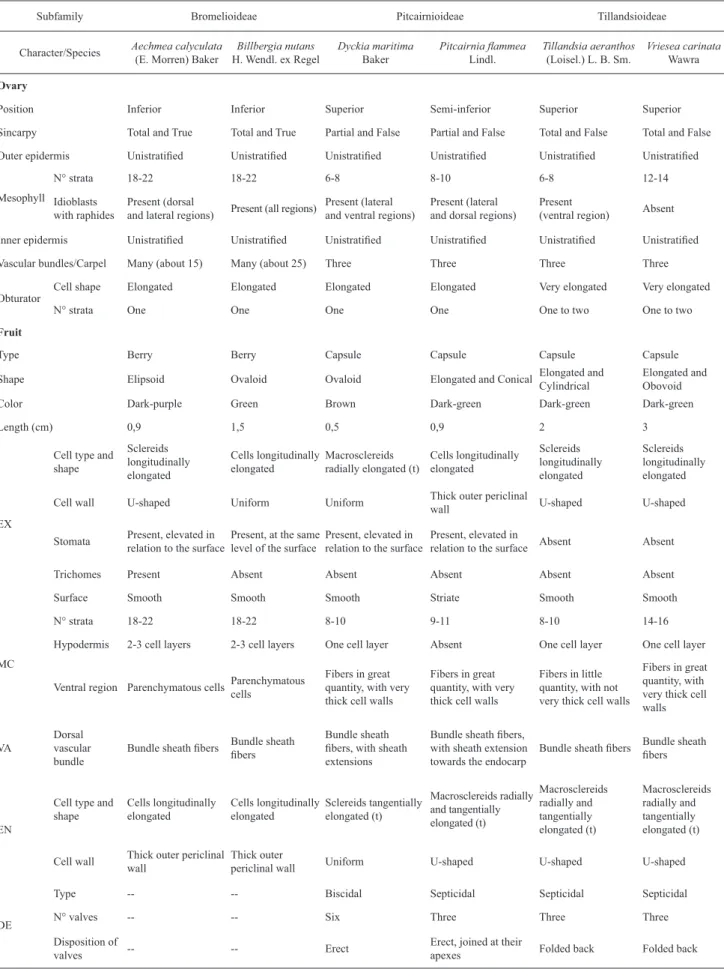
The morphological and anatomical characters referring to the ovary and fruit of all species analyzed are compiled in Tab. 1, in order to summarize and compare the data presented in this study.
Discussion
Many investigators have emphasized that pericarp growth already begins in the fl oral bud, mainly by cell multiplication, and it stops gradually until anthesis (Nitsch 1953). In the ontogenesis of the infrutescence of Ananas comosus, cell division is apparently complete before the fl ower opens, so that the increase in tissue volume is derived from cell enlargement (Okimoto 1948), which also occurs in all species analyzed in this study.
Acta bot. bras. 24(3): 765-779. 2010.
771
Figures 44-57. Mature fruit of Pitcairnioideae, mostly in transverse section. 44-50. D. maritima. 44. General view of mature fruit. 45. Fruit wall, showing thickening of hypodermis cell wall (arrow) and dehiscence zone (broad arrow). 46. Dehiscence lines wrapped in fi bers in the central region of the fruit. 47. Exocarp. 48. Exocarp, in paradermal section. 49. Endocarp. 50. Endocarp, in paradermal section. 51-57. P. fl ammea. 51. General view of the mature fruit. 52. Fruit wall. 53. Dorsal vascular bundle. 54. Exocarp. 55. Exocarp, in paradermal section. 56. Endocarp. 57. Endocarp, in paradermal section. db: dorsal vascular bundle, dl: dehiscence line, en: endocarp, ex: exocarp, fb: fi bers, hy: hypodermis, mc: mesocarp, se: bundle sheath extension, sf: bundle sheath fi bers. Bars= 50 μm (Fig. 45-50, 52-57); 400 μm (Fig. 44, 51).
fusion is superfi cial or not, distinguishing the tissue limit between carpels. In this study, the terms “false” and “true” have a morphological connotation of tissue distinction between carpels, and should not be confounded with the functional signifi cance of the syncarpy studied by Carr & Carr (1961), which distinguishes the “pseudosyncarpic”
gynoecium from the “eusyncarpic” one as regards the fertilization process. The study performed by Sajo et al.
Acta bot. bras. 24(3): 765-779. 2010.
773
Figures 58-71. Mature fruit of Tillandsioideae, mostly in transverse section. 58-65. T. aeranthos. 58. General view of mature fruit. 59. Fruit wall, showing the compressed cell layers (arrow). 60. Fibers. 61. Exocarp and hypodermis with subtle cell wall thickening (arrow). 62. Exocarp, in paradermal section. 63. Endocarp. 64. Endocarp, in paradermal section. 65. Cell wall lamellation of the endocarp, in SEM. 66-71. V. carinata. 66. General view of mature fruit. 67. Fruit wall, with compressed cell layers (arrow). 68. Exocarp and hypodermis, the latter with unequal cell wall thickening (arrow). 69. Exocarp, in paradermal section. 70. Endocarp. 71. Endocarp, in paradermal section. db: dorsal vascular bundle, en: endocarp, ex: exocarp, hy: hypodermis, mc: mesocarp, sf: bundle sheath fi bers. Bars= 5 μm (Fig. 65); 50 μm (Fig. 59-64, 67-71); 400 μm (Fig. 58, 66).
Pitcairnioideae and Puyoideae a partially apocarpic one, just as had been identifi ed for Puya spathacea (Puyoideae), as partially syncarpic, by Kulkarni & Pai (1982).
In this study, the species presented different degrees of carpel fusion, typical of each subfamily, which agrees with the data of Sajo et al. (2004a), but adds important information
Table 1. Comparison of morphoanatomical characters involving the ovary and mature fruit among the six species studied. DE: dehiscence, EN: endocarp, EX: exocarp, MC: mesocarp, VA: vascularization, (t): in the transverse direction.
Subfamily Bromelioideae Pitcairnioideae Tillandsioideae
Character/Species Aechmea calyculata
(E. Morren) Baker
Billbergia nutans
H. Wendl. ex Regel
Dyckia maritima
Baker
Pitcairnia fl ammea
Lindl.
Tillandsia aeranthos
(Loisel.) L. B. Sm.
Vriesea carinata
Wawra
Ovary
Position Inferior Inferior Superior Semi-inferior Superior Superior
Sincarpy Total and True Total and True Partial and False Partial and False Total and False Total and False
Outer epidermis Unistratifi ed Unistratifi ed Unistratifi ed Unistratifi ed Unistratifi ed Unistratifi ed
Mesophyll
N° strata 18-22 18-22 6-8 8-10 6-8 12-14
Idioblasts with raphides
Present (dorsal
and lateral regions) Present (all regions)
Present (lateral and ventral regions)
Present (lateral and dorsal regions)
Present
(ventral region) Absent
Inner epidermis Unistratifi ed Unistratifi ed Unistratifi ed Unistratifi ed Unistratifi ed Unistratifi ed
Vascular bundles/Carpel Many (about 15) Many (about 25) Three Three Three Three
Obturator
Cell shape Elongated Elongated Elongated Elongated Very elongated Very elongated
N° strata One One One One One to two One to two
Fruit
Type Berry Berry Capsule Capsule Capsule Capsule
Shape Elipsoid Ovaloid Ovaloid Elongated and Conical Elongated and
Cylindrical
Elongated and Obovoid
Color Dark-purple Green Brown Dark-green Dark-green Dark-green
Length (cm) 0,9 1,5 0,5 0,9 2 3
EX
Cell type and shape Sclereids longitudinally elongated Cells longitudinally elongated Macrosclereids radially elongated (t)
Cells longitudinally elongated Sclereids longitudinally elongated Sclereids longitudinally elongated
Cell wall U-shaped Uniform Uniform Thick outer periclinal
wall U-shaped U-shaped
Stomata Present, elevated in
relation to the surface
Present, at the same level of the surface
Present, elevated in relation to the surface
Present, elevated in
relation to the surface Absent Absent
Trichomes Present Absent Absent Absent Absent Absent
Surface Smooth Smooth Smooth Striate Smooth Smooth
MC
N° strata 18-22 18-22 8-10 9-11 8-10 14-16
Hypodermis 2-3 cell layers 2-3 cell layers One cell layer Absent One cell layer One cell layer
Ventral region Parenchymatous cellsParenchymatous
cells
Fibers in great quantity, with very thick cell walls
Fibers in great quantity, with very thick cell walls
Fibers in little quantity, with not very thick cell walls
Fibers in great quantity, with very thick cell walls
VA
Dorsal vascular bundle
Bundle sheath fi bers Bundle sheath
fi bers
Bundle sheath fi bers, with sheath extensions
Bundle sheath fi bers, with sheath extension towards the endocarp
Bundle sheath fi bers Bundle sheath
fi bers
EN
Cell type and shape Cells longitudinally elongated Cells longitudinally elongated Sclereids tangentially elongated (t) Macrosclereids radially and tangentially elongated (t) Macrosclereids radially and tangentially elongated (t) Macrosclereids radially and tangentially elongated (t)
Cell wall Thick outer periclinal
wall
Thick outer
periclinal wall Uniform U-shaped U-shaped U-shaped
DE
Type -- -- Biscidal Septicidal Septicidal Septicidal
N° valves -- -- Six Three Three Three
Disposition of
valves -- -- Erect
Erect, joined at their
Acta bot. bras. 24(3): 765-779. 2010.
775
verifi cation may help understand the evolutionary process undergone by the reproductive structures.
The obturator is a type of transmitting tissue which is differentiated in the carpel margins and is closely associated with the micropyle of the ovules (Tilton & Horner 1980). Sajo et al. (2004b) cited the presence of this tissue in the placental region, but did not describe it. In this study, the morphology of the obturator was verifi ed in the family, and in Bromelioideae and Pitcairnioideae the cells are not very elongated, with a dome-shaped, outer periclinal face, and in Tillandsioideae, the cells are very elongated, forming uni- or bistratifi ed tissue. In the six species studied, the development of cell wall labyrinth was observed in the outer periclinal face of the cells of this tissue. These descriptions and comparisons are completely new for Bromeliaceae, showing the common occurrence of this tissue in its different taxa, and the existence of morphological variability of the character.
The defi nition of pericarp layers varies according to the author’s interpretation. Roth (1977) says that the delimitation of the exocarp, mesocarp and endocarp can be lato or stricto sensu. In lato sensu, exocarp and endocarp can be composed by their respective epidermises and subepidermal tissues, while in stricto sensu, both layers originate only from their epidermises. In this study a stricto sensu defi nition was adopted, since it represents the separation of the tissues from the point of view of their origin.
In this study the fruit of Aechmea calyculata and
Billbergia nutans was classifi ed as a berry, which consists of an indehiscent fruit with a pericarp that is not differentiated internally by a rigid endocarp, according to the concept of Spjut (1994). Some authors classify the fl eshy fruit originating from an inferior ovary (sometimes superior, with hypanthium) as pomaceous fruit, which are divided into specifi c subtypes of families or genera (Hertel 1959; Souza 2006). Considering that the fruits of the species analyzed here, as well as all Bromelioideae, originate in the inferior ovary, and that, in the category of the pomaceous fruits, they cannot be classifi ed within their subtypes, it would be plausible to create a subtype that is representative of these fruits. For this, however, it would be necessary to perform the anatomical study of a wide number of species which would represent that particular subfamily more completely. Therefore, this study follows the traditional classifi cation for Bromelioideae fruits, considered berries.
The dry fruit of the remaining species was determined as being of the capsule type, i.e., a simple fruit, dry or fleshy, from a superior or inferior ovary, bicarpellary to pluricarpellary, pluri-ovulate and dehiscent through longitudinal slits (Souza 2006). The capsules are classifi ed based on their dehiscence pattern, which is defi ned as the exposure or release of seeds through a naturally produced opening in the pericarp (Spjut 1994).
The U-shaped cell wall thickenings, a marked characteristic in the leaf structure of Bromeliaceae (Braga 1977; Aoyama & Sajo 2003; Proença & Sajo 2004; Proença
& Sajo 2007), result from the thickening of the inner periclinal wall and of both the anticlinal walls, showing conspicuous stratification, and they have already been recorded in cells of the epicarp or endocarp of species of Asteraceae, Caryophyllaceae, Lamiaceae, Ranunculaceae, Boraginaceae, Valerianaceae and Plumbaginaceae, all eudicotyledons (Roth 1977). This pattern of thickening was detected in the exocarp and/or endocarp of four of the species examined in this study, showing that it is not exclusive to eudicotyledon fruits.
A well developed hypodermis, adjacent to the exocarp, is characteristic of many fruits, including most berries already studied (Roth 1977), and it can be observed in berries of
Aechmea calyculata and Billbergia nutans, which presented 2-3 cell layers in their hypodermis. Dyckia maritima,
Tillandsia aeranthos and Vriesea carinata also presented a hypodermis in their fruits, but it was composed only of a single inconspicuous cell layer, and the cell wall thickenings were limited to the outer periclinal walls.
Different forms of sclereids occur most commonly in the fl eshy tissue of berries and drupes, while fi bers prevail in the endocarp of drupes, in nuts and in capsules, and play an important role in the dehiscence mechanism of the latter (Roth 1977). This pattern was also identifi ed in the fruits of Bromeliaceae, with the presence of sclereids in the mesocarp of berries and fi bers in the mesocarp of capsules.
In the species with capsular fruits, besides the fi bers in the mesocarp, sclereids occur in the endocarp, and sometimes in the exocarp. These sclereids, characterized as such by their shape, differential thickening of the cell wall and type of pit, are of different sizes and shapes; some of them are not very elongated, and others are very elongated, similar to fi bers in a paradermal section, as in the endocarp of Pitcairnia fl ammea. In this case, the macrosclereids can be considered fi briform.
The stomata are characterized, in the vegetative organs, by guard cells that are usually located below the epidermal surface in the genera Aechmea and Billbergia and elevated in the genus Dyckia (Tomlinson 1969). Other evaluations confi rmed the patterns of Billbergia and Dyckia (Proença & Sajo 2007), but differed concerning the genus Aechmea, revealing stomata located at the same level (Aoyama & Sajo 2003) or at a level lower than that of the epidermal cells (Proença & Sajo 2004). In this study, it was found that stomata are elevated in relation to the surface in D. maritima, corroborating the data from the literature for the genus; and elevated for A. calyculata, and, at the same level, for B. nutans, which goes against the existing data for the genera. In all species, the stomata are located in individual furrows, as described by Proença & Sajo (2007) for some species, and they have guard cells above the under-arching subsidiary cells, as found by Tomlinson (1969). The orientation of the stomata in the leaves of monocotyledons is often parallel to the longer axis of the organ (Croxdale 1998), a pattern which coincides with the fruits of all species analyzed here.
to those present in the leaves may occur in infl orescence axes and in reproductive organs. In relation to structure, in Bromelioideae, these trichomes normally have two basal cells and a shield never organized into distinct disc and wing, and in the genus Aechmea there are 3-4 or more stalk cells. According to Braga (1977), the peltate scales of Aechmea
have a rounded shield, regular or irregular. In this study, the fruit of A. calyculata presented peltate scales that agreed with the descriptions above, except for their asymmetrical and extremely irregular shield – very similar to the starred scales common to the genera Cottendorfi a and Navia (Robinson 1969; Tomlinson 1969). In this sense, Strehl (1983) remarks that there is very great variation of scale morphology in Bromelioideae, and there are forms similar to those of the Pitcairnioideae and Tillandsioideae.
According to Benzing (2000), besides the known functions of water and nutrient absorption from the atmosphere, refl ection of solar radiation and reduction of transpiration, the peltate trichomes can also attract dispersers, due to the formation of dense indumenta which refl ect the dim light from the fl eshy fruits. Roth (1977) ascribes to trichomes in fruits the roles of mechanical protection and reducing transpiration, when lost at fruit maturity, or of facilitating dispersal by wind or by animals, when they persist in the mature fruit. Considering the permanence of the peltate scales until the complete development of the fruit and the dispersal mechanism of subfamily Bromelioideae – which includes Aechmea calyculata – determined as zoochory, possibly the trichomes of this species are related to the attraction of dispersers. For Aechmea, there are no records of chiropterochory, which reduces the probability of the refl ection of dim light acting as an attractant to the disperser. In this way, if the peltate scales are morphological adaptations to the disperser animal, the mechanism by which attraction truly occurs is not known.
Secondary sculpture of the fruit surface, which refers to the micro-ornamentation of the cuticle, is striate in
Pitcairnia fl ammea and smooth in the rest of the species. In a taxonomic context, the cuticular sculptures can constitute excellent diagnostic characters, but their signifi cance for delimitation of categories above the species level is very limited. As an adaptation to the environment, it is assumed that the surface area augmented by the sculpture increases the energy exchange with the colder surrounding air, controlling the temperature of the surface under insolation (Barthlott 1981). Considering that P. fl ammea is a heliophilous species (Reitz 1983) and that the respective genus does not have the CAM character (Crayn et al. 2004), typical of many bromeliads from arid environments, it is possible that the secondary sculpture observed helps to control temperature.
According to Fahn & Zohary (1955), two factors are necessary for dehiscence: the presence of crossed sclerenchymatous tissues and/or crossed cellulose micelles; the presence of a separation tissue. The morphology of all capsules studied confi rmed the presence of these structures
through the occurrence of the dehiscence lines and fi bers and sclereids at different orientations.
In loculicidal capsules, the mechanical layers responsible for fruit opening generally reside in the region of the endocarp, the mesocarp, or in both, as shown in representatives of Melianthaceae (Doweld 2001), Pedaliaceae (Day 2000), Meliaceae (Pinto et al. 2003), Bignoniaceae (Souza et al.
2005) and Malvaceae (Souza 2006). In Dyckia maritima, on the contrary, the main mechanical tissues are located in the exocarp and in the mesocarp. This differentiated disposition of the tissues can be the result of the combined occurrence of loculicidal dehiscence with the septicidal.
In this study, the term septicidal was used according to authors Spjut (1994) and Barroso et al. (1999), which includes openings in two distinct regions (septa and ventral sutures), since it was considered more appropriate to adopt the concept of dehiscence from the ecological point of view, which presupposes the opening of the locule followed by exposure of the seeds. However, from this point of view, the term septicidal, in its etymology (septi = septa; cidal = opening), does not refl ect its real concept completely.
As for the structure of the septicidal capsules, in
Prestonia coalita (Apocynaceae) Gomes (2008) observed the presence of the sclerenchymatous exocarp with inverted U-shaped cell walls, mesocarp with a ring of longitudinal fibers and endocarp with groups of longitudinal fibers adjacent to the dehiscence zone. This structural pattern shares similarities with the septicidal capsules of Bromeliaceae described in this study, which have an exocarp and endocarp, or only the endocarp, with U-shaped cell wall thickening and longitudinal fi bers close to the dehiscence lines.
Morra et al. (2002) found that the septicidal capsule of
Tillandsia tricholepis presents an exocarp with thin cell walls, mesocarp with 2-3 cell layers and endocarp with cells similar to fi brosclereids, with thickened and lignifi ed anticlinal walls and inner periclinal wall. In this study, the mature pericarp of Tillandsia aeranthos was characterized as having an exocarp with unequally thickened cell walls, in a U-shape, a mesocarp with 8-10 cell layers and endocarp constituted of macrosclereids, also with U-shaped cell walls. In this way, there is a clear interspecifi c variation of characters relative to the mature fruit, and this type of comparison and fi nding have never occurred before for the family.
Acta bot. bras. 24(3): 765-779. 2010.
777
carinata (Tillandsioideae) are septicidal, the capsule of
Pitcairnia fl ammea (Pitcairnioideae) is septicidal, but with a differentiated shape and opening from the previous ones, and that of Dyckia maritima (Pitcairnioideae) is biscidal. These data corroborate the evolutionary considerations mentioned previously, since Tillandsioideae presents traits considered basal, Pitcairnioideae shows new traits, such as the loculicidal dehiscence, besides the septicidal one, and Bromelioideae presents the fruit structure as a berry, derived and differentiated from the previous ones (capsules).
As well as Tillandsioideae and Pitcairnioideae, all the other subfamilies that were not analyzed in this study present fruit of the capsule type, which probably is a plesiomorphy of the family, taking into account a parsimonious view of the types of fruits, in the light of the phylogeny of Givnish
et al. (2007), which positions Bromelioideae (with baccate fruits) as the latest-divergent group.
Considering the issues presented, this study shows that the probable evolution of the fruits from dehiscent capsules to indehiscent berries in Bromeliaceae occurred through the reduction of sclerenchymatous strata. Of the three subfamilies analyzed, Tillandsioideae presented capsules with conspicuous sclerenchymatous strata in the exocarp, endocarp and mesocarp, Pitcairnioideae presented capsules with these strata in the exocarp or endocarp and in the mesocarp, and fi nally, Bromelioideae presented berries with rigid strata in the exocarp and hypodermis. Furthermore, the two species of Bromelioideae showed the existence of cell rows similar to the dehiscence lines of the other species, which certainly constitute relicts of the dehiscence apparatus. This tendency towards the reduction of the sclerenchymatous strata, leading to fruit indehiscence, was also shown in Fabaceae in the study by Fahn & Zohary (1955).
Based on the morphological and anatomical characters of this study, the species that differed most among themselves within the same subfamily were D. maritima and P. fl ammea. This refl ects the current taxonomic reviews which position the genera Dyckia and Pitcairnia in two distinct tribes – Dyckieae and Pitcairnieae, respectively (Robinson & Taylor 1999). The similarity between A. calyculata and B. nutans is appropriate to the phylogeny of Faria et al. (2004), in which the clade where the former species is inserted is positioned as the sister group of the clade which includes Billbergia. On the other hand, the great similarity between T. aeranthos and
V. carinata agrees with the close phylogenetic relationship between the respective genera, inserted in a same clade within Tillandsioideae (Givnish et al. 2007). Among the subfamilies, the morphological approach of Tillandsioideae and Pitcairnioideae, in which the capsular fruit character is retained, as already discussed, is clearly perceived.
In relation to the chemical compounds, there are many raphides in all parts of the plant in Bromeliaceae (Tomlinson 1969) and they occur in many idioblasts in the infrutescence of Ananas comosus, in the carpellary and non-carpellary tissues (Okimoto 1948). In this study, the calcium oxalate
raphides occur in the ovary, non-carpellary tissues and/ or fruits of all species, except in V. carinata, expressing distribution patterns differentiated according to species. In this case, it is believed that the presence of raphides is related mainly to the calcium storage function which can be used for the ontogenesis of the fruit itself or redirected to other structures such as the obturator, ovule and seed, considering the association of raphides with the placenta in some species and their decline or disappearance in the mature fruit. The starch grains are easily identifi ed in the carpellary and non-carpellary tissues of Ananas comosus, and they become less conspicuous as the cells expand, and disappear when the fruit matures (Okimoto 1948; Smith & Harris 1995), a pattern which is also observed in all species analyzed here. The silica bodies, detected in the exocarp of Aechmea calyculata
in this study, are very common in Bromeliaceae, and almost invariably occur as spherical bodies in the epidermis of leaves and stems (Tomlinson 1969).
The mucilages, complex polymers of high molecular weight, may serve as a food reserve or for water retention in general cases (Fahn 1979). In Ziziphus species (Rhamnaceae), it was found that mucilage seems not to benefit water relations in the tissues, and may be a signifi cant source of carbohydrates and solutes for survival during dry periods (Clifford et al. 2002). Dyckia maritima is a xerophyte that contains mucilage in the cells around the stomata of the developing fruit, which would point to a role in water retention. However, Dyckia presents the CAM character (Crayn et al. 2004), which is already an effi cient strategy against desiccation. Thus, it is assumed that it would not be necessary to have another mechanism against water loss, and mucilage in D. maritima may play a reserve role, as described for Ziziphus spp.
Based on the morphoanatomical description of fruit ontogenesis, the present study enabled the identifi cation of characters that are important to distinguish subfamilies and genera, and, with the help of literature, to delimit species. The following characters of the ovary and fruit stand out as useful in the taxonomy of Bromeliaceae: type of syncarpy; position of the ovary; shape of cells and number of cell strata of the obturator; type, shape, size and color of the fruit; shape, size and cell wall thickening of the exocarp and endocarp; number of cell layers of the mesocarp; quantity and cell wall thickening of the fi bers that border the dehiscence lines; number of vascular bundles; presence of sheath extension in the vascular bundles; presence of hypodermis, and also the number of cell layers, cell shape and cell wall thickening; presence of trichomes in the exocarp; presence of stomata in the exocarp; type of dehiscence; disposition of the valves.
of the same taxonomic level. The presence of characters common to the family was detected, such as calcium oxalate raphides, silica bodies, U-shaped cell wall thickenings, guard cells superposed on the under-arching subsidiary cells and peltate scales. The U-shaped cell wall thickenings, previously reported only for eudicotyledon fruits, begin to be considered structures also belonging to monocotyledon fruits. And, fi nally, macrosclereids are considered marked structures of the pericarp of Bromeliaceae capsules.
As for the evolution of the fruits in the family, the characters observed reproduce the information about the phylogeny of fruits in angiosperms, so that the most basal subfamily presents characters treated as basal for fruits and so on; the differences and similarities among the taxa studied refl ect the taxonomic reviews and current phylogenies of Bromeliaceae; the evolution of the fruits shows a tendency to indehiscence; and, adding the data of this study to known morphological information, the capsule character is presumably plesiomorphic.
Acknowledgements
To the Plant Anatomy Laboratory at Universidade Federal do Rio Grande do Sul (UFRGS), for technical support; to CAPES, for granting an MSc. scholarship to the fi rst author; to CNPq, for the productivity in research grant given to the second author; to Fundação Zoobotânica do Rio Grande do Sul and to Jardim Botânico de Porto Alegre, for permission to collect from the Bromeliaceae Collection and, specifi cally, to the Curator, Dr. Andréia Carneiro and to the employees for their help during our visits.
References
Angiosperm Phylogeny Group II. 2003. An update of the Angiosperm Phylogeny Group classifi cation for the orders and families of fl owering plants: APG II. Botanical Journal of the Linnean Society 141: 399-436.
Aoyama, E. M. & Sajo, M. G. 2003. Estrutura foliar de Aechmea Ruiz & Pav. Subgênero Lamprococcus (Beer) Baker e espécies relacionadas (Bromeliaceae). Revista Brasileira de Botânica 26(4): 461-473. Barfuss, M.H.J.; Samuel, R.; Till, W. & Stuessy, T.F. 2005. Phylogenetic
relationships in subfamily Tillandsioideae (Bromeliaceae) based on DNA sequence data from seven plastid regions. American Journal of Botany 92(2): 337-351.
Barroso, G.M.; Morim, M.P.; Peixoto, A.L. & Ichaso, C.L.F. 1999. Frutos e sementes: morfologia aplicada à sistemática de dicotiledôneas.
Viçosa, Editora UFV.
Barthlott, W. 1981. Epidermal and seed surface characters of plants: systematic applicability and some evolutionary aspects. Nordic Journal of Botany 1(3): 345-355.
Benzing, D.H. 2000. Bromeliaceae: Profi le of an adaptive radiation.
Cambridge, Cambridge University Press.
Braga, M.M.N. 1977. Anatomia foliar de Bromeliaceae da Campina. Acta Amazonica 7(3): Suplemento.
Carr, S.G.M. & Carr, D.J. 1961. The functional signifi cance of syncarpy.
Phytomorphology 11(3): 249-256.
Chamberlain, C.J. 1932. Methods in plant histology. Chicago, University of Chicago Press.
Clifford, S.C.; Arndt, S.K.; Popp, M. & Jones, H.G. 2002. Mucilages and polysaccharides in Ziziphus species (Rhamnaceae): localization, composition and physiological roles during drought-stress. Journal of Experimental Botany 5(366): 131-138.
Crayn, D.M.; Winter, K. & Smith, J.A.C. 2004. Multiple origins of crassulacean acid metabolism and epiphytic habit in the Neotropical family Bromeliaceae. Proceedings of the National Academy of Sciences 101(10): 3703-3708.
Croxdale, J. 1998. Stomatal patterning in monocotyledons: Tradescantia as a model system. Journal of Experimental Botany 49: 279-292. Day, J.S. 2000. Anatomy of capsule dehiscence in sesame varieties. Journal
of Agricultural Science 134: 45-53.
Decraene, L.P.R.; Hong, S. & Smets, E. 2000. Systematic signifi cance of fruit morphology and anatomy in tribes Persicarieae and Polygoneae (Polygonaceae). Botanical Journal of the Linnean Society 134: 301-337.
Doweld, A.B. 1998. Carpology, Seed Anatomy and Taxonomic Relationships of Tetracentron (Tetracentraceae) and Trochodendron (Trochodendraceae). Annals of Botany 82: 413-443.
Doweld, A.B. 2001. The systematic relevance of fruit and seed structure in Bersama and Melianthus (Melianthaceae). Plant Systematics and Evolution 227: 75-103.
Fahn, A. 1979. Secretory tissues in plants. London, Academic Press. Fahn, A. & Zohary, M. 1955. On the pericarpial structure of the legumen,
its evolution and relation to dehiscence. Phytomorphology 5: 99-111. Faria, A.P.G.; Wendt, T. & Brown, G.K. 2004. Cladistic relationships of
Aechmea (Bromeliaceae, Bromelioideae) and allied genera. Annals of the Missouri Botanical Garden 91: 303-319.
Gabriel, B.L. 1982. Biological electron microscopy. New York, Van Nostrand Reinhold Company.
Gerrits, P.O. & Smid, L. 1983. A new less toxic polymerization system for the embedding of soft tissue in glycol methacrylate and subsequent preparing of serial sections. Journal of Microscopy 132: 81-85. Gersterberger, P. & Leins, P. 1978. Rasterelektronmikroskopische
Untersuchungen an Blütenknospen von Physalis philadelphica (Solanaceae). Anwendung einer neuen Präparationsmethode. Berichte der Deutsche Botanische Geselschaft 91: 381-387.
Givnish, T.J.; Millam, K.C.; Berry, P.E. & Sytsma, K.J. 2007. Phylogeny, adaptive radiation and historical biogeography of Bromeliaceae inferred from ndhF sequence data. Aliso 23: 3-26.
Gomes, S.M. 2008. Morfo-anatomia de frutos secos em espécies de Apocynaceae: signifi cado ecológico e evolutivo. Acta Botanica Brasilica 22(2): 521-534.
Hertel, R.J.G. 1959. Contribuições para a fi tologia teórica II. Algumas concepções na carpologia. Humanitas 4(4): 11-48.
Horres, R.; Zizka, G.; Kahl, G.; Weising, K. 2000. Molecular Phylogenetics of Bromeliaceae: Evidence from trnL (UAA) Intron Sequences of the Chloroplast Genome. Plant Biology 2(3): 306-315.
Johansen, D.A. 1940. Plant Microtechnique. New York, McGraw-Hill. Kulkarni, R.A. & Pai, R.M. 1982. The fl oral anatomy of Puya spathacea
Mez. (Bromeliaceae) with special reference to nectaries. Proceedings of Indian Academy of Sciences 91(6): 472-478.
Lersten, N.R. 2004. Flowering Plant Embryology. Iowa, Blackwell Publishing.
Luther, H.E. 2008. An alphabetical list of bromeliad binomials. Sarasota, The Bromeliad Society International.
McDowell, E.M. & Trump, B.R. 1976. Histological fi xatives for diagnostic light and electron microscopy. Archives of Pathology & Laboratory Medicine1000:405-414.
Moon, H. & Hong, S. 2006. Nutlet morphology and anatomy of the genus Lycopus (Lamiaceae: Mentheae). Journal of Plant Research 119(6): 633-644.
Morra, L.; Dottori, N. & Cosa, M.T. 2002. Ontogenia y anatomía de semilla y fruto en Tillandsia tricholepis (Bromeliaceae). Boletín de la Sociedad Argentina de Botánica37(3-4): 193-201.
Nitsch, J.P. 1953. The physiology of fruit growth. Annual Review of Plant Physiology 4: 199-236.
O’Brien, T.P. & McCully, M.E. 1981. The study of plant structure: principles and selected methods. Melbourne, Termacarphi Pty. Okimoto, M.C. 1948. Anatomy and histology of the pineapple infl orescence
and fruit. Botanical Gazette 110(2): 217-231.
Pinto, D.D.; Mourão, K.S.M.; Souza, L.A. & Moscheta, I.S. 2003. Morfo-anatomia do fruto e da semente em desenvolvimento de Guarea macrophylla Vahl. (Meliaceae). Acta Científi ca Venezolana 54: 238-246.
Acta bot. bras. 24(3): 765-779. 2010.
779
Proença, S.L. & Sajo, M.G. 2007. Anatomia foliar de bromélias ocorrentes em áreas de cerrado do Estado de São Paulo, Brasil. Acta Botanica Brasilica 21(3): 657-673.
Ranker, T.A.; Soltis, D.E.; Soltis, P.S.; Gilmartin, A.J. 1990. Subfamilial Phylogenetic Relationships of the Bromeliaceae: Evidence from Chloroplast DNA Restriction Site Variation. Systematic Botany 15(3): 425-434.
Reitz, R. 1983. Bromeliáceas e a Malária-Bromélia endêmica. Pp. 1-559. In: Flora Ilustrada Catarinense. Itajaí, Herbário Barbosa Rodrigues. Robinson, H. 1969. A monograph on foliar anatomy of the genera Connelia,
Cottendorfi a, and Navia (Bromeliaceae). Pp. 1-41. In: Smithsonian Contributions to Botany, n. 2. Washington, Smithsonian Institution Press.
Robinson, H. & Taylor, D.C. 1999. The status of the pitcairnioid genera of the Bromeliaceae. Harvard Papers in Botany 4(1): 195-202. Roth, I. 1977. Fruits of Angiosperms. Berlin, Gebrüder Borntraeger. Sajo, M.G.; Rudall, P.J. & Prychid, C.J. 2004a. Floral anatomy of
Bromeliaceae, with particular reference to the evolution of epiginy and septal nectaries in commelinid monocots. Plant Systematics and Evolution 247: 215-231.
Sajo, M.G.; Prychid, C.J. & Rudall, P.J. 2004b. Structure and development of the ovule in Bromeliaceae. Kew Bulletin 59: 261-267.
Smith, L.B. & Downs, R.J. 1974. Pitcairnioideae (Bromeliaceae). Pp.1-662. In:Flora Neotropica Monograph, monograph 14, part 1. New York, Hafner Press.
Smith, B.G. & Harris, P.J. 1995. Polysaccharide Composition of Unlignifi ed Cell Walls of Pineapple [Ananas comosus (L.) Merr.] Fruit. Plant Physiology 107: 1399-1409.
Smitt, U.W.; Jäger, A.K.; Adsersen, A. & Gudiksen, L. 1995. Comparative studies in phytochemistry and fruit anatomy of Thapsia garganica and T. transtagana, Apiaceae (Umbelliferae). Botanical Journal of the Linnean Society 117: 281-292.
Souza, L.A. 2006. Anatomia do fruto e da semente. Ponta Grossa, Editora UEPG.
Souza, L.A.; Iwasaki, M.C. & Moscheta, I.S. 2005. Morphology of the Pericarp and Seed of Tabebuia chrysotricha (Mart. ex DC.) Standl. (Bignoniaceae). Brazilian Archives of Biology and Technology 48
(3): 407-418.
Spjut, R.W. 1994. A Systematic Treatment of Fruit Types. New York, Memoirs of the New York Botanical Garden.
Strehl, T. 1983. Forma, distribuição e fl exibilidade dos tricomas foliares usados na fi logenia das bromeliáceas. Iheringia, Série Botânica 31: 105-119. Tilton, V.R. & Horner, H.T. 1980. Stigma, style, and obturator of
Ornithogalum caudatum (Liliaceae) and their function in the reproductive process. American Journal of Botany 67: 1113-1131. Tobe, H.; Suzuki, M. & Fukuhara, T. 1992. Pericarp Anatomy and Evolution
in Coriaria (Coriariaceae). Botanical Magazine Tokyo 105: 289-302. Tomlinson, P.B. 1969. Commelinales – Zingiberales. Pp. 193-294. In:
Metcalfe C.R. (ed.). Anatomy of the Monocotyledons, v. 3. Oxford, Clarendon Press.