Comparative effects of organic and
inorganic mercury on
in vivo
dopamine
release in freely moving rats
1Departamento de Fisiologia, Centro de Ciências Biológicas, Universidade Federal do Pará, Belém, PA, Brasil
2Departamento de Biología Funcional e Ciencias da Salud, Universidad de Vigo, Vigo, España
L.R.F. Faro1, K.J.A. Rodrigues1, M.B. Santana1, L. Vidal1, M. Alfonso2 and R. Durán2
Abstract
The present study was carried out in order to compare the effects of administration of organic (methylmercury, MeHg) and inorganic (mercury chloride, HgCl2) forms of mercury on in vivo dopamine
(DA) release from rat striatum. Experiments were performed in con-scious and freely moving female adult Sprague-Dawley (230-280 g) rats using brain microdialysis coupled to HPLC with electrochemical detection. Perfusion of different concentrations of MeHg or HgCl2 (2
µL/min for 1 h, N = 5-7/group) into the striatum produced significant increases in the levels of DA. Infusion of 40 µM, 400 µM, or 4 mM MeHg increased DA levels to 907 ± 31, 2324 ± 156, and 9032 ± 70% of basal levels, respectively. The same concentrations of HgCl2
in-creased DA levels to 1240 ± 66, 2500 ± 424, and 2658 ± 337% of basal levels, respectively. These increases were associated with significant decreases in levels of dihydroxyphenylacetic acid and homovallinic acid. Intrastriatal administration of MeHg induced a sharp concentra-tion-dependent increase in DA levels with a peak 30 min after injec-tion, whereas HgCl2 induced a gradual, lower (for 4 mM) and delayed
increase in DA levels (75 min after the beginning of perfusion). Comparing the neurochemical profile of the two mercury derivatives to induce increases in DA levels, we observed that the time-course of these increases induced by both mercurials was different and the effect produced by HgCl2 was not concentration-dependent (the effect was
the same for the concentrations of 400 µM and 4 mM HgCl2). These
results indicate that HgCl2 produces increases in extracellular DA
levels by a mechanism differing from that of MeHg. Correspondence
L.R.F. Faro
Departamento de Fisiologia Centro de Ciências Biológicas Universidade Federal do Pará Campus Universitário do Guamá Rua Augusto Correa, 01 66075-110 Belém, PA Brasil
E-mail: lfaro@ufpa.br or lilian.faro@uol.com.br
Research supported by Junta de Galicia and University of Vigo (Spain). L.R.F. Faro was the recipient of a CNPq fellowship.
Received November 18, 2006 Accepted May 14, 2007
Key words
•Methylmercury •Mercury chloride •Dopamine release in vivo •Microdialysis
•Mercury determination •Striatum
Mercury is a global pollutant considered to be a persistent bioaccumulative and toxic chemical. In its elemental inorganic form (Hg0), mercury is transported in the
atmos-phere and transformed to mercuric mercury (water soluble), that is capturedby the air or rain, reaching the oceans and rivers. In this
form, mercury may be transformed into a stable and soluble organic form, methylmer-cury (MeHg), after deposition (1).
are more toxic to living organisms than the inorganic forms (2), probably, because of the high lipid solubility of MeHg. Thus, MeHg penetrates the blood-brain barrier and cell membranes more readily than the inor-ganic forms (3).
The mechanism by which MeHg pro-duces neurotoxicity is poorly understood. MeHg is very reactive and has high affinity for -SH groups; this affinity is the main factor responsible for its effects but also contributes to the various mechanisms by which MeHg expresses its neurotoxicity. The MeHg-induced alterations in the nervous system may be due to the ability of the compound to disrupt synaptic transmission. It has been shown that exposure to MeHg stimulates the spontaneous release of dopa-mine, glutamate, acetylcholine, and amino acids in vitro (4-9), and dopamine in vivo
(10).
The inorganic forms of mercury (HgCl, HgCl2) have different degrees of
neurotox-icity. HgCl2 exerts a well-known inhibitory
effect on membrane transport (11) that may cause, for example, selective inhibition of glutamate uptake by mouse astrocytes (9,12) and by cerebral cortex slices from young rats (13). Although limited studies have been reported on this form of mercury, stimula-tion of the spontaneous release of neuro-transmitters such as dopamine and aspartate has also been observed (14).
Because MeHg and HgCl2 are
neuro-toxic to nervous system with an influence on neurotransmission, we decided to study their effects on the dopaminergic striatal system. Thus, we compared the intrastriatal adminis-tration of HgCl2 and of MeHg in order to
characterize the effects of the two mercury derivatives on the in vivo release of dopa-mine (DA), the main striatal neurotransmit-ter, and its main metabolites, dihydroxy-phenylacetic acid (DOPAC) and homoval-linic acid (HVA), from the striatum of con-scious and freely moving rats using a micro-dialysis technique coupled to HPLC with
electrochemical detection.
Female adult Sprague-Dawley rats (weighing 230-280 g) were used in the ex-periments. Animals were housed under con-trolled conditions of temperature (22 ± 2ºC) and photoperiod (light:dark cycle, 14 h:10 h), with free access to food and water. All ex-periments were performed in accordance with the Guidelines of the European Union Coun-cil (86/609/EU) and the Spanish regulations (BOE 67/8509-12, 1988) for the use of labo-ratory animals.
MeHg and HgCl2 (99%) were purchased
from Sigma (St. Louis, MO, USA) and were dissolved in the perfusion fluid and applied locally to the striatum via a dialysis probe. All other chemicals were of analytical grade. For microdialysis sampling, animals were anesthetized ip with chloral hydrate (400 mg/kg) and placed in a stereotaxic apparatus (Narishige SR-6, Tokyo, Japan) for the im-plantation of a guide cannula. A microdialy-sis probe (CMA/12,CMA Microdialymicrodialy-sis In-struments, Solna, Sweden), 3-mm membrane length, was implanted through the guide cannula into the left striatum at the following coordinates from bregma: A/P +2.0, L +3.0, V +6.0 mm. After the experiments, rats were sacrificed with an overdose of chloral hy-drate and their brains were fixed with 10% formalin via intracardiac perfusion. Coronal sections (30 µm) were stained with cresyl violet and examined to determine the exact location of the dialysis probe.
Continuous perfusion with Ringer solu-tion (147 mM NaCl, 4 mM KCl, 3.4 mM CaCl2, pH 7.4) was performed using a CMA/
102 infusion pump (CMA/Microdialysis) at a flow rate of 2 µL/min. The experiments were carried out over a period of 4 h on awake, conscious, and freely moving ani-mals, with sampling of the striatal dialysates every 15 min (30 µL).
The experiments were carried out 24 h after surgery using different concentrations of MeHg or HgCl2 (40 µM, 400 µM, and 4
ap-plied locally to the striatum via a dialysis probe. After 4 basal perfusates (60 min) carried out to obtain a stable output of DA and metabolites, the striatum was perfused with the different HgCl2 concentrations for
60 min. The perfusate was then switched back to the unmodified perfusion medium and the measurements were continued for an additional period of 120 min.
The samples obtained with the microdi-alysis procedure (30 µL) were collected with a CMA/142 microsampler (CMA/Microdi-alysis) and DA, DOPAC and HVA levels were quantified by HPLC with electrochemi-cal detection. The samples obtained by the microdialysis procedure were injected into a Hewlett-Packard Series 1050 liquid chro-matograph (Boston, MA, USA), using a Rheodyne 7125 injection valve (Cotati, CA, USA). The isocratic separation of DA, DOPAC, and HVA was carried out using Spherisorb ODS-1 reverse-phase columns (10-µm particle size; Deeside,UK) by the method of Durán et al. (15). The eluent, pH 4.0, was prepared as follows: 70 mM KH2PO4, 1 mM octanesulfonic acid, 1 mM
EDTA, and 5% methanol. The flow rate was 1 mL/min. The substances were detected with an ESA Coulochem 5100A electro-chemical detector (Boston, MA, USA) at a potential of +400 mV. DA, DOPAC, and HVA were separated in a run time of 15 min. The data were corrected for recovery for every microdialysis probe, which was simi-lar for the different probes and substances analyzed (17% for DA, 24% for DOPAC, and 24% for HVA). The mean substance concentrations in the three samples before HgCl2 administration were considered as
basal levels. These basal levels were consid-ered to be 100% in order to compare the different response of DA and metabolites after HgCl2 administration. Data are reported
as the mean ± SEM of 4-5 experiments and expressed as percentage of basal levels.
Statistical analysis of the results was per-formed by repeated measures ANOVA and
the Student-Newman-Keuls multiple range test, with P ≤ 0.005 set as the level of signif-icance.
The basal output of dialysate DA and its metabolites from the striatum (mean ± SEM) was considered as the mean of substance con-centrations in the three samples before mer-cury perfusion as follows: DA = 0.15 ± 0.01 (N = 30), DOPAC = 16.60 ± 0.49 (N = 30), and HVA = 10.95 ± 1.13 ng/15 min (N = 30).
Different doses of MeHg produced a con-centration-related increase in the striatal out-put of DA (Figure 1A), with maximum
in-Figure. 1. Effects of intrastriatal perfusion of different concentrations (4 mM, 400 µM, 40 µM) of MeHg (A) or HgCl2 (B) on extracellular dopamine (DA) levels in rat striatum. The arrow
denotes the infusion of mercury for 60 min. Data are reported as mean ± SEM of 4-5 experiments per group, expressed as a percentage of basal levels (100%). Basal levels (0.15 ± 0.01 ng/15 min) were the mean substance concentrations in the three samples before HgCl2 perfusion. *P < 0.05 compared to basal levels (Student-Newman-Keuls
The highest concentrations of HgCl2 test
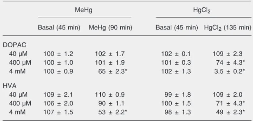
(400 µM and 4 mM) caused a significant decrease in extracellular levels of DOPAC and HVA in the striatum 90 and 105 min after the beginning of HgCl2 administration
(Table 1). The lowest concentration of HgCl2
(40 µM) had no significant effect on the acidic metabolites of DA.
This comparative study demonstrated that intrastriatal administration of different con-centrations of MeHg produced significant increases in the release of DA from rat stri-atal tissue, and they were associated with significant decreases in the extracellular lev-els of DOPAC and HVA. The administra-tion of all concentraadministra-tions of HgCl2 assessed
also induced significant increases in striatal DA levels, with decreases in the extracellu-lar levels of DOPAC and HVA.
The concentrations of mercury used in the present study were similar to those used in other in vitro studies (14,16). Both MeHg and HgCl2 were administered in situ through
a dialysis probe at a flow of 2 µL/min using a Ringer medium. Under these conditions, about 17% of mercury crossed the mem-brane and only submicromolar concentra-tions of mercury were achieved at the striatal site of action.
To exclude the possibility of excitotoxic-ity promoted by mercury administration, at the end of the intrastriatal administration of mercury we perfused a Ringer solution with a high concentration of KCl (75 mM) for 30 min. In this hyperkalemic situation the DA levels increased to 1562 ± 296% compared to basal values (data not shown). This effect was not significantly different from that ob-served with mercury administration and al-lowed us to confirm the functional integrity of the DA synapses.
Our results indicate that MeHg-induced
in vivo DA release seems to be concentra-tion-dependent. In contrast, regarding the effects observed after intrastriatal MeHg administration on extracellular DA release, the effect of HgCl2 administration was not
creases 30 min after the beginning of MeHg perfusion. Maximum values of 907 ± 31, 2324 ± 156, and 9032 ± 70% of basal levels, respectively, were achieved. DA returned to basal values 105 min (40-µM dose), 135 min (40-µM dose), and 165 min (40-µM dose) after MeHg administration.
The highest dose of MeHg assessed (4 mM) caused a significant decrease in extra-cellular levels of DOPAC and HVA in the striatum 45 min after the beginning of MeHg application (a 65 ± 2.3 and 53 ± 2.2% de-crease compared to basal levels, respective-ly; Table 1). The remaining concentrations of MeHg (40 and 400 µM) had no significant effect on the acidic metabolites of DA.
Like MeHg, the different concentrations of HgCl2 used produced increases in the
striatal output of DA (Figure 1B) but the maximum DA release was less than ob-served with MeHg. The data (Figure 1B) show that 40 µM, 400 µM, and 4 mM HgCl2
produced a maximum increase 75 min after the beginning of HgCl2 perfusion (1240 ±
66, 2500 ± 424, and 2658 ± 337% of basal levels, respectively). DA concentration re-turned to basal values 120 min after HgCl2
administration.
Table 1. Effects of MeHg or HgCl2 on extracellular DOPAC and HVA levels from rat
striatum.
MeHg HgCl2
Basal (45 min) MeHg (90 min) Basal (45 min) HgCl2 (135 min)
DOPAC
40 µM 100 ± 1.2 102 ± 1.7 102 ± 0.1 109 ± 2.3
400 µM 100 ± 1.0 101 ± 1.9 101 ± 0.3 74 ± 4.3*
4 mM 100 ± 0.9 65 ± 2.3* 102 ± 1.3 3.5 ± 0.2*
HVA
40 µM 109 ± 2.1 110 ± 0.9 99 ± 1.8 109 ± 2.0
400 µM 106 ± 2.0 90 ± 1.1 100 ± 1.5 71 ± 4.3*
4 mM 107 ± 1.5 53 ± 2.2* 98 ± 1.3 49 ± 2.3*
Data are reported as means ± SEM for 4-5 experiments, expressed as a percentage of basal levels (100%). The basal output of dialysate was considered as the mean substance concentrations in the three samples before mercury perfusion as follows: dihydroxyphenylacetic acid (DOPAC) = 16.60 ± 0.49 ng/15 min (N = 30) and homovan-illic acid (HVA) = 10.95 ± 1.13 ng/15 min (N = 30).
dose-dependent, with the increases in DA levels induced by HgCl2 being the same for
400 µM and 4 mM HgCl2 (~2500%).
Another difference between the effects of the two forms of mercury was the time needed to produce the maximal increase in extracellular DA levels, which was 30 min after the beginning of administration for MeHg and 75 min for HgCl2. Comparing the
results obtained with MeHg and HgCl2, we
observed that the increases in extracellular DA levels induced by HgCl2 were lower (for
the 4-mM concentration) and were delayed by 45 min when compared with the increases in DA release induced by the organic form of mercury.
At least three main mechanisms can be proposed to explain the increase in extracel-lular DA levels induced by mercury: 1) this chemical acts by releasing DA from its
stor-age vesicles, 2) it inhibits monoamine oxi-dase, 3) it inhibits DA reuptake, which is the main mechanism of elimination of the neu-rotransmitter in the synaptic cleft. In previ-ous papers, we have reported that MeHg increases the in vivo DA release from rat striatum and that this increase occurs through an action on the DA membrane transporter inhibiting the reuptake of this neurotrans-mitter (10,17).
The intrastriatal administration of differ-ent concdiffer-entrations of MeHg or HgCl2
pro-duced significant increases in the release of DA from rat striatal tissue, which were asso-ciated with significant decreases in the ex-tracellular levels of DOPAC and HVA (in response to the highest concentrations). We suggest that these results are different mech-anisms of action of the organic and ionic forms of mercury on DA release.
References
1. Guimaraes JRD, Ikingura J, Akagi H. Methyl mercury production and distribution in river water-sediment systems investigated through radiochemical techniques. Water Air Soil Pollut 2000; 124: 113-124. 2. Gochfeld M. Cases of mercury exposure, bioavailability, and
ab-sorption. Ecotoxicol Environ Saf 2003; 56: 174-179.
3. Anonymous. Heavy metal poisoning: mercury and lead. Ann Intern Med 1972; 76: 779-792.
4. McKay SJ, Reynolds JN, Racz WJ. Effects of mercury compounds on the spontaneous and potassium-evoked release of [3H]dopamine
from mouse striatal slices. Can J Physiol Pharmacol 1986; 64: 1507-1514.
5. Saijoh K, Inoue Y, Smith K. Effects of methylmercury chloride and mercury chloride (II) on release and uptake of 3H-dopamine in
guinea pig striatal slices. Toxicol In Vitro 1987; 1: 233-237. 6. Reynolds JN, Racz WJ. Effects of methylmercury on the
spontane-ous and potassium-evoked release of endogenspontane-ous amino acids from mouse cerebellar slices. Can J Physiol Pharmacol 1987; 65: 791-798.
7. Minnema DJ, Cooper GP, Greenland RD. Effects of methylmercury on neurotransmitter release from rat brain synaptosomes. Toxicol Appl Pharmacol 1989; 99: 510-521.
8. Kalisch BE, Racz WJ. The effects of methylmercury on endogenous dopamine efflux from mouse striatal slices. Toxicol Lett 1996; 89: 43-49.
9. Juarez BI, Martinez ML, Montante M, Dufour L, Garcia E, Jimenez-Capdeville ME. Methylmercury increases glutamate extracellular levels in frontal cortex of awake rats. Neurotoxicol Teratol 2002; 24:
767-771.
10. Faro LR, do Nascimento JL, San Jose JM, Alfonso M, Duran R. Intrastriatal administration of methylmercury increases in vivo dopa-mine release. Neurochem Res 2000; 25: 225-229.
11. Bondy SC, Anderson CL, Harrington ME, Prasad KN. The effects of organic and inorganic lead and mercury on neurotransmitter high-affinity transport and release mechanisms. Environ Res 1979; 19: 102-111.
12. Gasso S, Sunol C, Sanfeliu C, Rodriguez-Farre E, Cristofol RM. Pharmacological characterization of the effects of methylmercury and mercuric chloride on spontaneous noradrenaline release from rat hippocampal slices. Life Sci 2000; 67: 1219-1231.
13. Moretto MB, Funchal C, Santos AQ, Gottfried C, Boff B, Zeni G, et al. Ebselen protects glutamate uptake inhibition caused by methyl mer-cury but does not by Hg2+. Toxicology 2005; 214: 57-66.
14. Atchison WD, Hare MF. Mechanisms of methylmercury-induced neurotoxicity. FASEB J 1994; 8: 622-629.
15. Durán R, Alfonso M, Arias B. Determination of biogenic amines in rat brain dialysates by high-performance liquid chromatography. J Liq-uid Chromatogr Related Tech 1998; 21: 2799-2811.
16. Siriois JE, Atchison WD. Effects of mercurials on ligand- and volt-age-gated ion channels: a review. Neurotoxicol 1996; 17: 63-84. 17. Faro LR, do Nascimento JL, Alfonso M, Duran R. Mechanism of
