0103 - 5053 $6.00+0.00
A
r
ti
c
le
*e-mail: [email protected]
Total and Methyl Mercury in Different Species of Molluscs from Two Estuaries in
Rio de Janeiro State
Helena A. Kehrig,*,a Monica Costa,b Isabel Moreirac and Olaf Malma
a
Universidade Federal do Rio de Janeiro, CCS, Instituto de Biofísica Carlos Chagas Filho, Laboratório de Radioisótopos Eduardo Penna Franca, 21949-900 Rio de Janeiro-RJ, Brazil
b
Universidade Federal de Pernambuco, CTG, Departamento de Oceanografia, Recife-PE, Brazil
c
Pontifícia Universidade Católica, Departamento de Química, Rio de Janeiro-RJ, Brazil
Mercúrio total e metilmercúrio foram avaliados em três espécies de moluscos (Perna perna
-mexilhão, Crassostrea rhizophorae - ostra, Anomalocardia brasiliana - vôngole) provenientes de
dois estuários no estado do Rio de Janeiro, impactados por matéria orgânica e metais pesados. O mexilhão foi a espécie que apresentou melhor capacidade para acumular o mercúrio dentre as espécies de molusco estudadas. Foi observada uma diferença significante nas concentrações de mercúrio entre os organismos de mexilhão, fêmeas e machos (81 ± 1 µg kg-1 e 70 ± 5 µg kg-1 em
peso seco respectivamente), com comprimento de concha semelhante. Entretanto, não foi observada diferença significante na percentagem de metilmercúrio entre fêmeas (64%) e machos (63%) de mexilhão. Apesar de possuírem o hábito alimentar semelhante, o mexilhão apresentou concentrações de mercúrio total e metilmercúrio superiores (76 ± 7 µg kg-1 e 48 ± 5 µg kg-1 peso seco) às da ostra
(19 ± 4 µg kg-1 e 6 ± 1 µg kg-1 peso seco). Este fato pode estar relacionado com diferenças na
capacidade em selecionar o tamanho de partículas e os alimentos ingeridos; também, estar refletindo a maior habilidade do mexilhão em concentrar e excretar o metilmercúrio dos seus tecidos, bem como refletindo as condições ambientais.
The paper assesses methyl and total mercury concentrations in three mollusc species (Perna perna – common mussel, Crassostrea rhizophorae – mangrove oyster, Anomalocardia brasiliana
- clam) from two estuaries in Rio de Janeiro State, both impacted by organic matter and heavy metals. Mussels showed higher capacity to accumulate mercury compared to the other mollusc species (oyster, clam). A significant difference was observed between mercury concentration in the female mussel organisms and in males (81 ± 1 µg kg-1 dry wt. and 70 ± 5 µg kg-1 dry wt.
respectively) with similar total shell length. No significant difference was observed among the % MeHg in the female (64%) and male (63%) mussel organisms. Even though the feeding habits of the molluscs are similar; mussels presented higher mercury and methylmercury concentrations in their soft tissues (76 ± 7 µg kg-1 dry wt. and 48 ± 5 µg kg-1 dry wt.) than
oysters (19 ± 4 µg kg-1 dry wt. and 6 ± 1 µg kg-1 dry wt.). This is possible related to their
capacity to select particle size and the composition of the ingested food they assimilate, and also reflects the greater ability of mussels to concentrate and excrete methylmercury and also to reflect their environmental conditions.
Keywords: bivalve molluscs, Rio de Janeiro estuaries, methylmercury, total mercury
Introduction
Shallow estuarine and near-shore marine waters have become increasingly degraded over recent years. In spite of efforts to improve natural resource management, industrial plants continue to release toxic compounds into
the environment, via liquid effluents and atmospheric
The presence and behaviour of mercury in aquatic systems is of great interest and importance since it is the only heavy metal which bioaccumulates and biomagnifies through the aquatic food chain.2 Methylmercury, the most
abundant organic form of mercury in the environment, has been recognised as a serious pollutant of aquatic ecosystems. However only limited information about the manner in which it spreads through the tropical estuarine and marine food chains is available. Methylmercury is largely responsible for the accumulation of mercury in organisms (bioaccumulation) and the transfer of mercury from one trophic level to another (biomagnification).
The trophic transfer of trace elements along marine food webs has been increasingly recognized as an important process influencing metal bioaccumulation and geochemical cycling.3 Lying in the second trophic level, molluscs have
the ability to accumulate both essential and non-essential trace elements from the aquatic environment in which they live.4 Filter-feeding bivalves, such as mussels and oysters,
can be expected to accumulate less methylmercury than predatory fish, and hence apparently present less of a risk to human consumers.5 These bivalves, which are widely
distributed and easily collected, are often proposed as good bioindicators of water quality, as they reflect, in a semi-quantitative manner, levels of environmental pollution.6,7
They can assimilate mercury from ingesting suspended particulate material,8 but trace metal assimilation efficiencies
depend on the inorganic and organic chemical composition of the particulate material.9 The bioavailability and the
chemical species, specially as free ions, influence on the trace metal toxicity and its bioaccumulation by organisms in the estuarine environment. Mercury and methylmercury bioaccumulation is different from that of other metals because uptake of free ions metal via facilitated transport is not the only important mechanism.10 Wright and Mason10 suggested
that mercury and methylmercury accumulation in the presence of large organic compounds occurs through other mechanisms of uptake besides passive diffusion. Dissolved organic matter (DOM) interacts very strongly with mercury, affecting its speciation, solubility, mobility and toxicity in the aquatic ecosystems. DOM reduces the bioavailability of both inorganic and methyl mercury such that bioaccumulation factors decrease with increasing organic content of the exposure medium.10,11
Methylmercury is more likely to accumulate in the soft (edible) tissues of the bivalves.12 However, the
adductor muscle is preferable as a biological indicator for monitoring the concentration of methylmercury pollution in water, as this tissue has an efficient uptake system, and is easy to sample.13 The concentrations of
mercury and methylmercury accumulated by marine
molluscs are a function not only of the water quality, but also of seasonal factors, temperature, salinity, diet, spawning and individual variation.14
Few data are available for comparative purposes on a worldwide scale,5,15-18 and very few data concerning
mercury and methylmercury distribution through coastal food webs are available for tropical estuaries.16,19-26 Some
estuaries in the Southeast Brazilian coast at Rio de Janeiro State, Sepetiba Bay and Guanabara Bay, have been the object of a number of environmental investigations, but very few studies have been done dealing with mercury distribution in their aquatic biota.23,27-29 However, there is
a relatively good understanding of the responses of the northern hemisphere Mytilus (mussel) and Crassostrea
(oyster) species to pollution, although relatively little work has been published on equivalent species in the southern hemisphere. Few methylmercury measurements are reported in the literature for marine bivalves from coastal regions worldwide. However, this lack of information is mainly for oyster and clam.
The present study comparatively evaluated the total mercury (THg) and methylmercury (MeHg) concentrations and the ratios of MeHg to THg in the soft tissues of three species of marine bivalves, Perna perna (common mussel), Crassostrea rhizophorae (mangrove oyster) and Anomalocardia brasiliana (clam) from Sepetiba Bay. The
study also compared the results in Perna perna species from
Sepetiba Bay with other organisms collected at Guanabara Bay, another estuary in the same region traditionally more contaminated than the first one. The objectives were to do comparisons, choose a bivalve as a “sentinel organism” for mercury in Sepetiba Bay and also to evaluate the influence of some parameters to explain the different mercury concentrations in different types of mollusc bivalves and between individuals of the same species. The concentrations of total mercury in the water samples (dissolved and particulate) from different sites within Guanabara Bay were also assessed and used for calculation of the bioaccumulation factor.
The filter feeding molluscs, which are present in high abundance and are widely distributed on the Southeast Brazilian coast, are those most frequently consumed by human population. These organisms are characteristic of tropical areas in the Southern Atlantic.
Experimental
Sampling and sampling areas
In August 2000, specimens of Perna perna (common
rocky substrate inside Sepetiba and Guanabara Bays. Specimens of Crassostrea rhizophorae (mangrove oyster)
were collected the same month inside Sepetiba Bay from mangrove roots and Anomalocardia brasiliana (clam)
from the upper 2 cm of the sediment. In order to avoid possible size specific effects, preference was given to collecting individuals of the same species with similar sizes (Table 1). Water samples were collected only from Guanabara Bay.
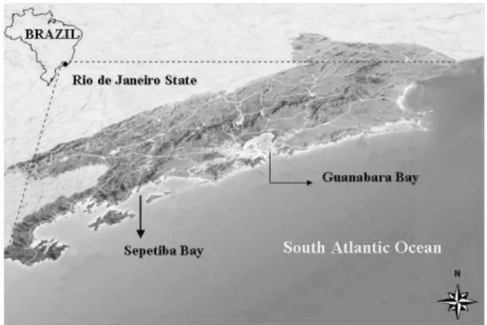
The two estuarine areas can be considered among the most important estuaries for fish and shellfish production along the Southeast Brazilian coast (Figure 1). Both estuaries present different sources or magnitudes of mercury inputs, being subject to different environmental impacts.
Sepetiba Bay (23°S, 44°W, 470 km2), a shallow bay
approximately 60 km south of Rio de Janeiro city, is an area with increasing industrial activity and growing pollution problems. The bay receives inputs from untreated domestic and industrial sewage from a densely populated area (1.1 million inhabitants) with around 400 industrial plants, basically metallurgical, and a harbour. It is also an important fishing ground
and a tourist interest centre. It is an ecosystem impacted by organic matter and heavy metals. Mercury emissions to the Sepetiba Bay environment are mostly from diffuse sources, particularly from an electric power plant, steel and iron production, waste incineration and leaching from a large land fill.30
Guanabara Bay, (22°S, 43°W, 381 km2) is surrounded
by mangroves and its watershed includes several small rivers and channels that contribute to the freshwater inputs.31 The
bay receives untreated domestic and industrial sewage from a densely populated area, and the second largest industrialised region in Brazil, with around 10,000 industrial plants, two harbours, shipyards and oil terminals.32 In some
areas of the bay, its ecosystem is heavily impacted by organic matter, oil and heavy metals, including Hg, whose main consequences are elevated concentrations of toxic metals and hydrocarbons in sediments and changes in the pelagic and benthonic communities.33-35 An important point
source of mercury for this estuary is a chlor-alkali plant located at the most polluted region of its watershed, on the north-western side. The bay is among the most productive marine ecosystems in Rio de Janeiro State, presenting a high phytoplankton density and also high nutrients concentrations (C, N, P) that result in high primary production waters, with an average net primary production (NPP) of 0.17 mol C m-2 day-1.34,35
A total of 100 mollusc specimens, Perna perna (40), Crassostrea rhizophorae (30) and Anomalocardia brasiliana – clam (30), were collected from two sampling
stations at Sepetiba Bay (Ilha da Madeira and Ilha Guaíba). A total of 41 mollusc specimens of Perna perna from
rocky shores and 15.0 litters of their surrounding water were collected at three points at the Southern end of Guanabara Bay (Marina da Glória, Boa Viagem Beach, Rio-Niterói Bridge). These sampling points present different water quality records and also, were subjected to different sorts and degrees of environmental impacts.
Figure 1. Study Areas in Rio de Janeiro State (Sepetiba Bay and
Guanabara Bay).
Table 1. Average of the biological parameters of the bivalve species (total shell length) sampled in August 2000 inside Sepetiba and Guanabara Bays
Total shell length of the bivalve species / mm (min. – max.)
Sampling Perna perna Crassostrea rhizophorae Anomalocardia brasiliana
Point common mussel mangrove oyster clam
Sepetiba Ilha Guaíba 52 45 32
Bay (51-66) (31-47) (28-35)
Ilha da Madeira – 35 –
Guanabara Marina da Glória 63 (30-42)
Bay (59-68)
Boa Viagem 62
(55-66) – –
Ponte Rio-Niterói 78
All the samples of filter-feeding molluscs were prepared according to FAO guidelines.36 They were sized
and kept for 24 h in estuarine water from the collection site in order to eliminate faeces and pseudofaeces. The fresh weight of the soft tissues of the molluscs was obtained after absorbing the excess of water by laying the organism upon a clean sheet of ash free filter paper for over 15 min. Then they were homogenised and freeze dried in separate groups of around 10 organisms. After freeze drying, the soft tissues of the mollusc species lost around 77% of water from their content.
The water samples of the surface estuarine water were collected in double-bagged bottles that were transported to the sampling side while partially filled with dilute trace metal grade HCl (Merck p.s.) were collected while wearing arm length poly gloves. Before each sample was collected, the HCl (Merck p.s.) was emptied (downstream of collection) and the bottle rinsed three times with ambient water. The sample bottle was then filled, immediately recapped, double bagged, and stored in the cooler for transport back to the laboratory.37 The suspended particulate
material was removed by filtering the estuarine water through a 0.45 µm Whatman GF/C filter (Millipore) that had been pre-cleaned by heating in a furnace overnight at 400 °C using acid-cleaned glassware and other filtering apparatus37 and then was processed using the technique
described by Sato et al.13 Three filters were collected per
sample. The filtration apparatus was rinsed with clean acid and then Milli-Q-water between samples. The filter was lifted with Teflon-coated tweezers and placed in an acid-cleaned plastic Petri dish, which was then sealed with Teflon tape. The filters were stored in air-tight PVC containers at below -10 °C until analysis. Total mercury concentrations in the filtrate and suspended particulates were determined.
Instrumentation
Biological and water (dissolved and particulate) samples were analysed for total mercury with a cold vapour atomic absorption spectrometer with a Flow Injection Mercury System (FIMS) – FIAS 400 (Perkin Elmer) with auto sampler AS90 (Perkin Elmer). The carrier gas was argon (99.998%) at a flow rate of 75 mL min-1. For methylmercury analysis,
the chromatographic system used was a 14 B Shimadzu gas chromatograph (GC) with a pulsed current 63Ni
electron-capture detector-ECD (Kyoto, Japan) equipped with a Shimadzu C-R6A Chromatopac integrator and a GC silane-treated glass column of 1 m × 3 mm i.d. (GL Sciences Japan) with Hg-20A as stationary phase on 60-80 mesh Uniport HP (GL Sciences, Japan). On the top of column, nearest the injection port, 0.2 g of NaCl crystals were added to enhance
the methylmercury detection. This method is based on the fact that methylmercury dithizonate in the final solution in toluene is converted into its chloride form as soon as it passes through the NaCl on the top of the columns.38 The column
oven, detector and injector temperature were maintained at 150 °C, 250 °C and 180 °C, respectively. The carrier gas was nitrogen (99.999%) at a flow rate of 40 mL min-1.
The total carbon content in the suspended particulate material was determinated by a Shimadzu TOC-5000A with a Shimadzu solid sample module-SSM-5000A analyzer (Kyoto, Japan).
Analytical methodologies
Total mercury analysis in filtrate estuarine water. Filtered
estuarine water (2.0 L) was mixed well with 10 mL of H2SO4
(Merck p.a.) and 5 mL of 0.5% KMnO4 (Merck p.a.)
solution, and allowed to stand for 5 min. The sample was neutralized with 20 mL of 10 mol L-1 NaOH (Merck p.a.)
and 5 mL of 10% hydroxylamine hydrochloride (Merck p.a.) and allowed to stand for 20 min. After addition of 5 mL of 10% EDTA tetrasodium salt (Dojindo P.A.), the mercury was extracted with 10 mL of 0.01% dithizone (Merck p.a.) in toluene (Tedia ABSOLV) purified with an equal volume of 0.1 mol L-1 NaOH (Merck p.a.) just before
use. An aliquot (5 mL) of the organic layer was transferred into a 50 mL centrifuge tube and dried using a rotary evaporator. The residue in the tube was acid digested for total mercury analysis following the same procedure as that used for suspended particulate and biological samples.39
Total mercury analysis in suspended particulates and in soft tissues of the molluscs. Suspended particulate and dried
soft tissues (0.05 g) was acid digested with 3 mL of
H2SO4:HNO3 (1:1v/v) (Merck p.a.) and 1 mL of
concentrated H2O2 (Merck p.a.) in a 50 mL centrifuge tube at 60 °C in water bath for 45 min. After addition of 5 mL of 5% KMnO4 (Merck p.a.) solution, the digested sample allowed to stand for overnight. Total mercury concentrations in the acid digested solution were determined by CVAAS (FIMS –system) with sodium borohydride (Merck p.a.) as a reducing agent.39 Recovery of Hg in internal standard
experiments was between 90 and 105%.39
Methylmercury analysis in soft tissues of the molluscs.
Dried tissue (0.05 g) was digested with 10 mL of 1 mol L-1
alcoholic potassium hydroxide solution in a 50 ml screw-capped centrifuge tube at 100 °C in water bath for 45 min. The digested sample was slightly acidified with 10 mL of 1 mol L-1 HCl (Merck p.a.). After washing with 5 mL of
n-hexane (Tedia ABSOLV) the methylmercury was extracted with 10 mL of 0.05% dithizone (Merck p.a.) in toluene (Tedia ABSOLV) purified with an equal volume of 0.1 mol L-1 NaOH
just before to use. The organic layer was then washed twice with 5 mL of 1 mol L-1 NaOH to remove the excess dithizone.
An aliquot (5 mL) of the organic layer was back extracted with 2 mL of 0.01% Na2S in 0.1 mol L-1 NaOH/ethanol
(1:1v/v). The excess sulphite ions from the methylmercury solution were eliminated with continuous bubbling (50 mL min-1) with N
2 gas and some drops of 1 mol L-1 HCl for a
further 5 min. To the sample solution, 2 mL of Walpole’s Buffer (pH 3.0) was added. Walpole’s Buffer was made with 600 mL of Milli Q water + 200 mL of 1 mol L-1 CH
3COONa + 200
mL of 1 mol L-1 HCl. The methylmercury from this inorganic
layer was re-extracted with 1 mL of 0.05% purified dithizone-toluene. The organic layer was then washed twice with 2 mL of 1 mol L-1 NaOH to remove the excess dithizone and
subsequently with 5 mL of distilled water and acidified with a few drops of 1 mol L-1 HCl followed by GC-ECD.38,40
The precision and accuracy of the analytical methods were determined using certified material from the
International Atomic Energy Agency: IAEA MA-1 (copepod homogenate) and IAEA 142 (mussel homogenate). The results for total mercury in IAEA MA-1 (N=18) were 0.29 ± 0.03 µg g-1. The CRM has a certified THg value of 0.28 ± 0.02 µg g-1. Our routine methylmercury results for the mussel reference sample IAEA 142 (N=10) were 46.9 ± 1.7 µg kg-1; the CRM MeHg value is 47.7 ± 4.3 µg kg-1. The overall
reproducibility for the analysis period was determined from the results obtained using certified samples. The coefficient of variation (SD/average) was less than 10%.
After we verified the normal distribution of each data set, an analysis of variance followed by a Post-Hoc test (Duncan test) were used to compare the concentration in
the soft tissues of the three species of marine bivalves
from Sepetiba Bay. Mean comparisons in Perna perna
species and also among Perna perna and Crassostrea rhizophorae were carried out using the Student’s “t” test.
Results and Discussion
In this study, total mercury (THg) and methylmercury (MeHg) concentrations, and the percentage of methylmercury (% MeHg) in the soft tissues of the three species of marine bivalves presented data that did not differ greatly from the measurements reported in the literature for marine bivalves from coastal regions worldwide (Table 2).
Table 2. Total mercury concentrations in field collections of bivalve species collected at various coastal regions worldwide
Bivalve species Total mercury (µg g-1 dry wt.) Place reference
(min. – max.)
mussel 0.1 – 0.4 worldwide 57
mussel average 2.70 Southwest Pacific 57
mussel 0.07 - 0.13 US coast 15
oyster 0.08 – 0.10 US coast 15
mussel 0.11 - 0.16 French coast 5
oyster 0.20 – 0.27 French coast 5
mussel average 0.53 Northern Spain 17
oyster average 0.44 Northern Spain 17
mussel 0.030 – 1.73 Western Mediterranean 42
mussel 0.015 – 0.017 Greenland 54
mussel 0.11 – 0.76 Ghanaian coast 16
oyster 0.03 – 0.47 Ghanaian coast 16
mussel 0.036 – 2.60 Moroccan coast 26
oyster < 0.001 – 0.013 Gulf and Gulf of Oman 18
clam average 0.32 Gulf and Gulf of Oman 18
mussel 0.011 – 0.19 (wet wt) Bohai Sea - China 58
oyster 0.016 – 0.064 (wet wt) Bohai Sea - China 58
oyster 0.27 – 2.21 Northeastern Brazilian coast 20
mussel 0.051 – 0.22 Southern Brazilian coast 25
mussel 0.023 – 0.054 (wet wt) Southeastern Brazilian coast(Guanabara Bay) 27
0.017 – 0.056 (wet wt) 28
0.011 – 0.056 (wet wt) 29
mussel 0.053 – 0.24 Guanabara Bay this study
0.068 - 0.083 Sepetiba Bay
oyster 0.015 – 0.023 Sepetiba Bay
In Sepetiba Bay, the average of THg concentrations in the soft tissues appears to be higher in Perna perna
(N=40) (75.5 ± 7.1 µg kg-1 dry wt.) than in Crassostrea rhizophorae (N=30) and Anomalocardia brasiliana
(N=30) (18.5 ± 3.6 µg kg-1 dry wt. and 1.1 ± 0.1 µg kg-1 dry wt. respectively) (Figure 2). The three species presented significant differences (F= 181.86; p= 4 × 10⁻³) between total mercury concentrations in their soft tissues. A post Hoc test showed that mercury concentrations in
Perna perna were significantly higher than in Crassostrea rhizophorae and Anomalocardia brasiliana (p < 0.04).
The soft tissues of all specimens of the common mussels and the mangrove oysters showed percentages of MeHg ranging from 31.9% to 64.5%, with an average of 53.2% (N=70). These results are considered similar to the other data reported for marine moluscs.5,16,27-29,41
Furthermore, the study of Pastor et al.42 measuring a
different suite of metals in the mussel Mytilus
galloprovincialis from a polluted area in the Western
Mediterranean (Spain), showed that the average of total mercury and methylmercury concentrations were similar to our results. Their results for total mercury and methylmercury ranged from 30 to 1730 µg THg kg-1dry wt. and 30-1310 µg MeHg kg-1 dry wt. respectively.
Furthermore, concentrations of total mercury in different organs of the mussel, Mytilus galloprovincialis,
collected at Minamata Bay, Japan, were found to be less than 50 µg kg-1 in most samples and the methylmercury to total mercury ratios ranged from 78 to 100% mainly in the adductor muscle.13
In species of Mytilus , metals are likely to be absorbed
both from solution and from ingested phytoplankton and other suspended particles.43 The common mussel, Perna
perna, which selects its ingested food by the particulate
size, from 1 µm to 4 mm, showed a higher capacity to
accumulate mercury than the other bivalves (mangrove oyster, clam).
The clams indicated of more specific restricted regions, since they are usually found in relatively saline areas of estuaries, and tend to be more common in sediments containing a high proportion of sand. They are most likely to absorb metals from solution and from suspended particles.44
A significant difference (t =10.48; p <1 × 10⁻³) was observed between the average of THg concentrations in the common mussel organisms (N=40) and in the mangrove oysters (N=10) collected at Ilha Guaíba (Sepetiba Bay) (74.3 ± 6.7 µg kg-1 dry wt. and 21.6 ± 1.3 µg kg-1 dry wt. respectively).
Common mussels (N=40) and mangrove oysters (N=10) collected at Ilha Guaíba presented a significant difference (t = 5.59; p < 0.05) between the average of MeHg concentrations (48.1 ± 5.1 µg kg-1 dry wt. and 6.9 ± 0.4 µg kg-1 dry wt. respectively) and the ratios of MeHg to THg were 63.9% and 32.0% respectively.
Our results concerning the ratios of methylmercury to total mercury (% MeHg) were higher than those reported for Perna perna collected at Guanabara Bay.28,29 In these
previous studies, the methylmercury to total mercury ratios
ranged from 28.7% to 35.1% and 28.7 to 46.2%.28,29
Furthermore, Mikac et al.45 reported a methylmercury to
total mercury ratio of about 40% in mussels from Krka Estuary (Croatia).
In a previous study along the French Coast, the average concentration of MeHg in oyster soft tissues (Crassostrea gigas) did not differ significantly from that in the soft
tissues of the mussel (Mytilus spp) (67 ± 6 vs. 62 ± 7 µg kg-1 dry wt.) when all sampling points were combined.5
The study of Franco et al.17 with heavy metals in molluscs
from the Basque Coast (Spain) showed that the total mercury concentrations presented no significant differences between the two mollusc species, Mytilus sp.
and Crassostrea angulata. In another study in a tropical
area (Ghana), methylmercury concentrations in Perna perna soft tissues differed significantly from that found
in oyster soft tissues.16
Filter-feeding bivalves such as mussels and oysters may exhibit distinct capacities for trace metals accumulation, metabolism and excretion.46 The assimilation efficiencies
of the trace metals in the bivalves (Mytilus edulis, Crassostrea virginica) are directly related to the proportion
of each element in the cytoplasmic fraction of ingested phytoplankton, indicating that > 80% of the element in a prey alga’s cytoplasm was assimilated.46
Normally, the common mussel Perna perna
preferentially feeds on organic detritus, silt and
Figure 2. Average of THg and MeHg concentrations (µg kg-1) and the
nanozooplankton;47,48 while the mangrove oyster,
Crassostrea rhizophorae is more selective in respect to
the size and quality of particles ingested (2 µm to 10 µm), and prefers to feed on phytoplankton.48
Ruelas-Inzunza and Páes-Ozuna49 compared the
bioavailability of trace metals using different filter-feeder molluscs (mussels, oysters) from a subtropical coastal lagoon and concluded that there was different accu-mulation patterns for the biological groups and elements studied. This may be a function of the preference for food particle sizes and composition as well as the detoxifying mechanism that each species possesses.
The epifaunal species, Crassostrea gigas (oyster) and Mytylus edulis (mussel), showed significant differences
in the process of filtering particles, demonstrating a high capacity to selectively ingest organic matter.50 However,
when compared to M. edulis, C. gigas was not as efficient
either in the net selection of organic matter or in digesting and/ or assimilating ingested organics.
In the study of the correlations between metal uptake in the soft tissue of Perna perna and gill filament pathology
after exposure to mercury, Gregory et al.51 concluded that
these mollusc species presented efficient capacities to accumulate metal, even though they were also able to rapidly depurate their soft tissues. This resilience suggests that caution should be applied in using Perna perna, and probably other
mussel species as biomonitors over long time frames.51
Recently, new data have shown that uptake of heavy metals by Crassostrea rhizophorae and their accumulation
can take long periods to achieve equilibrium and depuration seems to be relatively small,52 questioning the
traditional use of the oyster as a sentinel organism.53
The average of THg concentrations in the soft tissues appears to be higher in Crassostrea rhizophorae collected
at Ilha Guaíba (N=10) (21.6 ± 1.3 µg kg-1 dry wt.) than in the same species from Ilha da Madeira (N=40) (14.5 ± 1.1 µg kg-1 dry wt.). They presented significant differences (t= 7.18; p < 0.002). The oyster from Ilha Guaíba presented a shell length slightly higher than the ones from Ilha da Madeira (Table 1). However, oysters from both sampling station presented similar percentages of methylmercury, around (32%).
The soft tissues of the mangrove oyster, Crassostrea rhizophorae, from a mercury polluted area on the
north-eastern Brazilian coast presented higher mercury concentrations (ranging from 270 to 2210 µg kg-1 dry wt.)20
than the organismsof the same species collected at
Sepetiba Bay.
A significant difference (t = 4.07; p < 0.01) was observed between the average of THg concentrations in the female common mussel organisms (N=20) and in
males (N=20) with similar total shell length (81.0 ± 1.6 µg kg-1 dry wt. and 70.0 ± 5.2 µg kg-1 dry wt. respectively).
No significant difference was observed between the% MeHg in the female and male mussel organisms (64.5% and 63.0%). Nevertheless, in previous studies with Perna perna from Guanabara Bay, no statistically significant
difference between the sexes was detected for the average size, weight or mg Hg kg-1.27
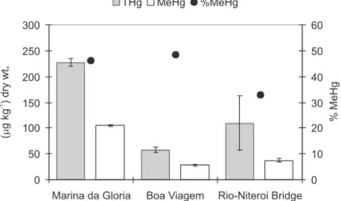
In Guanabara Bay, the average of THg and MeHg concentrations in the soft tissues of all specimens of Perna perna (N=41) were 126.3 ± 77.2 µg THg kg-1 dry wt. and 52.1 ± 38.1 µg MeHg kg-1 dry wt., ranging from 58.2 to 227.2 µg THg kg-1 dry wt. and from 21.0 to 105.0 mg MeHg kg-1 dry wt. respectively. The general average of the
percentage of MeHg in the mussel soft tissues was 40.1%, ranging from 28.7% to 48.4%. These values varied according to the sampling point and water quality of Guanabara Bay. Our results can be considered consistent with previous data from other studies with Perna perna
from Guanabara Bay.27-29 Costa et al.27 concluded that the
THg content in the common mussels from Guanabara Bay has probably kept constant for the last 10 years, with possible isolated fluctuations attributed to occasional changes in water quality (after strong and persistent rain events that are typical from Rio de Janeiro for instance).
In our study, mercury and methylmercury concentrations in Perna perna from Guanabara Bay are similar to the few
measurements reported in the literature for this same marine bivalve species worldwide.16,21,25,26
The average of MeHg to THg ratios in the mussel soft tissues found during the present study can be considered consistent with the data from the study of Joiris et al.16
with Perna perna, in different tropical estuaries from
Ghana-Africa.
The specimens of Perna perna collected at the three
different sampling point inside Guanabara Bay presented significant differences (F= 216.89; p < 1 × 10⁻³) between total mercury concentrations in their soft tissues. “Duncan test” showed that mercury concentrations of Perna perna
from Marina da Glória were significantly higher than from Ponte Rio Niterói and Boa Viagem (p < 0.02). However, common mussels from Marina da Glória and Boa Viagem presented similar shell lengths that were smaller than the ones from Ponte Rio Niterói (Table 1).
The common mussels from Marina da Glória (N=11) presented the highest concentrations of THg and MeHg on a dry weight basis (227.2 ± 6.9 µg kg-1) and (105.0 ± 1.7 µg kg-1) and also the ratios of MeHg to THg (46.2%) in their
Bridge sampling point (5 litres) (0.7 ± 0.08 ng L-1) and 3
times higher than in Boa Viagem Beach (5 litres) (1.6 ± 0.2 ng L-1) (Figure 4a). Furthermore, Marina da Glória sampling
point is located in a region of the bay with the poorest water quality and most limited water circulation of the three sites compared here, and also receives large amounts of untreated domestic sewage. This sampling point also presented the highest THg concentration and the total carbon content (% C) in the suspended particulate material (380.0 µg kg-1 wet wt.. and 7.33% respectively) (Figure 4b).
The suspended particulate material from Rio-Niterói Bridge and Boa Viagem Beach, which present better water quality and circulation, presented similar total carbon content (5.77%, 5.38% respectively), and also THg concentrations (60.7 mg kg-1 wet wt.. and 71.0 mg kg-1
wet wt., respectively) (Figure 4b). There was a positive significant correlation (r2=0.953) between the THg content
and the total carbon content (% C) in the suspended particulate material from the three sampling points.
Meanwhile, the common mussels collected at Boa Viagem Beach presented the lowest THg and MeHg concentrations (58.2 ± 5.1 µg THg kg-1, 28.2 ± 1.5 µg MeHg kg-1) in the dry soft tissues, when compared to those
found in the others sampling points (Figure 3).
The bioaccumulation factor (BAF) observed relating to the THg accumulation by the common mussels collected at Rio-Niterói Bridge from the water (41 × 103) was 4.5 times higher than those from Marina da Glória (8.7 × 10³) and 5.5 times higher than those from Boa Viagem Beach (7.5 × 10³) (Figure 5). One of the factors that can affect the BAF is the total mussel shell length, which is proportional to their age. At Rio-Niterói Bridge, its size is longer (around 80 mm) than at Marina da Glória and Boa Viagem Beach (63 mm, 62 mm respectively). In a previous study in different sampling points at Greenland, the mercury concentration in the soft tissues of the blue mussel (Mytilus edulis) increased with the shell length.54 However, total
mercury concentration in the soft tissues of Mytilus galloprovincialis decreased proportionally with an increase
of tissue weight, water content, size and age.45,55
Average comparisons using the Student’s t-test verified that the data obtained in this study in the soft tissues of
Perna perna from Sepetiba Bay (75.5 ± 7.1 µg THg kg-1 and 48.2 ± 5.1 µg MeHg kg-1) and from Guanabara Bay (126.3 ± 77.2 µg THg kg-1 and 52.1 ± 38.1 µg MeHg kg-1) showed no significant difference (t=1.305, p=0.340
and t=0.208, p=0.842 respectively).
Figure 3. Average of THg and MeHg concentrations (µg kg-1) and the
ratios of MeHg as THg in the dry soft tissues of the common mussel (Perna perna) from different sampling points in Guanabara Bay.
Figure 4a. Average of dissolved THg concentration in estuarine water
samples from different sampling points in Guanabara Bay.
Figure 4b. Average of THg concentration (µg kg-1) and% of Total
Car-bon (% C) in the suspended particulate material from different sampling points in Guanabara Bay.
Figure 5. Relation between the THg accumulated by the common
Conclusions
In this study the values of THg and MeHg con-centrations found in the molluscs could not be considered high. Probably, the organisms respond to the different environmental conditions, mesotrophic in Sepetiba Bay and eutrophic conditions of Guanabara Bay, which receives a very high load of suspended material that may be more significant than the industrial wastes. The availability and distribution of particulate organic matter in estuarine and marine waters is expected to markedly influence the biological availability of mercury,56
especially for those who feed on seston.
Even though, the feeding habits of the molluscs Perna perna and Crassostrea rhizophorae are similar THg and
MeHg concentrations in the soft tissues were substantially higher in the common mussel than in the mangrove oyster. This is possibly related to their capacity to select, or filter, the particle size and the composition of the ingested food they assimilate, and also reflects the greater ability of the mussels to concentrate and excrete methylmercury in their tissues. Common mussels preferentially ingest higher particle size (organic detritus, silt and nanozooplankton) than mangrove oysters (phytoplankton). Probably, the high THg and MeHg contents in mussels’ soft tissues are related to their capacity to ingest food from higher trophic levels than oysters (biomagnification of MeHg). Due to the high concentrations of total mercury and methylmercury found in the soft tissues of Perna perna, mussels proved to be
better biomonitors of tropical environments, demonstrating a greater capacity to accumulate THg and MeHg compared to the other filter feeding molluscs (oysters and clams), thus reflecting the water quality in which they live. The large difference in observed concentrations of mercury and methylmercury is also the result of differences in residence times in the contaminated areas.
References
1. WHO; Environmental Health Criteria, World Health Organization: Geneva, 1989, p. 86.
2. Lindqvist, O.; Johnasson, K.; Aastrup, M.; Andersson, A.; Bringmark, L.; Hovsenius, G.; Håkanson, L.; Iverfeldt, Å.; Meili, M.; Timm, B.; Water, Air, Soil Pollut.1991, 55, 1. 3. Fisher, N. S.; Reinfelder, J. R. In Metal Speciation and
Bioavailability in Aquatic Systems; Tessier, A.; Turner, D. R., eds.; John Wiley & Sons: Chichester, 1995.
4. Dallinger, R.; Rainbow, P. S. In Ecotoxicology of Metals in Invertebrates; Dallinger, R., ed.; CRC Press: Boca Raton, 1993. 5. Claisse, D.; Cossa, D.; Bretaudeau-Sanjuan, J.; Touchard, G.;
Bombled, B.; Mar. Pollut. Bull.2001, 42, 329.
6. Boyden, C. R.; Phillips, D. J. H.; Mar. Ecol.-Prog. Ser.1981, 5, 29.
7. O’Conner, T. P.; Mar. Environ. Res.1996, 41, 183.
8. Gagnon, C.; Fisher, N. S.; Environ. Sci. Technol.1997, 31, 993.
9. Luoma, S. N.; Fisher, N. S. In Ecological Risk Assessments of Contaminated Sediments; Ingersoll, C. G.; Dillon, T.; Biddinger, G. R., eds.; SETAC Spec Publ. Ser.: Pensacola, 1997. 10. Wright, D. A.; Mason, R. P.; Int. J. Environ. Pollut.2000, 13, 226. 11. Ravichandran, M.; Chemosphere2004, 55, 319.
12. Boening, D. W.; Chemosphere2000, 40, 1335.
13. Sato, M.; Haraguchi, K.; Ando, T.; Tomiyasu, T.; Akagi, H.; Environ. Sci.1997, 5, 225.
14. Cossa, D.; Oceanol. Acta1989, 12, 417.
15. Beliaeff, B.; O’Connor, T. P.; Claisse, D.; Environ. Monit. Assess.1998, 49, 87.
16. Joiris, C. R.; Holsbeek, L.; Otchere, F. A.; Mar. Pollut. Bull.
2000, 40, 457.
17. Franco, J.; Borja, A.; Solaun, O.; Pérez, V.; Mar. Pollut. Bull.
2002, 44, 973.
18. de Mora, S.; Fowler, S. W.; Wyse, E.; Azemard, S.; Mar. Pollut. Bull.2004, 49, 410.
19. Sbriz, L.; Aquino, M. R.; Alberto de Rodriguez, N. M.; Fowler, S. W.; Sericano, J. L.; Mar. Pollut. Bull.1998, 12, 971. 20. Meyer, U.; Hagen, W.; Medeiros, C.; Mar. Biol. 1998, 131,
113.
21. Otchere, F. A.; Joiris, C. R.; Holsbeek, L.; Sci. Total Environ.
2003, 304, 369.
22. Joiris, C. R.; Azokwu, M. I.; Otchere, F. A.; Ali, I. B.; Sci. Total Environ.1998, 224, 181.
23. Kehrig, H. A.; Malm, O.; Moreira, I.; Sci. Total Environ.1998, 213, 263.
24. Sant’Anna Jr., N.; Costa, M.; Akagi, H.; Environ. Sci. Pollut. Res.2001, 8, 280.
25. Baraj, B.; Niencheski, L. F.; Corradi, C.; Water, Air, Soil Pollut.
2003, 145, 205.
26. Banaoui, A.; Chiffoleau, J. F.; Moukrim, A.; Burgeot, T.; Kaaya, A.; Auger, D.; Rozuel, E.; Mar. Pollut. Bull.2004, 48, 385. 27. Costa, M.; Paiva, E.; Moreira, I.; Sci. Total Environ.2000, 261,
69.
28. Kehrig, H. A.; Costa, M.; Moreira, I.; Malm, O.; Environ. Sci. Pollut. Res.2001, 8, 275.
29. Kehrig, H. A.; Costa, M.; Moreira, I.; Malm, O.; Mar. Pollut. Bull.2002, 44, 1018.
30. Marins, R. V.; Lacerda, L. D.; Paraquetti, H. H. M.; Paiva, E. C.; Villas Boas, R. C.; Bull. Environ. Contam. Toxicol.1998,
61, 57.
31. JICA; The Study on Recuperation of the Guanabara Bay Ecosystem: Draft Final Report, 1994.
33. Valentin, J. L.; Tenenbaum, D. R.; Bonecker, A. C. T.; Bonecker, S. L. C.; Nogueira, C. R.; Villac, M. C. In Ecologia de Ambientes Costeiros do Estado do Rio de Janeiro; Silva, S. H. G.; Lavrado, H. P., eds.; Oecologia Brasiliensis, PPGE-UFRJ: Rio de Janeiro, 1999.
34. Carreira, R. S.; Wagener, A. L. R.; Readman, J. W.; Fileman, T. W.; Macko, S. A.; Veiga, A.; Mar. Chem.2002, 79, 202. 35. Rebello, A. L.; Ponciano, C. R.; Melges, L. H.; An. Acad. Bras.
Cienc.1988, 60, 419.
36. FAO/SIDA, Manual de Métodos de Investigación del Medio Ambiente Acuático, Doc. Téc. Pesca, FAO, 1983.
37. Lawson, N. M.; Mason, R. P.; Laporte, J. M.; An. Acad. Bras. Cienc.2001, 35, 501.
38. Akagi, H.; Nishimura, H. In Advances in Mercury Toxicology; Suzuki T.; Nobumassa I.; Clarkson T. W., eds.; Plenum Press, 1991.
39. Bastos, W. R.; Malm, O.; Pfeiffer, W. C.; Cleary, D.; Ciênc. Cult.1998, 50, 255.
40. Kehrig, H. A.; Malm, O.; Appl. Organomet. Chem.1999, 13, 687.
41. Odzäk, N.; Zvonaric, T.; Kljakovic Gaspic, Z.; Horvat, M.; Baric, A.; Sci. Total Environ.2000, 261, 61.
42. Pastor, A.; Hernández, F.; Peris, M. A.; Beltrán, J.; Sancho, J. V.; Castillo, M. T.; Mar. Pollut. Bull.1994, 28, 50.
43. George, S. G.; Pirie, J. S.; J. Mar. Biol. Assoc. U.K.1980, 60, 575.
44. Bryan, G. W.; Langston, W. J.; Hummerstone, L. G.; Burt, G. R., In A guide to the Assessment of Heavy Metals Contamination in Estuaries Using Biological Indicators; Marine Biological Association of the United Kingdom: Devon, 1985.
45. Mikac, N.; Kwokal, Z.; Martincic, D.; Branica, M.; Sci. Total Environ.1996, 184, 173.
46. Reinfelder, J. R.; Wang, W. X.; Luoma, S. N.; Fisher, N. S.; Mar. Biol.1997, 129, 443.
47. Foster-Smith, R. L.; J. Mar. Biol.1975, 55, 411. 48. http:// www.lcmm.ufsc.br, acessed on May 2004.
49. Ruelas-Inzunza, J. R.; Páez-Osuna, F.; Environ. Pollut.2000, 107, 437.
50. Hawkins, A. J. S.; Bayne, B. L.; Bougrier, S.; Heral, M., Iglesias, J. I. P.; Navarro, E.; Smith, R. F. M.; Urrutia, M. B.; J. Exp. Mar. Biol. Ecol.1998, 19, 87.
51. Gregory, M. A.; Marshall, D. J.; George, R. C.; Anandraj, A.; McClurg, T. P.; Mar. Pollut. Bull. 2002, 45, 114.
52. Wallner-Kersanach, M.; Theede, H.; Eversberg, U.; Lobo, S.; Arch. Environ. Contam. Toxicol.2000, 38, 40.
53. Rebelo, M. F.; do Amaral, M. C. R.; Pfeiffer, W. C.; Mar. Pollut. Bull.2003, 46, 1341.
54. Riget, F.; Dietz, R.; Johansen, P.; Asmund, G.; Sci. Total Environ.2000, 245, 3.
55. Saavedra, Y.; González, A.; Fernández, P.; Blanco, J.; Sci. Total Environ.2004, 318, 115.
56. Window, H. L.; Kendall, D. R. In The Biogeochemistry of Mercury in the Environment; Nriagu, J. O. ed., Elsevier: Amsterdam, 1979.
57. Kennish, M. J. In Practical Handbook of Estuarine and Marine Pollution; Kennish, M. J., ed., CRC Press: New York, 1997. 58. Liang, L.; Shi, J.; He, B.; Jiang, G.; J.Agric. Food Chem. 2003,
51, 7373
Received: May 5, 2006