Effe cts o f ae ro bic e xe rcise training
o n he art rate variability during
wake fulne ss and sle e p and
cardio re spirato ry re spo nse s o f
yo ung and middle -age d he althy me n
1Laboratório de Fisioterapia Cardiovascular, Departamento de Fisioterapia, and 2Departamento de Estatística, Universidade Federal de São Carlos,
São Carlos, SP, Brasil
3Departamento de Fisiologia e Biofísica, Instituto de Biologia,
Universidade Estadual de Campinas, Campinas, SP, Brasil
4Laboratório de Fisiologia do Exercício, Faculdade de Educação Física,
Universidade Estadual de Campinas, Campinas, SP, Brasil
5Divisão de Cardiologia, Departamento de Clínica Médica,
Hospital das Clínicas, Faculdade de Medicina de Ribeirão Preto, Universidade de São Paulo, Ribeirão Preto, SP, Brasil
A.M. Catai1,3,
M.P.T. Chacon-Mikahil3,4,
F.S. Martinelli3,4,
V.A.M. Forti4, E. Silva1,
R. Golfetti4, L.E.B. Martins4,
J.S. Szrajer4, J.S. Wanderley4,
E.C. Lima-Filho4, L.A. Milan2,
J.A. Marin-Neto5,
B.C. Maciel5
and L. Gallo-Junior5
Abstract
The purpose of the present study was to evaluate the effects of aerobic physical training (APT) on heart rate variability (HRV) and cardiores-piratory responses at peak condition and ventilatory anaerobic thresh-old. Ten young (Y: median = 21 years) and seven middle-aged (MA = 53 years) healthy sedentary men were studied. Dynamic exercise tests were performed on a cycloergometer using a continuous ramp proto-col (12 to 20 W/min) until exhaustion. A dynamic 24-h electrocardio-gram was analyzed by time (TD) (standard deviation of mean R-R intervals) and frequency domain (FD) methods. The power spectral components were expressed as absolute (a) and normalized units (nu) at low (LF) and high (HF) frequencies and as the LF/HF ratio. Control (C) condition: HRV in TD (Y: 108, MA: 96 ms; P<0.05) and FD - LFa, HFa - was significantly higher in young (1030; 2589 ms2/Hz) than in
middle-aged men (357; 342 ms2/Hz) only during sleep (P<0.05);
post-training effects: resting bradycardia (P<0.05) in the awake condition in both groups; V.O2 increased for both groups at anaerobic threshold
(P<0.05), and at peak condition only in young men; HRV in TD and FD (a and nu) was not significantly changed by training in either groups. The vagal predominance during sleep is reduced with aging. The resting bradycardia induced by short-term APT in both age groups suggests that this adaptation is much more related to intrinsic alter-ations in sinus node than in efferent vagal-sympathetic modulation. Furthermore, the greater alterations in V. O2 than in HRV may be related
to short-term APT.
Co rre spo nde nce
A.M. Catai
Laboratório de Fisioterapia Cardiovascular - Núcleo de Pesquisa em Exercício Físico, Departamento de Fisioterapia, UFSCar
Rodovia Washington Luís, km 235 13565-905 São Carlos, SP Brasil
E-mail: mcatai@ power.ufscar.br
Research supported by CAPES, FAPESP (No. 91/4754-9), CNPq (No. 300528/85-0), and FAEP-UNICAMP.
Received July 17, 2001 Accepted April 1, 2002
Ke y words
•Heart rate variability
•Power spectral density analysis
•Anaerobic threshold
•Aerobic exercise training
Intro ductio n
Heart rate variability (HRV) is mainly caused by efferent autonomic modulation of the sinus node. For many years this variable has been expressed only as mean values and standard deviations, i.e., a measure in the time domain representation. However, a non-invasive contribution by each division of autonomic modulation to HRV is possible when this variable is represented in its fre-quency domain, i.e., the power spectral den-sity analysis. Today, it is well accepted that under specific experimental conditions the power spectrum is a tool of great value for assessing the neural mechanisms controlling heart rate (HR) (1).
Analysis of HRV in the frequency do-main obtained from mathematical process-ing of the R-R intervals in the electrocardio-gram recordings obtained under resting con-ditions can discriminate two main spectral components: a high frequency one (ranging from 0.15 to 0.40 Hz) and a low frequency one (ranging from 0.04 to 0.15 Hz), consid-ered to be markers of parasympathetic and sympathetic control, respectively (1,2). How-ever, Skyschally et al. (3) have suggested that low frequency is influenced by both vagal and sympathetic activity.
Measurement of HRV may be useful as a noninvasive method to assess in man the autonomic nervous system modulation un-der several physiological conditions such as awake and sleeping situations, different body positions, physical training, and also in patho-logical conditions (1,4-6). Thus, the HRV expressed both in the time and frequency domains is reduced with age (4,7) due to the dominance of the sympathetic over the para-sympathetic balance in this particular condi-tion (4,7). This observacondi-tion is relevant since the reduction of HRV with aging is related to higher cardiovascular morbidity and mortal-ity rates (7,8).
There are conflicting reports in the litera-ture concerning the effects of aerobic
train-ing on HRV under resttrain-ing conditions. While some studies have reported an increase in the magnitude of this variable in the time domain (9), in the frequency domain others have reported absence of modifications (10), and an increase (11) or decrease (12) of sympathovagal balance in the sinus node.
The effect of age on physical working capacity has also been the subject of many studies (13,14) that have shown that maxi-mal aerobic capacity, measured as V. O2 max,
reaches a maximum value around the age of 30 years and decreases progressively there-after. Concerning V. O2 at the anaerobic
thresh-old, the literature has also shown a decline of this parameter with advancing age (13) and there are studies indicating the occurrence of significant changes in aerobic capacity and autonomic changes in HR after aerobic train-ing in middle-aged subjects (15,16).
On the basis of these considerations, the purpose of the present study was to evaluate the effects of 3-month aerobic physical train-ing on the efferent autonomic cardiac con-trol that modulates the HR response at rest in awake and sleeping conditions and on the oxygen uptake at ventilatory anaerobic threshold and peak conditions during dy-namic exercise in young and middle-aged men.
Mate rial and Me thods
Subje cts
None of the subjects studied was taking any type of medication. Two different age groups were compared: young group (N = 10), age range of 19 to 29 years (median = 21), and middle-aged group (N = 7), age range of 50 to 59 years (median = 53). The subjects were informed about the experimental procedures and all signed an informed consent form to participate in the present study, which was approved by the Ethics Committee of the State University of Campinas. All individu-als were evaluated during the same time of day at an experimental room temperature of 23ºC and relative air humidity between 50 and 60%. Before the day of the experiment the subjects were taken to the experimental room for familiarization with the procedures and the equipment to be used. Each subject had been oriented to avoid caffeinated and alcoholic beverages, to refrain from smok-ing and not to perform moderate or heavy exercise on the day before the application of protocols I and II or during the 24-h period for the Holter test. On each experimental day, before conducting the programmed pro-tocols, the volunteers were interviewed and examined to confirm the state of good health, the occurrence of a normal night sleep, and to confirm that the control conditions (HR and systemic blood pressure) were in the normal range.
Pro to co ls
Protocol I. All subjects were studied in
the resting condition (supine and seated) and during two dynamic exercise tests in the seated position on a cycloergometer, using a continuous protocol on different days sepa-rated by a 2-7-day interval, as follows:
a) Clinical and diagnostic evaluation. The main purpose of this procedure was to in-clude in the study only healthy men, exclud-ing any subject with evidence of silent is-chemic heart disease or other pathologic abnormalities of the cardiovascular system. A 12-lead standard ECG recording was
ob-tained at rest in the supine position. The exercise protocol consisted of 3-min step power increments of 25 W, with a rotation frequency of 60 rpm maintained throughout the test. The exercise tests ended when the subjects presented one or more of the fol-lowing conditions: 1) clear evidence of physi-cal exhaustion, 2) reaching the age-predicted maximal HR, and 3) inability to maintain a standard cycling frequency due to muscular fatigue. During the protocol the subjects were monitored using the thoracic CM5 lead. An ECG tracing (CM5, aVF and V2) was ob-tained during the last 10 s of each power level. Arterial pressure was measured by the auscultatory method using a mercury sphyg-momanometer during the last 15 s of each power level.
b) Functional capacity evaluation: oxy-gen uptake test. The subjects performed an oxygen uptake test using a progressive incre-mental exercise protocol. This protocol con-sisted of a 3-min warm-up at 4 W followed by a continuous power increase set at a value of 12 to 20 W up to physical exhaustion. The choice of the power value increment for each subject, i.e., 12, 15, 17 or 20 W/min, was based on the responses presented in the previous clinical test described above (pro-tocol I-a). A braked electromagnetic cyclo-ergometer equipped with a microprocessor (model Corival 400, Quinton, Seattle, WA, USA) allowed the precise application of in-dividualized power ramp values. At the peak of effort each subject attributed a rating of perceived exertion based on Borg’s scale (17), that varied from 0 to 10 units.
(V. ), CO2 production (V
.
CO2) and oxygen
uptake (V. O2) at each power were plotted
as a function of time; V. , V.CO2 and V
. O2
peaks were selected as the highest values reached during the incremental exercise pro-tocol.
In all subjects, anaerobic threshold (in V. O2) was measured when the V
.
and V. CO2
began to increase non-linearly as compared to V. O2 (18). This was determined by visual
analysis of V., V. O2 and V
.
CO2 curve
re-sponses. Three different observers measured the anaerobic threshold values (in V. O2) in
all exercise tests. Using this procedure, anaerobic threshold (in V. O2) could be
meas-ured with a difference of about 2%. In the present study anaerobic threshold was ex-pressed as absolute (ml/min) and normalized values, i.e., corrected for body weight (ml kg-1 min-1) and as percentage of peak V. O
2.
Absolute HR values at anaerobic thresh-olds and under peak conditions were ob-tained with an ECG recording system (Funbec, São Paulo, SP, Brazil). The signals were recorded in real time after analog to digital conversion and the R-R intervals (pe-riods expressed in milliseconds between R-R peak waves of ECG signal) were calcu-lated on a beat-to-beat basis using a specific software (19). The HR values are reported as averages at 10-s intervals.
Protocol II: 24-h Holter electrocardio-gram. At least 48 h after the previous test (I-b), the subjects were submitted to a 24-h Holter recording. The main purpose of this test was to assess the contribution of the autonomic nervous system to the control of HR before and after physical training, by measuring HRV using time and frequency domain methods. ECG signals (leads CM2 and CM5) were recorded using a 24-h Holter tape recorder (Del Mar Avionics, Irvine, CA, USA).
At the beginning of the Holter recording, the volunteers were asked to rest in the su-pine position for 60 min. After this time, they were instructed on how to proceed
through-out the recording period. After a 24-h re-cording, the volunteers returned to the labo-ratory to finish the procedure.
The reading and analysis of the ECG recording were done using a Holter Manage-ment System (model 750 A Innovator, Del Mar Avionics). A complete automated 24-h report and a visual inspection by the re-searcher were performed to make sure that the cardiac rhythm was sinusal and that there was no abnormality in atrioventricular elec-trical conduction. Then, the average HR and R-R interval, with the respective standard deviations (time domain), were measured under awake (initial 60 min - 2:00-3:00 pm) and sleeping conditions (central 6 h - i.e., without the first and last sleep hours - 0:00-6:00 am). Following the next step of analy-sis, the highest stationary sections of R-R intervals on the monitor display were se-lected for analysis of HRV as a criterion required for correct application of frequency domain analysis, i.e., fast Fourier transform (1).
The data of R-R intervals during a period of resting in the supine position in the awake (2:00-3:00 pm) and sleeping states (0:00-6:00 am) were analyzed in short-term re-cordings which included four consecutive nonoverlapping windows of 256 s each. The data of R-R intervals during the sleep period were analyzed after an initial sleeping time of approximately 160 min for the young group and 180 min for the middle-aged group. The selected time domain parameters studied were the mean R-R interval and the corresponding standard deviation. For fre-quency domain analysis, the power spectral components are reported at low (0.04 to 0.15 Hz) and high (0.15 to 0.4 Hz) frequencies obtained using the fast Fourier transform in absolute and normalized units. The low/high frequency ratio of absolute power was also measured (5,20). The absolute low and high frequencies are reported as ms2/Hz while
frequency component by the total power, after subtracting from it the power of the component with a range frequency between 0 and 0.03 Hz, i.e., very low frequency, and multiplying this ratio by 100 (1,20).
Protocol III: aerobic exercise training.
The program was conducted for 3 months on a field track and included stretching for 10 min followed by walking and/or jogging for 40 min, three times a week at a prescribed HR that corresponded to 70 to 85% of peak HR obtained during a continuous dynamic exercise test performed in a laboratory envi-ronment (protocol I-b: functional capacity evaluation). This intensity training range was also based on metabolic profile, since 75% of peak HR in each volunteer corre-sponded or was very close to the HR at the anaerobic threshold obtained previously dur-ing the oxygen uptake test. Thus, in each training session the subjects were submitted to progressive intensities of exercise (walk-ing and/or jogg(walk-ing) at HR values that were below, equal to and above those related to anaerobic threshold. During the exercise pro-gram the subjects used pulse monitors (mo-del Vantage XL, Polar, Port Washington, NY, USA) to ensure they were exercising at the appropriate intensity. The aerobic train-ing intensity was adjusted durtrain-ing a 7-day interval based on rest and exercise HR meas-ured with the above specified pulse monitor, compared to the previous control period (pro-tocol I-b).
Statistical analysis
The data are reported as medians, quartiles (1st and 3rd) and minimum and maximum values using the Tukey box-plot. Due to non-Gaussian distribution and/or inhomogeneity of variance of variable values, nonparamet-ric tests were selected for statistical analysis. Thus, the Mann-Whitney and Wilcoxon non-parametric tests were used for intergroup and intragroup comparisons, respectively, with the level of significance set at 5%.
Re sults
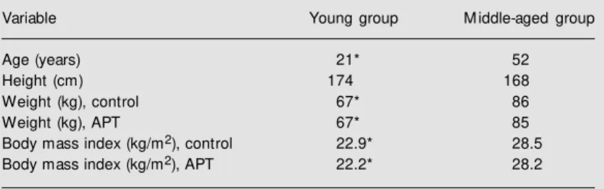
The physical characteristics of the young and middle-aged subjects are shown in Table 1. Under control conditions, and after aero-bic training, median age, weight and body mass index were higher in the middle-aged than in the young group (P<0.05); only me-dian height was similar for the two groups. After aerobic training, the intragroup differ-ences in weight and body mass index were not statistically significant.
Exe rcise conditions
Responses to dynamic exercise:
anaero-bic threshold and peak oxygen uptake.
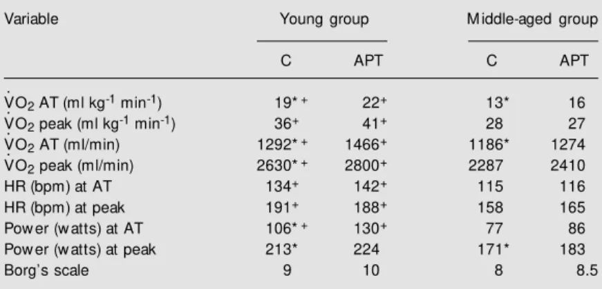
Un-der control conditions, anaerobic threshold and V. O2 reported as absolute oxygen uptake
and normalized values for body weight were lower (P<0.05) in middle-aged than in young men (Table 2). Again, under control condi-tions the values of HR at anaerobic threshold and peak effort, as well as power at anaero-bic threshold were significantly lower in middle-aged men than young men (Table 2). After 3 months of aerobic physical train-ing the absolute and normalized values of V. O2 at anaerobic threshold increased
signifi-cantly (P<0.05) for both groups; under peak conditions the same occurred for V. O2
(abso-lute) only for the young group and for the power values for both groups. However, un-der peak conditions the inter- and intragroup
Table 1. Comparison of the anthropometric data for the subjects before (control) and after three months of aerobic physical training (APT).
Variable Young group M iddle-aged group
Age (years) 21* 52
Height (cm) 174 168
Weight (kg), control 67* 86
Weight (kg), APT 67* 85
Body mass index (kg/m2), control 22.9* 28.5
Body mass index (kg/m2), APT 22.2* 28.2
Holte r ECG analysis: R-R inte rval and its
variability in awake and sle e ping conditions
Time domain index of HRV: before and
after exercise training. In the awake resting
supine position the mean R-R interval and its standard deviation did not differ between groups before or after training (Table 3). The HR values were lower in the sleeping than in the awake resting supine condition (the in-verse for R-R interval) for both groups (P<0.05). Aerobic physical training induced significant (P<0.05) bradycardia (increase in average R-R interval) of comparable mag-nitude for both groups studied in the awake resting supine position (Table 3).
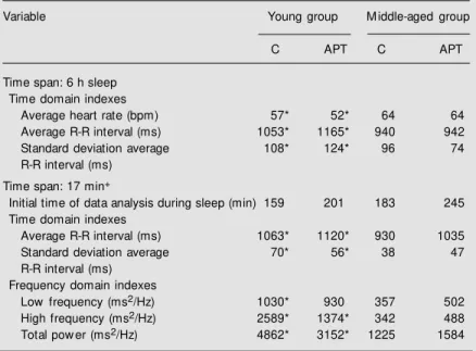
Table 4 shows that the mean R-R interval and standard deviation throughout the 6-h central sleep were significantly (P<0.05) higher in the young than in the middle-aged group under control conditions as well as after physical training. The initial time of sleep analysis for short-term time and fre-quency domains did not differ significantly between groups, being 159 min for the young group and 183 min for the middle-aged group. In relation to the effect of physical training on sleep, there were no statistically signifi-cant changes in average HR, average R-R intervals or standard deviation of average R-R intervals in either group studied.
Frequency domain index of HRV before
and after exercise training. Tables 3 and 4
show the HRV in the frequency domain expressed as absolute values in the resting supine position in the awake and sleeping condition, respectively. In the awake control condition no statistical difference could be found in the low frequency, high frequency or total power component between groups, as reported in absolute values. However, after aerobic physical training only the young group presented a significantly higher abso-lute low frequency power component com-pared to the control condition (P<0.05); in-tergroup analysis showed that in the post-training condition the young group presented
Table 2. Cardiorespiratory variables measured during dynamic exercise before (con-trol, C) and after three months of the aerobic physical training (APT).
Variable Young group M iddle-aged group
C APT C APT
V. O2 AT (ml kg-1 min-1) 19*+ 22+ 13* 16
V. O2 peak (ml kg-1 min-1) 36+ 41+ 28 27
V. O2 AT (ml/min) 1292*+ 1466+ 1186* 1274
V. O2 peak (ml/min) 2630*+ 2800+ 2287 2410
HR (bpm) at AT 134+ 142+ 115 116
HR (bpm) at peak 191+ 188+ 158 165
Pow er (w atts) at AT 106*+ 130+ 77 86
Pow er (w atts) at peak 213* 224 171* 183
Borg’s scale 9 10 8 8.5
Data are reported as medians. Young group, N = 10; middle-aged group, N = 7. AT, anaerobic threshold; HR, heart rate.
* P<0.05 for intragroup comparisons (Wilcoxon test).
+P<0.05 for intergroup comparisons (M ann-Whitney test).
differences in effort perception (Borg’s scale) before and after training were not statisti-cally significant (Table 2).
Table 3. Comparison of heart rate variability during the resting supine aw ake condition (2:00-3:00 pm) before (control, C) and after three months of aerobic physical training (APT).
Variable Young group M iddle-aged group
C APT C APT
Time span: 60 min w ake Time domain indexes
Average heart rate (bpm) 69* 60 72* 62
Average R-R interval (ms) 880* 1003 845* 976
Standard deviation average R-R interval (ms) 85 97 61 64
Time span: 17 min#
Time domain indexes
Average R-R interval (ms) 869* 1010 833* 1000
Standard deviation average R-R interval (ms) 83 92 51 55 Frequency domain indexes
Low frequency (ms2/Hz) 818* 1048+++++ 687 513
High frequency (ms2/Hz) 277 429 265 253
Total pow er (ms2/Hz) 1821 2870 2601 2942
Heart rate variability w as determined using time and frequency domain methods. Data are reported as medians. Young group, N = 10; middle-aged group, N = 7. #Four
consecutive w indow s of 256 s each.
* P<0.05 for intragroup comparisons (Wilcoxon test).
a significantly higher absolute low frequency power component than the middle-aged one (P<0.05).
On the other hand, during the control sleeping condition, the absolute high fre-quency component, an index of vagal tone, was 7.6 times higher (P<0.05) in the young than in the middle-aged group (median = 2589 and 342 ms2/Hz, respectively). This
magnitude decreased 2.8 times after training (P<0.05). The absolute low frequency com-ponent during sleep was significantly higher (P<0.05) in the young than in the middle-aged group only in the control condition (1030 and 357 ms2/Hz) and not in the
aero-bic physical training condition (930 and 502 ms2/Hz). Also, the total power was
signifi-cantly greater (P<0.05) in the young than in the middle-aged group in the control condi-tion (4862 and 1225 ms2/Hz, respectively)
and in the post-training condition (3152 and 1584 ms2/Hz, respectively).
The outstanding finding is that the aero-bic training did not change any frequency domain component (absolute low and high frequencies) when intragroup comparisons were considered.
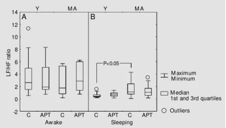
When the content of each frequency band was reported as normalized units (normal-ized high and low frequencies) and as a ratio of absolute values (low/high frequency) the differences were statistically significant only in the sleeping control condition, since nor-malized low frequency and the low/high fre-quency ratio were lower and normalized high frequency was higher in the young than in the middle-aged group. After aerobic train-ing the differences were also not statistically significant for inter- or intragroup compari-sons (Figures 1 and 2).
D iscussio n
In the present study, aerobic exercise training caused an increase in aerobic capac-ity, i.e., oxygen uptake and transport, indi-cated by a significant increase in V. O2
-anaero-Table 4. Comparison of heart rate variability during sleep (0:00-6:00 am), during control conditions (C) and after three months of aerobic physical training (APT).
Variable Young group M iddle-aged group
C APT C APT
Time span: 6 h sleep Time domain indexes
Average heart rate (bpm) 57* 52* 64 64
Average R-R interval (ms) 1053* 1165* 940 942
Standard deviation average 108* 124* 96 74
R-R interval (ms)
Time span: 17 min+
Initial time of data analysis during sleep (min) 159 201 183 245 Time domain indexes
Average R-R interval (ms) 1063* 1120* 930 1035
Standard deviation average 70* 56* 38 47
R-R interval (ms) Frequency domain indexes
Low frequency (ms2/Hz) 1030* 930 357 502
High frequency (ms2/Hz) 2589* 1374* 342 488
Total pow er (ms2/Hz) 4862* 3152* 1225 1584
Heart rate variability w as measured using time and frequency domain methods. Data are reported as medians. Young group, N = 10; middle-aged group, N = 7. +Four
consecutive w indow s of 256 s each.
* P<0.05 for intergroup comparisons (M ann-Whitney test).
Figure 1. Low and high frequency pow er spectral components in normalized units (nu), obtained during aw ake (A) and sleeping (B) conditions, in the young (Y) and middle-aged (M A) groups, during control (C) conditions and after aerobic physical training (APT). Data are reported as box-plots. P<0.05 obtained by the M ann-Whitney test.
P
o
w
e
r
s
p
e
c
tr
u
m
(
n
u
)
100
80 Y
C APT
Low frequency
Y
C APT
High frequency
C APT
C APT
M A Y M A
Y M A
M A
60
40
20
0
P
o
w
e
r
s
p
e
c
tr
u
m
(
n
u
)
100
80
60
40
20
0 B A
C APT
Low frequency
C APT
High frequency
C APT
C APT
M aximum M inimum M edian
1st and 3rd quartiles
P<0.05
P<0.05
bic threshold in both groups and in peak V. O2
for the young group only, as well as an increase in power peak for both groups. This increase in aerobic capacity has been docu-mented in other studies (10,15,16,21,22) for both anaerobic threshold and peak V. O2. The
reasons for the middle-aged group not to increase the peak V. O2 may not be due
exclu-sively to the experimental design used, i.e., a longitudinal study with short-time aerobic training. Some possible explanations for this difference are: 1) anthropometric character-istics and the number of subjects studied; 2) type of experimental protocols used, i.e., continuous exercise on a cycloergometer, in relation to the type of exercise training, i.e., walking and jogging on a field track. On the other hand, the perception of effort at peak level, evaluated with Borg’s scale (17), was not modified by exercise training despite the increase in aerobic power, suggesting that during exercise the volunteers were pushed to their limits.
In addition, our data showed that the V.O2-anaerobic threshold when reported as
absolute and normalized (ml kg-1 min-1)
val-ues proved to be more sensitive than peak V.O2 in detecting aerobic capacity changes
induced by short-term aerobic training in both age groups. It should be emphasized
that anaerobic threshold has also the advan-tage of being a parameter measured directly under submaximal testing conditions inde-pendently of a required voluntary motiva-tion effort by the subjects, i.e., without the need for vocal reinforcement by the re-searcher aiming to extend the exercise to the power value needed to obtain the V. O2 max
or peak V. O2 measurement (16,18).
The HR during the central 6 h of sleep in both conditions was significantly lower in the young than in the middle-aged group. These data agree with the study of Crasset et al. (23), which documented that the R-R interval did not differ between young and older subjects during awake periods but was higher in the young than in the older subjects during both rapid eye movement (REM) and non-REM sleep. Goldsmith et al. (24) have also reported higher R-R interval values dur-ing the night in young men as an expression of vagal predominance during sleep.
Under awake control conditions, the in-tergroup differences of HRV in both the time and frequency domains found in our study were not statistically significant. Neverthe-less, during the sleeping situation there were significant differences in parasympathetic modulation between young and middle-aged men. With respect to high frequency power, the higher values found in the young than in middle-aged group support the interpreta-tion that young men exhibit a higher para-sympathetic activity during sleep. Our re-sults were similar to those obtained by Jensen-Urstad et al. (25) who reported a higher high frequency power value in younger than in older subjects during sleep. Crasset et al. (23) also reported that R-R variability was higher in the young subjects than in older volunteers during the awake and sleeping con-ditions both in the REM and non-REM stages. As a whole, the above findings of an age-dependent difference in R-R intervals and HRV during the sleep condition indicate that the occurrence of vagal predominance in this physi-ological condition is decreased by aging.
Figure 2. Ratio of the low versus high frequency pow er spectral components (LF/HF ratio), obtained during aw ake (A) and sleeping (B) conditions, in the young (Y) and middle-aged (M A) groups, during control (C) conditions and after aerobic physical training (APT). Data are reported as box-plots. P<0.05 obtained by the M ann-Whitney test.
M aximum M inimum
M edian
1st and 3rd quartiles
L
F
/H
F
r
a
ti
o
14
12
10
8
6
4
2
0
-2 C APT
Aw ake
C APT
Sleeping
C APT
C APT Outliers
P<0.05
Y M A Y M A
It should be emphasized that the decrease in vagal tonus in the sinus node has also been reported in pathological conditions such as myocardial infarction, and is associated with an increased risk of new cardiac events and sudden death (8). However, it is not known if the age-dependent decrease in vagal tonus has the same risk effect on the heart of healthy middle-aged men, as documented in the present study.
A limitation of our study was that the 24-h ECG recording was not accompanied by polysomnographic sleep recording. Despite this, the established criterion was to analyze intervals with the highest stationary periods of ECG recording during sleep since this condition most likely occurs in the non-REM stages - when there is a shift of cardiac sympathovagal balance, with a correspond-ing increase in parasympathetic over sympa-thetic stimulation in the sinus node (23,26, 27).
However, it should be emphasized that sleep is a peculiar physiological condition and the mechanisms controlling the high frequency component of R-R variability are unclear. The literature reports that the high frequency component is mediated not only by direct modulation of vagal efferent activ-ity, resulting from baroreceptor responses conveyed to respiratory and blood pressure centers in the central nervous system (28), but also by mechanical effects on the sinus node related to phasic changes in venous return caused by respiratory movement (29). In spite of these considerations, the re-duction of HRV with age in man has been well documented in the literature (4,7,25). However, the mechanisms responsible for this physiological response are unknown. Byrne et al. (4) suggested that age per se, and not the reduction in aerobic capacity or the increase in fat usually associated with the aging process, plays the major role in de-creasing HRV in older subjects. Also, it has been shown that the decline in HRV with aging is mainly, but not exclusively, due to a
decline in parasympathetic tonus (23,30). Concerning the effects of aerobic train-ing on HRV, several studies have found HRV modifications in this physiological con-dition (7,15,24,31). Particularly important is the investigation conducted by Goldsmith et al. (24) who studied and compared 24-h HRV in aerobically trained and untrained healthy young men and observed that para-sympathetic activity is substantially greater in trained than in untrained men, during both waking and sleeping hours.
In the present study, although several significant cardiorespiratory adaptations re-lated to oxygen uptake were induced by dynamic training in both groups, they were not accompanied by significant changes in resting HR and HRV (time and frequency domains) during the sleeping condition. How-ever, in the awake condition a resting brady-cardia was observed after training in both groups studied, without concomitant changes in time or frequency domain HRV. So, the resting bradycardia observed in this study was not accompanied by an increase of the high frequency component, suggesting a non-significant participation of vagal modulation in this adaptive response. In this regard, our results are similar to those of Boutcher and Stein (10) who reported significant increases in both absolute and relative peak V. O2
HRV and a reduction in nocturnal HR. Also, the same investigators have shown that a sustained increase in HRV lasted over a one-year period for those who maintained a steady training level.
Thus, the studies discussed above sup-port the fact that in short-term aerobic physi-cal training different mechanisms may be responsible for the resting bradycardia in-duced by aerobic training. It should be men-tioned that this adaptation is a well-docu-mented response reported for both man (16,25,32) and other species (33).
The resting HR is modulated by a bal-ance between sympathetic and parasympa-thetic tone with a predominance of the latter (16,34). On this basis, some reports state that an increased vagal tonus is the main mech-anism for the bradycardia induced by aero-bic physical training (35). Goldsmith et al. (24) reported that the bradycardia exhibited by endurance-trained individuals is attrib-uted, at least in part, to greater parasympa-thetic activity. However, several other stud-ies have failed to demonstrate differences in vagal tone between trained and untrained subjects (6,32,36,37). Yet, others have indi-cated a decrease in sympathetic activity in the sinus node (38) or both an increase in vagal activity and a decrease in sympathetic activity (39). On the other hand, studies on animals (33) and on humans (6,32,36,40) have suggested that this bradycardia is mainly due to a reduction in intrinsic HR.
Within this context, our data suggest that, at least in men, resting bradycardia induced by short-term aerobic training seems to be mediated by adaptations much more related to intrinsic alterations in the sinus node than to changes in efferent vagal-sympathetic modulation of the sinus node, because the resting bradycardia observed was not ac-companied by an increase of the high fre-quency component and/or a decrease of the low frequency component that would ex-press a higher vagal modulation and/or a low sympathetic modulation of this structure.
Our findings are consistent with previous studies conducted on animals (33) and mainly on humans (6,32,36,37,40) under carefully designed protocols using less invasive or noninvasive procedures associated with bet-ter quantitative methods. Nevertheless, Negrão et al. (33) did not exclude the possi-bility of a decreased resting firing rate of the vagus after training when they observed im-pairment of vagal function evaluated by re-flex bradycardia and electrical vagal stimu-lation.
The absence of significant changes in HRV associated with an increase in aerobic capacity induced by aerobic training, documented in the present study, may be related to the fact that the experimental design was directed to evaluate the cardiorespiratory adaptation in short-term training. Our data support the re-sults of other studies that documented no HRV change after aerobic exercise training in young (40) and middle-aged men (10).
The results of the present investigation indicate that the vagal predominance during sleep in men is reduced with age. Again, the resting bradycardia induced by short-term aero-bic training in both young and middle-aged men is much more related to intrinsic alter-ations in the sinus node than to changes in efferent vagal-sympathetic modulation. Fur-thermore, the greater alterations in aerobic capacity than in HRV in both groups may be related to the magnitude of different time-dependent responses of each cardiorespira-tory variable induced by the training stimulus.
Ackno wle dgm e nts
Re fe re nce s
1. Task Force of the European Society of Cardiology and the North American Soci-et y of Pacing and Elect rophysiology (1996). Heart rate variability: standards of measurement, physiological interpreta-tion and clinical use. Circulation, 93: 1043-1065.
2. Akselrod S, Gordon D, Ubel FA, Shannon DC, Berger AC & Cohen RJ (1981). Pow er spectrum analysis of heart rate fluctua-tion: a quantitative probe of beat-to-beat cardiovascular control. Science, 213: 220-222.
3. Skyschally A, Breuer H-WM & Heusch G (1996). The analysis of heart rate variabil-ity does not provide a reliable measure-ment of cardiac sympathetic activity. Clini-cal Science, 91 (Suppl): 102-104. 4. Byrne EA, Fleg JL, Vaitkevicius PV, Wright
J & Porges SW (1996). Role of aerobic capacity and body mass index in the age-associated decline in heart rate variability.
Journal of Applied Physiology, 81: 743-750.
5. Pagani M , Lombardi F, Guzzetti S, Rimoldi O, Furlan R, Pizzinelli P, Sandrone G, M alfatto G, Dell’Orto S, Piccaluga E, Turiel M , Baselli G, Cerutti S & M alliani A (1986). Pow er spectral analysis of heart rate and arterial pressure variabilities as a marker of sympatho-vagal interaction in man and conscious dog. Circulation Research, 59: 178-193.
6. M art inelli FS, Chacon-M ikahil M PT, Golfetti R, M artins LEB, Lima-Filho EC & Gallo Jr L (2000). Heart rate variability in cyclists and sedentary young men at rest and during head-up tilt. Physiologist, 43: A14-A17, 338 (Abstract).
7. De M eersman RE (1993). Heart rate vari-ability and aerobic fitness. American Heart Journal, 125: 726-731.
8. Bigger Jr JT, Fleiss JL, Steinman RC, Rolnitzky LM , Kleiger RE & Rottman JN (1992). Frequency domain measures of heart period variability and mortality after myocardial infarction. Circulation, 85: 164-171.
9. Sacknoff DM , Gleim GW, Stachenfeld N & Coplan NL (1994). Effect of athletic training on heart rate variability. American Heart Journal, 127: 1275-1278.
10. Boutcher SH & Stein P (1995). Associa-tion betw een heart rate variability and training response in sedentary middle-aged men. European Journal of Applied Physiology, 70: 75-80.
11. Furlan R, Piazza S, Dell’Orto S, Gentile E, Cerutti S, Pagani M & M alliani A (1993).
Early and late effects of exercise and ath-letic training on neural mechanisms con-trolling heart rate. Cardiovascular Re-search, 27: 482-488.
12. Shin K, M inamitani H, Onishi S, Yamazaki H & Lee M (1997). Autonomic differences betw een athletes and nonathletes: spec-tral analysis approach. M edicine and Sci-ence in Sports and Exercise, 29: 1482-1490.
13. Inbar O, Oren A, Scheinow itz M , Rotstein A, Dlin R & Casaburi R (1994). Normal cardiopulmonary responses during incre-mental exercise in 20- to 70-yr-old men.
M edicine and Science in Sports and Exer-cise, 26: 538-546.
14. Jackson AS, Beard EF, Wier LT, Ross RM , Stuteville JE & Blair SN (1995). Changes in aerobic pow er of men, ages 25-70 yr.
M edicine and Science in Sports and Exer-cise, 27: 113-120.
15. Stein PK, Ehsani AA, Domitrovich PP, Kleiger RE & Rottman JN (1999). Effect of exercise training on heart rate variability in healthy older adults. American Heart Journal, 138 (Part 1): 567-576.
16. Chacon-M ikahil M PT, Forti VAM , Catai AM , Szrajer JS, Golfetti R, M artins LEB, Lima-Filho EC, Wanderley JS, M arin Neto JA, M aciel BC & Gallo-Junior L (1998). Cardiorespiratory adaptations induced by aerobic training in middle-aged men: the importance of a decrease in sympathetic stimulation for the contribution of dynam-ic exercise tachycardia. Brazilian Journal of M edical and Biological Research, 31: 705-712.
17. Borg G (1998). Borg’s Perceived Exertion and Pain Scales. Human Kinetics, Cham-paign, IL, USA.
18. Wasserman K, Hansen JE, Sue DY, Whipp BJ & Casaburi R (1994). Principles of Ex-ercise Testing and Interpretation. 2nd edn. Lea & Febiger, Philadelphia, PA, USA. 19. Silva E, Catai AM , Trevelin LC, Guimarães
JO, Silva Jr LP, Silva LM P, Oliveira L, M ilan LA, M artins LEB & Gallo Junior L (1994). Design of a computerized system to evaluate the cardiac function during dynamic exercise. Physics in M edicine and Biology, 33: 409 (Abstract). 20. M alliani A (1995). Association of heart rate
variability components w ith physiological regulatory mechanisms. In: M alik M & Camm AJ (Editors), Heart Rate Variability. Futura Publishing Company, Armonk, NY, USA, 173-188.
21. Davis JA, Frank M H, W hipp BJ & Wasserman K (1979). Anaerobic
thresh-old alterations caused by endurance train-ing in middle-aged men. Journal of Ap-plied Physiology: Respiratory, Environ-mental and ExercisePhysiology, 46: 1039-1046.
22. Gregoire J, Tuck S, Yam am ot o Y & Hughson RL (1996). Heart rate variability at rest and exercise: influence of age, gender, and physical training. Canadian Journal of Applied Physiology, 21: 455-470.
23. Crasset V, M ezzetti S, Antoine M , Lin-kow ski P, Degaute JP & van de Borne P (2001). Effects of aging and cardiac de-nervation on heart rate variability during sleep. Circulation, 103: 84-88.
24. Goldsmith RL, Bigger Jr JT, Steinman RC & Fleiss JL (1992). Comparison of 24-hour parasympathetic activity in endur-ance-trained and untrained young men.
Journal of the American College of Cardi-ology, 20: 552-558.
25. Jensen-Urstad K, Storck N, Bouvier F, Ericson M , Lindblad LE & Jensen-Urstad M (1997). Heart rate variability in healthy subjects is related to age and gender.
Acta Physiologica Scandinavica, 160: 235-241.
26. Scholz UJ, Bianchi AM , Cerutti S & Kubicki S (1997). Vegetative background of sleep: spectral analysis of the heart rate variabil-ity. Physiology and Behavior, 62: 1037-1043.
27. Elsenbruch S, Harnish M J & Orr WC (1999). Heart rate variability during w ak-ing and sleep in healthy males and fe-males. Sleep, 22: 1067-1071.
28. Piepoli M , Sleight P, Leuzzi S, Valle F, Spadacini G, Passino C, Johnston J & Ber-nardi L (1997). Origin of respiratory sinus arrhythmia in conscious humans. An im-portant role for arterial carotid barorecep-tors. Circulation, 95: 1813-1821. 29. Bernardi L, Keller F, Sanders M , Reddy
PS, Griffith B, M eno F & Pinsky M R (1989). Respiratory sinus arrhythmia in the denervated human heart. Journal of Ap-plied Physiology, 67: 1447-1455. 30. Shannon DC, Carley DW & Benson H
(1987). Aging of modulation of heart rate.
American Journal of Physiology, 253 (Part 2): H874-H877.
31. Schuit AJ, van Amelsvoort LGPM , Verheij TC, Rijneke RD, M aan AC, Sw enne CA & Schouten EG (1999). Exercise training and heart rate variability in older people. M edi-cine and Science in Sports and Exercise, 31: 816-821.
Guz A (1982). Sympathetic and parasym-pathetic cardiac control in athletes and nonathletes at rest. Journal of Applied Physiology: Respiratory, Environmental and ExercisePhysiology, 52: 1652-1657. 33. Negrão CE, M oreira ED, Santos M CLM ,
Farah VM A & Krieger EM (1992). Vagal function impairment after exercise train-ing. Journal of Applied Physiology: Respi-ratory and Environmental Exercise, 72: 1749-1753.
34. Lakatta EG (1995). Cardiovascular system. In: M asoro EJ (Editor), Handbook of Phys-iology. A Critical Comprehensive Presen-tation of Physiological Know ledge and Concepts. Oxford University Press, New York, NY, USA, 413-474.
35. Smith M L, Hudson DL, Graitzer HM & Raven PB (1989). Exercise training brady-cardia: the role of autonomic balance.
M edicine and Science in Sports and Exer-cise, 21: 40-44.
36. M aciel BC, Gallo Junior L, M arin Neto JA, Lima Filho EC, Terra Filho J & M anço JC (1985). Parasympathetic contribution to bradycardia induced by endurance train-ing in man. Cardiovascular Research, 19: 642-648.
37. Perrault H, Gagnon M C, Johnson D, M okrane A & Nadeau RA (1996). An en-hanced vagal influence does not explain training-induced bradycardia. Physiologist, 39: A20 (Abstract).
38. Williams RS, Eden RS, M oll M E, Lester
RM & Wallace AG (1981). Autonomic mechanisms of training bradycardia: beta-adrenergic receptors in humans. Journal of Applied Physiology: Respiratory, Envi-ronmental and Exercise Physiology, 51: 1232-1237.
39. Ekblom B, Kilbom A & Soltysiak J (1973). Physical training, bradycardia, and auto-nom ic nervous system . Scandinavian Journal of Clinical and Laboratory Investi-gation, 32: 251-256.