Cardiorespiratory adaptations induced
by aerobic training in middle-aged
men: the importance of a decrease in
sympathetic stimulation for the
contri-bution of dynamic exercise tachycardia
1Laboratório de Fisiologia do Exercício, Faculdade de Educação Física,
and 2Departamento de Fisiologia e Biofísica, Instituto de Biologia,
Universidade Estadual de Campinas, Campinas, SP, Brasil
3Divisão de Cardiologia, Departamento de Clínica Médica,
Faculdade de Medicina de Ribeirão Preto, Hospital das Clínicas, Universidade de São Paulo, Ribeirão Preto, SP, Brasil
M.P.T. Chacon-Mikahil1,2,
V.A.M. Forti1, A.M. Catai1,2,
J.S. Szrajer1, R. Golfetti1,
L.E.B. Martins1, E.C. Lima-Filho1,
J.S. Wanderley1,
J.A. Marin-Neto3, B.C. Maciel3
and L. Gallo-Jr.3
Abstract
We investigated the effects of aerobic training on the efferent auto-nomic control of heart rate (HR) during dynamic exercise in middle-aged men, eight of whom underwent exercise training (T) while the other seven continued their sedentary (S) life style. The training was conducted over 10 months (three 1-h/sessions/week on a field track at 70-85% of the peak HR). The contribution of sympathetic and para-sympathetic exercise tachycardia was determined in terms of differ-ences in the time constant effects on the HR response obtained using a discontinuous protocol (4-min tests at 25, 50, 100 and 125 watts on a cycle ergometer), and a continuous protocol (25 watts/min until exhaustion) allowed the quantification of the parameters (anaerobic threshold, V
.
O2 AT; peak O2 uptake, V.
O2 peak; power peak) that reflect
oxygen transport. The results obtained for the S and the T groups were: 1) a smaller resting HR in T (66 beats/min) when compared to S (84 beats/min); 2) during exercise, a small increase in the fast tachycardia (∆0-10 s) related to vagal withdrawal (P<0.05, only at 25 watts) was observed in T at all powers; at middle and higher powers a significant decrease (P<0.05 at 50, 100 and 125 watts) in the slow tachycardia (∆1-4 min) related to a sympathetic-dependent mechanism was ob-served in T; 3) the V
.
O2 AT (S = 1.06 and T = 1.33 l/min) and V.
O2 peak
(S = 1.97 and T = 2.47 l/min) were higher in T (P<0.05). These results demonstrate that aerobic training can induce significant physiological adaptations in middle-aged men, mainly expressed as a decrease in the sympathetic effects on heart rate associated with an increase in oxygen transport during dynamic exercise.
Correspondence L. Gallo-Jr. Divisão de Cardiologia Departamento de Clínica Médica Hospital das Clínicas, FMRP, USP 14049-900 Ribeirão Preto, SP Brasil
Fax: 55 (016) 633-1144
Research supported by FAPESP (Nos. 89/1297-6, 91/4754-9) and FAEP-UNICAMP (No. 1055/91).
Received April 9, 1997 Accepted January 15, 1998
Key words
•Sympathetic modulation of heart rate
•Dynamic exercise •Aerobic training
Introduction
The cardiorespiratory system at rest and during exercise is usually studied by moni-toring variables such as heart rate (HR) and oxygen transport (V
.
O2). HR in particular isconsidered to be an important variable in the quantification of certain physiological prop-erties of the cardiovascular system. This vari-able has the advantage that it can be deter-mined readily with minimum error using noninvasive methods (1,2).
The ability to continuously monitor HR permits the use of dynamic exercise in func-tional evaluations of the two components of the cardiac autonomic nervous system, as suggested by the investigations conduced with or without pharmacological blockade on sedentary or trained men (3-6). Such studies suggest that tachycardia evoked by dynamic exercise is mediated by a biphasic mechanism which initially involves vagal withdrawal (very fast increase of HR), and increased delayed sympathetic activity par-ticipates as an additional component at higher powers (slow linear HR increase) (1,3,4).
Oxygen transport is a complex process that involves the participation of several physiological components such as the car-diorespiratory system, transport mechanisms (hemoglobin and myoglobin) and oxidative enzymes. The two parameters that best re-flect the functioning of this process are maxi-mal oxygen consumption (V
.
O2 max) andven-tilatory anaerobic threshold (AT). Since the direct measurement of V
.
O2 max is verydiffi-cult to obtain under normal and pathological conditions, the AT provides a useful means of measuring oxygen transport mainly be-cause it can be easily determined at sub-maximal powers by noninvasive methods. In addition, the AT allows a suitable separation of two important physiological states: 1) below the AT, the cardiorespiratory responses are linear and most variables reach a steady-state condition, and 2) above the AT, the respective responses assume a nonlinear
be-havior in which the variables do not fully stabilize (1,7,8).
Several studies have shown that maximal aerobic capacity reaches a maximum around the age of 30 years and that it decreases pro-gressively thereafter. Limitations related to a reduction in maximum cardiac output appear to play an important role in age-dependent changes in aerobic transport during exercise (9-13). While many studies have character-ized the cardiovascular responses in middle-aged men, the autonomic control of HR in this particular group during exercise has received much less attention (12-18).
In the present study we determined whether aerobic training induced significant adaptations in the sympathetic and parasym-pathetic control of HR during dynamic exer-cise in middle-aged men.
Material and Methods
Subjects
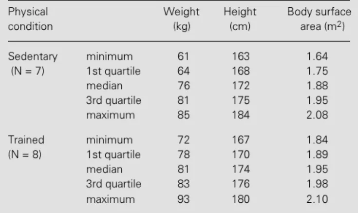
Fifteen men (46-60 years old; mean ± SD, 50.7 ± 3.6) were studied. The character-istics of the groups are shown in Table 1. All volunteers were in good health, and had no evidence of cardiovascular disease on the basis of clinical and laboratory evaluation that included the analysis of standard elec-trocardiograms and chest X-rays. The sub-jects were not taking any medication and led a relatively sedentary life style, although some of them occasionally participated in week-end sports. During the study, seven subjects continued to lead a sedentary life while the remaining eight underwent an aero-bic training program. All volunteers gave informed consent to participate in this inves-tigation. The study was approved by the Ethics Committee of the University Hospital of the State University of Campinas.
Aerobic training
10-month aerobic training program consisting of walking and jogging on a field track three times a week for 1 h. The intensity of exer-cise was set at 70-85% of the peak heart rate (HRpeak) obtained in a previous exercise session (sitting on a cycle ergometer) per-formed in the laboratory.
Experimental protocols
The subjects were studied at rest (supine position) and during dynamic exercise tests on a cycle ergometer. In all the protocols, the electrocardiogram (ECG) was continuously monitored using the precordial MC-5 lead. Before the tests, the subjects were allowed to familiarize themselves with the procedures and the equipment to be used. The sessions were held 2-3 h after a light meal at an environmental temperature of around 23oC.
The resting HR was obtained after 20 min of resting in the supine position. The HR values obtained here and immediately be-fore (1 min) and during exercise testing (in a sitting position) are reported as the mean determined at 10-s intervals by counting the number of QRS complexes directly from the ECG tracing using a calibrated ruler.
The dynamic exercise was always per-formed in the sitting position using an elec-tromagnetic braked cycle ergometer model Ciclo II (FUNBEC, São Paulo, SP, Brazil). Two exercise protocols were employed. In the discontinuous protocol workloads corre-sponding to 25, 50, 100 and 125 watts (4 min each) were used with a frequency of 60 rpm being maintained throughout each stage. Varying resting periods were allowed be-tween the different powers so that HR could return to control (basal) levels. The electro-cardiogram was recorded continuously from 30 s before the beginning to 1 min after the end of effort using a one-channel recording unit model ECG40A (FUNBEC, Brazil).
The main objective of this protocol was to evaluate the contribution of the sympa-thetic and parasympasympa-thetic systems to the
increase in HR induced by dynamic exercise and was based on the time constant differ-ences of the two divisions of the autonomic nervous system when modulating HR changes during dynamic exercise. This method was validated in previous studies in which the HR response induced by exercise was evaluated before and after selective phar-macological blockade of the sympathetic and parasympathetic systems (1,3,4).
The continuous protocol was performed by progressively increasing power steps un-til physical exhaustion. The objective of this protocol was to evaluate aerobic capacity by measuring the AT (7,19) at submaximal work loads and the V
.
O2 peak at physicalexhaus-tion. After a 2-min warm-up period (at 5 watts), the power was increased by 25 watts each minute (steps) until the volunteers be-came physically exhausted. The researcher closely monitored the signals from the sub-ject (facial expression, blood pressure and abnormal ECG recordings) and always ver-bally encouraged the volunteer to maximize his performance. The test exercise ended when the subject was unable to maintain the standard cycling frequency. To avoid syn-cope after reaching peak power, the subjects continued the exercise at a lower frequency and power for approximately 2 min.
During both experimental protocols, the subjects breathed through a low-resistance, non-rebreathing, two-way valve model 2700
Table 1 - Anthropometric characteristics of the groups studied.
Physical Weight Height Body surface
condition (kg) (cm) area (m2)
Sedentary minimum 61 163 1.64
(N = 7) 1st quartile 64 168 1.75
median 76 172 1.88
3rd quartile 81 175 1.95
maximum 85 184 2.08
Trained minimum 72 167 1.84
(N = 8) 1st quartile 78 170 1.89
median 81 174 1.95
3rd quartile 83 176 1.98
test was used to compare the variables and parameters between the two studied groups. The level of significance was set at 5%.
Results
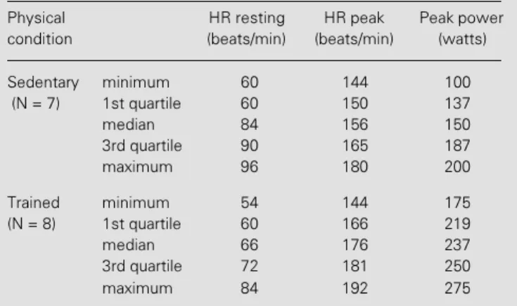
The resting HR and the HR and power peaks during exercise for the sedentary and trained groups are shown in Table 2. The trained group had lower median values than the sedentary group under resting conditions but the difference was not statistically sig-nificant. An inverse non-significant situa-tion was also observed for HRpeak. Under both conditions, the difference between the two groups was approximately 20 beats/min.
The increases in HR (∆HR) during
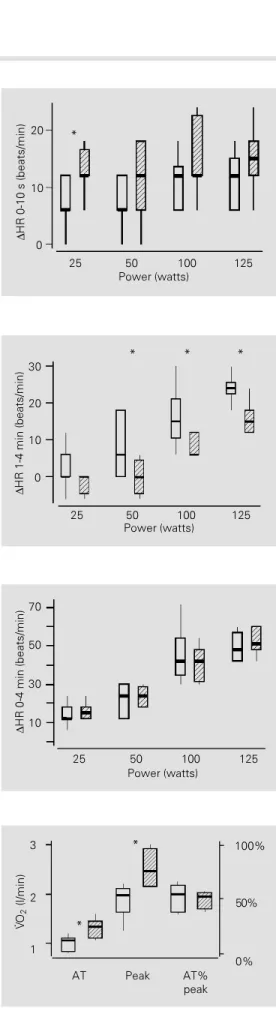
dy-namic exercise in the sedentary and trained groups are presented in Figures 1, 2 and 3.
In spite of the higher median values ob-served in the trained group during the 0-10-s interval (∆0-10 s), the difference was only significant at a power value of 25 watts (P = 0.05) (Figure 1). On the other hand, the 1-4 min median increases in HR (Figure 2) were, with one exception (25 watts, P = 0.24), lower in the trained than in the sedentary group at all power values: 50 watts (P = 0.04), 100 watts (P = 0.04) and 125 watts (P = 0.01). For the 0-4 min increases in HR (∆ 0-4 min), the median values of the two groups were similar at all powers studied (Figure 3). Figure 4 illustrates several parameters that reflect the magnitude of oxygen trans-port at submaximal (V
.
O2 AT) and peak (V.
O2peak) powers. The values for V
.
O2 AT(sed-entary, 1.06 l/min; trained, 1.33 l/min; P = 0.02) and V
.
O2 peak (sedentary, 1.97 l/min;trained, 2.47 l/min; P = 0.01) were signifi-cantly higher in the trained group. However, when the AT was expressed as a percentage of V
.
O2 peak (AT% V.
O2 peak) the results
were similar for both groups.
Discussion
The resting HR is modulated by a bal-Table 2 - Functional data of the subject groups studied.
Physical HR resting HR peak Peak power
condition (beats/min) (beats/min) (watts)
Sedentary minimum 60 144 100
(N = 7) 1st quartile 60 150 137
median 84 156 150
3rd quartile 90 165 187
maximum 96 180 200
Trained minimum 54 144 175
(N = 8) 1st quartile 60 166 219
median 66 176 237
3rd quartile 72 181 250
maximum 84 192 275
(Hans Rudolph, Kansas City, MO), and the expired gas was analyzed continuously us-ing a metabolic measurus-ing system (MMC
Horizon Systems, Sensor Medics, Yorba
Linda, CA). Average values for ventilation (V
.
), oxygen uptake (V.
O2), carbon dioxideoutput (V
.
CO2), and respiratory exchangera-tio (RER) were calculated automatically at 15-s intervals.
The peak of each variable was selected as the highest value reached during the incre-mental exercise period since, as a result of muscle fatigue, the true V
.
O2 max was neverattained. This was indicated by the lack of a plateau (saturation) in oxygen uptake during exercise (7).
Using the ventilatory and gas exchange responses during the last exercise test it was possible to obtain a measurement of AT for all subjects with this noninvasive method. These values, reported as V
.
O2 (l/min), weredetermined using the criterion of a nonlinear increase in V
.
and V.
CO2 in comparison to thelinear increase in V
.
O2 at submaximal workloads (7,19,20). The AT values related to V
.
O2 peak (AT% V.
O2 peak) were also
calcu-lated.
Statistical analysis
ance between sympathetic and parasympa-thetic tone, with the latter predominating. Although the resting HR does not change with increasing age, Lakatta (21) has re-ported a decrease in the respiratory variation of HR, probably caused by a reduction in parasympathetic and sympathetic modula-tion. In the presence of both sympathetic and parasympathetic blockade, the intrinsic si-nus node rate decreases significantly with age: at the age of 20 years, the average intrinsic HR is 104 beats/min compared with 92 beats/min in the 45-55-year-old group (22). In the middle-aged men studied here, aerobic training was expected to cause a greater reduction in resting HR than that actually observed; the absence of a statisti-cally significant difference in our data prob-ably reflects the small number of subjects studied. Regardless of these considerations, the magnitude of bradycardia found in middle-aged men was comparable to that reported by others (14,15). It should be em-phasized that this adaptation in HR is usually considered to be a good marker of adequate training (10,23-26) and, at least in men, an intrinsic mechanism seems to play a pre-dominant role in the respective adaptation (27-29).
Since in the resting state the cardiovascu-lar system usually operates below its full functional reserve, studies performed in this situation cannot adequately characterize the full extent of the regulatory mechanisms in-volved.
In the present investigation, we studied the HR responses during exercise and their relation to the autonomic control of HR us-ing a nonpharmacological method based on the time constant differences for the activa-tion of each efferent limb of the autonomic nerves acting on the sinus node (3,4,30,31). The fast tachycardia seen after the start of exercise occurs during the initial 10 s of activity at all levels of exercise and is medi-ated by a sudden reduction in vagal tone on the sinus node (3,31,32). The HR increase in
Figure 1 - Increases in HR (∆HR) during dynamic exercise at power values of 25, 50, 100 and 125 watts, in the 0-10-s interval (∆0-10 s) in the sedentary (open boxes, N = 7) and trained (hatched boxes, N = 8) groups. The data are reported as medi-ans, quartiles (1st and 3rd) and minimum and maximum values. *P<0.05.
Figure 2 - Increases in HR (∆HR) during dynamic exercise at power values of 25, 50, 100 and 125 watts, in the 1-4-min inter-val (∆1-4 min) in the sedentary (open boxes, N = 7) and trained (hatched boxes, N = 8) groups. The data are reported as medi-ans, quartiles (1st and 3rd) and minimum and maximum values. *P<0.05.
∆
HR 1-4 min (beats/min)
20
10
0
25 50 100 125
Power (watts)
*
30 * *
Figure 3 - Increases in HR (∆HR) during dynamic exercise at power values of 25, 50, 100 and 125 watts, in the 0-4-min inter-val (∆0-4 min) in the sedentary (open boxes, N = 7) and trained (hatched boxes, N = 8) groups. The data are reported as medi-ans, quartiles (1st and 3rd) and minimum and maximum values.
∆
HR 0-4 min (beats/min)
50
30
10
25 50 100 125
Power (watts) 70
100%
50%
0%
*
*
3
2
1
AT Peak AT%
peak
V
. O
2
(l/min)
Figure 4 - The anaerobic thresh-old (V
.
O2 AT), peak oxygen up-take (V.
O2 peak) and the AT re-lated to V.
O2 peak (AT% V.
O2 peak) values during a continu-ous protocol in the sedentary (open boxes, N = 7) and trained (hatched boxes, N = 8) groups. The data are reported as medi-ans, quartiles (1st and 3rd) and minimum and maximum values. *P<0.05.∆
HR 0-10 s (beats/min)
20
10
0
25 50 100 125
Power (watts)
the ∆0-10-s interval (vagal-dependent) found in our study differed little between groups, indicating simply a minor adaptation of va-gal mechanisms after training.
On the other hand, the slow increase in HR (∆1-4 min) during exercise was signifi-cantly lower in the trained group. This find-ing indicates that aerobic trainfind-ing can induce a substantial reduction in sympathetic stimu-lation during dynamic exercise at power lev-els above the anaerobic threshold (1,4). The nonsignificant difference in the ∆1-4 min increases in HR at 25 watts in both groups is explained by the fact that at this low intensity workload the exercise tachycardia predomi-nantly reflects parasympathetic withdrawal on the sinus node (1).
The absence of any change in total HR, i.e., from 0 to 4 min, reinforces the impor-tance of measuring the fractional contribu-tion of exercise tachycardia caused by each efferent division of the autonomic nervous system. Overall, our results for this age group are similar to those obtained for younger subjects (3,31).
In agreement with others (14,15), we ob-served small nonsignificant differences in HRpeak between groups. The values in both groups were lower than those found in younger subjects (9,33-36). This decrease in the HRpeak during exercise in older indi-viduals is usually independent of the degree of aerobic performance or of a concomitant disease, since a deficit of similar magnitude is seen in both healthy sedentary men and women as well as in older athletes (16,34,37, 38).
Numerous studies have described the changes in cardiovascular function occur-ring with age, particularly duoccur-ring acute exer-cise (9,13,15,21,36,39-41). Older individu-als show a lower aerobic capacity, and have several other noncardiovascular factors that can limit their physical performance, includ-ing orthopedic difficulties, alterations in body composition, a reduction in muscle mass and strength, a lower threshold for
neuromuscu-lar fatigue and so on (21).
It is well known that V
.
O2 max and V.
O2peak decline with age as reported in longitu-dinal and cross-sectional studies (9,39-41). The maximum cardiac output also declines progressively because of an associated re-duction in maximum HR (17,21). An impor-tant point to be considered is that the V
.
O2max or V
.
O2 peak can be improved withexer-cise training of sufficient duration, frequency, and intensity (15,17,18,34,42,43). The in-crease in V
.
O2 peak in our trained group wascomparable to that reported by others and can reflect an important ability for adapta-tion at all ages (14,15,17).
The major concern in exercise testing should be to determine whether the cardio-respiratory system is capable of providing the required amount of oxygen to the exer-cise-stressed muscles (7). However, for meth-odological reasons in many situations it is impossible to obtain the values of V
.
O2 maxthat are needed to characterize oxygen trans-port during maximal exercise. In such cir-cumstances, the anaerobic threshold is the best alternative to solve this problem.
The AT is defined as the power level of exercise above which aerobic energy pro-duction is supplemented by anaerobic mecha-nisms. In this condition, there is an increase in lactate and in the lactate/pyruvate ratio in muscle and arterial blood. AT shows a good correlation with V
.
O2 max and has proved tobe very useful in quantifying oxygen trans-port and its modification by physiological and pathological conditions (7).
Particularly important are the changes in AT values induced by aging and aerobic training. The absolute median AT values of our trained subjects were higher than those of the sedentary group, as also reported else-where (44,45). However, when AT was ex-pressed as a percentage of the V
.
O2 peak,there was no difference between groups. The reason for this finding is unclear.
to reach a steady state. Indeed, the full stabi-lization of cardiorespiratory variables is never reached during exercise above the AT (1). This nonstationary response of HR is related to the occurrence of sympathetic drive. The progressive increase in ∆1-4 min HR, with increasing power values reflects the propor-tional elevation of sympathetic stimulation at high workloads. In this context, our find-ings have shown a displacement of the AT toward higher workloads in middle-aged men similar to that occurring with the beginning and the intensification of sympathetic stimu-lation of the sinus node during exercise.
The present results have shown that aero-bic training induces significant physiologi-cal adaptations in the cardiorespiratory sys-tem of middle-aged men. The best markers of these adaptations were the smaller
sympa-thetic tachycardia (∆1-4 min) at comparable workloads and the improvement of oxygen transport, as documented by the increase in the anaerobic threshold and V
.
O2 peak duringdynamic exercise.
Acknowledgments
The authors are grateful to the Departa-mento de Clínica Médica and the Seção de Cardiologia, Faculdade de Ciências Médicas, Universidade Estadual de Campinas (UNICAMP), where the clinical examina-tion and standard ergometric and biochemi-cal tests were performed. We are also in-debted to Cleide Marques Antloga and Pedro Mikahil Neto for a final revision of the manu-script.
References
1. Gallo-Jr L, Maciel BC, Marin-Neto JA, Mar-tins LEB, Lima-Filho EC, Golfetti R, Chacon MPT & Forti VAM (1995). Control of heart rate during exercise in health and disease. Brazilian Journal of Medical and
Biological Research, 28: 1179-1184.
2. Miyamoto Y, Hiura T, Tamura T, Nakamura T, Higuchi J & Mikami T (1982). Dynamics of cardiac, respiratory and metabolic func-tion in men in response to step workload.
Journal of Applied Physiology, 52:
1198-1208.
3. Maciel BC, Gallo-Jr L, Marin-Neto JA, Lima-Filho EC & Martins LEB (1986). Au-tonomic nervous control of the heart rate during dynamic exercise in normal man.
Clinical Science, 71: 457-460.
4. Gallo-Jr L, Maciel BC, Marin-Neto JA & Martins LEB (1989). Sympathetic and parasympathetic changes in heart rate control during dynamic exercise induced by endurance training in man. Brazilian Journal of Medical and Biological
Re-search, 22: 631-643.
5. Chacon MPT, Forti VAM, Catai AM, Szrajer JS, Paschoal MA, Golfetti R, Mar-tins LEB, Maciel BC, Marin-Neto JA, Lima-Filho EC, Wanderley JS & Gallo-Jr L (1994). Cardiorespiratory adaptation to aerobic training in middle-aged men.
Physics in Medicine and Biology, 39a (Part
1): 115 (Abstract).
6. Forti VAM, Chacon MPT, Catai AM, Szrajer JS, Paschoal MA, Golfetti R, Mar-tins LEB, Maciel BC, Marin-Neto JA, Lima-Filho EC, Wanderley JS, Sztejnsznajd CA & Gallo-Jr L (1994). The effects of aerobic training on cardiovascular system in menopause. Physics in Medicine and
Bi-ology, 39a (Part 1): 113 (Abstract).
7. Wasserman K, Hansen JE, Sue DY, Whipp BJ & Casaburi R (1994). Principles of
Ex-ercise Testing and Interpretation. 2nd
edn. Lea & Febiger, Philadelphia. 8. Brooks GA (1991). Current concepts in
lactate exchange. Medicine and Science
in Sports and Exercise, 23: 895-906.
9. Robinson S (1938). Experimental studies of physical fitness in relation to age.
Arbeitsphysiologie, 10: 251-323.
10. Saltin B & Rowell LB (1980). Functional adaptations to physical activity and inac-tivity. Federation Proceedings, 39: 1506-1513.
11. Sutton JR (1992). V
.
O2 max. New con-cepts on an old theme. Medicine andSci-ence in Sports and Exercise, 24: 26-29.
12. Inbar O, Oren A, Scheinowitz M, Rotstein A, Dlin R & Casaburi R (1994). Normal cardiopulmonary responses during incre-mental exercise in 20- to 70-year-old men. Medicine and Science in Sports and Exer-cise, 26: 538-546.
13. Jackson A, Beard EF, Wier LT, Ross RM,
Stuteville JE & Blair SN (1995). Changes in aerobic power of men, ages 25-70yr. Medicine and Science in Sports and Exer-cise, 27: 113-120.
14. Hanson JS, Tabakin BS, Levy AM & Nedde W (1968). Long-term physical train-ing and cardiovascular dynamics in middle-aged men. Circulation, 38: 783-799.
15. Hagberg JM (1987). Effect of training on the decline of V
.
O2 max with aging.Fed-eration Proceedings, 46: 1830-1833.
16. Heath GW, Hagberg JM, Ehsani AA & Holloszy JO (1981). A physiological com-parison of young and older endurance ath-letes. Journal of Applied Physiology, 51: 634-640.
17. Posner JD, Gorman KM & Klein HS (1986). Exercise capacity in the elderly. American
Journal of Cardiology, 57: 52C-58C.
18. Ehsani AA (1987). Cardiovascular adapta-tions to exercise training in the elderly.
Federation Proceedings, 46: 1840-1843.
19. Beaver WL, Wasserman K & Whipp BJ (1986). A new method for detecting anaerobic threshold by gas exchange.
Journal of Applied Physiology, 60:
2020-2027.
21. Lakatta EG (1993). Cardiovascular regula-tory mechanisms in advanced age.
Physi-ological Reviews, 73: 413-467.
22. Jose AD (1966). Effect of combined sym-pathetic and parasymsym-pathetic blockade on heart rate and cardiac function in man.
American Journal of Cardiology, 18:
476-478.
23. Negrão CE, Forjaz CLM, Rondon MUPB & Brum PC (1996). Adaptação cardiovascu-lar ao treinamento físico dinâmico. In: Souza AGM & Mansur AJ (Editors),
Socesp Cardiologia. Vol. 2. Atheneu, São
Paulo.
24 Åstrand P-O & Rodahl K (1980). Tratado
de Fisiologia do Exercício. 2nd edn.
Interamericana, Rio de Janeiro.
25. Ekblom B, Kilbom A & Stoltysiak J (1973). Physical training, bradycardia and auto-nomic nervous system. Scandinavian Journal of Clinical and Laboratory
Investi-gation, 32: 251-256.
26. Lewis SF, Nylander E, Gad P & Areskog NH (1980). Non autonomic component in bradycardia of endurance trained men at rest and during exercise. Acta
Physi-ologica Scandinavica, 109: 297-305.
27. Katona PG, McLean M, Dighton DH, Davis HD & Guz A (1982). Sympathetic and para-sympathetic cardiac control in athletes and non-athletes at rest. Journal of Ap-plied Physiology: Respiratory,
Environ-mental and Exercise Physiology, 52:
1652-1657.
28. Maciel BC, Gallo-Jr L, Marin-Neto JA, Lima-Filho EC, Terra-Filho J & Manço JC (1985). Parasympathetic contribution to bradycardia induced by endurance train-ing in man. Cardiovascular Research, 19: 642-648.
29. Perrault H, Gagnon MC, Johnson D,
Mokrane A & Nadeau RA (1996). An en-hanced vagal influence does not explain training induced bradycardia. Physiologist, 39: 20 (Abstract).
30. Maciel BC, Gallo-Jr L, Marin-Neto JA, Maciel LMZ & Martins LEB (1988). Auto-nomic control of heart rate during dynam-ic exercise in human hyperthyroidism.
Clinical Science, 75: 209-215.
31. Gallo-Jr L, Morelo-Filho J, Maciel BC, Marin-Neto JA, Martins LEB & Lima-Filho EC (1987). Functional evaluation of sym-pathetic and parasymsym-pathetic system in Chagas disease using dynamic exercise.
Cardiovascular Research, 21: 922-927.
32. Fagraeus L & Linnarsson D (1976). Auto-nomic origin of heart rate fluctuations at the onset of muscular exercise. Journal of
Applied Physiology, 40: 679-682.
33. Landeree BR, Thomas TR, Ziogas G, Smith TD & Zhang Q (1995). %V
.
O2 max versus % HRmax regressions for six modes of exercise. Medicine and Sciencein Sports and Exercise, 27: 458-461.
34. Åstrand I (1960). Aerobic work capacity in men and women with special reference to age. Acta Physiologica Scandinavica, 49 (Suppl 169): 1-92.
35. Hammond HK & Froelicher VF (1985). Nor-mal and abnorNor-mal heart rate responses to exercise. Progress in Cardiovascular
Dis-eases, 27: 271-296.
36. Babcock MA, Paterson DH & Cunningham DA (1994). Effects of aerobic endurance training on gas exchange kinetics of older men. Medicine and Science in Sports and
Exercise, 26: 447-452.
37. Fleg JL, Schulman S, Gerstenblith G, Goldber A, Tankersley C, Becker L, Clulow J, Drinkwater D, Lakatta L & Lakatta EG (1988). Central versus peripheral
adapta-tions in highly trained seniors. Physiolo-gist, 31: 158 (Abstract).
38. Ehsani AA, Ogawa T, Miller TR, Spina RJ & Jilka SM (1991). Exercise training im-proves left ventricular systolic function in older men. Circulation, 83: 96-103. 39. Lakatta EG (1995). Cardiovascular system.
In: Masoro EJ (Editor), Handbook of Phys-iology. A Critical, Comprehensive Presen-tation of Physiological Knowledge and
Concepts. Section 11: Aging. Oxford
Uni-versity Press, New York.
40. Åstrand I, Åstrand P-O, Hallback I & Kilbom Å (1973). Reduction in maximal oxygen uptake with age. Journal of
Ap-plied Physiology, 35: 649-654.
41. Buskirk ER & Hodgson JL (1987). Age and aerobic power: the rate of change in men and women. Federation Proceedings, 46: 1824-1829.
42. Pollock ML, Foster C, Knapp D, Rod JL & Schmidt DH (1987). Effect of age and training on aerobic capacity and body com-position of master athletes. Journal of
Ap-plied Physiology, 62: 725-731.
43. Kasch FW, Boyer JL, VanCamp SP, Verity LS & Wallace J (1990). The effect of physi-cal activity and inactivity on aerobic power in older men (a longitudinal study).
Physi-cianSportsmedicine, 18: 73-81.
44. Davis JA, Frank MH, Whipp BJ & Wasserman K (1979). Anaerobic thresh-old alteration caused by endurance train-ing in middle-aged men. Journal of
Ap-plied Physiology, 46: 1039-1046.
45. Thomas SG, Cunningham DA, Thompson NJ & Rechnitzer PA (1985). Exercise train-ing and ventilation threshold in elderly.
Journal of Applied Physiology, 59: