ANTIFUNGAL COMPOUND PRODUCED BY THE CASSAVA
ENDOPHYTE
Bacillus pumilus MAIIIM4A
Flávia Mandolesi Pereira de Melo1,2*; Marli Fátima Fiore1; Luiz Alberto Beraldo de Moraes3;
Maria Estela Silva-Stenico1; Shirlei Scramin2; Manoel de Araújo Teixeira2,4; Itamar Soares
de Melo2
1
USP/CENA - Lab. de Biologia Celular e Molecular, Av. Centenário, 303, C.P. 96 - 13400-970 - Piracicaba, SP - Brasil.
2
Embrapa Meio Ambiente Lab. de Microbiologia Ambiental, Rod. SP 340, km 127,5, C.P. 69 13820000 Jaguariúna, SP - Brasil.
3
USP/FFCLRP - Depto. de Química, Av. Bandeirantes, 3900 - 14040-901 - Ribeirão Preto, SP - Brasil.
4
UNIVÁS/MG - Lab. de Microbiologia, Av. Prefeito Tuany Toledo, 470 - 37550-000 - Pouso Alegre, MG - Brasil. *Corresponding author <[email protected]>
ABSTRACT:In the search for new organisms and new secondary metabolites, a study was conducted to evaluate the diversity of endophytic bacteria from ethnovarieties of cassava cultivated by Brazilian Amazon Indian tribes and also to study the secondary metabolites produced by a Bacillus pumilus strain. Sixty seven cassava endophytic bacteria were subjected to 16S rRNA sequencing and FAME analysis. The bacterial profile revealed that 25% of all endophytic isolates belonged to the genus Bacillus. The isolate B. pumilus MAIIIM4a showed a strong inhibitory activity against the fungi Rhizoctonia solani, Pythium aphanidermatum and Sclerotium rolfsii. Secondary metabolites of this strain were extracted using hexane, dichloromethane and ethyl acetate. Extracts were subjected to bioautography and LC/MS analysis, which allowed the identification of pumilacidin, an antifungal compound produced by B. pumilus MAIIIM4a. The bacterial endophytic localization was confirmed by cassava cell tissue examination using scanning electron microscopy.
Key words: ESI-MS/MS, pumilacidin, endophytic bacteria, bioautography
COMPOSTO ANTIFÚNGICO PRODUZIDO PELO ENDÓFITO
DE MANDIOCA
Bacillus pumilus
MAIIIM4A
RESUMO: Na busca de novos organismos e novos metabólitos secundários, um estudo foi conduzido visando avaliar a diversidade de bactérias endofíticas de etnovariedades de mandioca cultivadas por tribos indígenas da Amazônia brasileira e também para estudar metabólitos secundários produzidos por Bacillus pumilus. Sessenta e sete bactérias endofíticas de mandioca foram identificadas através do seqüenciamento do gene 16S rRNA e por meio da análise de ácidos graxos (FAME). Essas análises revelaram que 25% de todos os endofíticos pertenciam ao gênero Bacillus. O isolado Bacillus pumilus MAIIIM4a apresentou forte ação inibitória contra os fitopatógenos Rhizoctonia solani, Pythium aphanidermatum e Sclerotium rolfsii. Os metabólitos secundários deste isolado foram extraídos do sobrenadante usando-se hexano, diclorometano e acetato de etila. Esses extratos foram utilizados nas análises de bioautografia e LC-MS, as quais permitiram a identificação do composto pumilacidina, um antifúngico produzido por B. pumilus MAIIIM4a. A localização das bactérias endofíticas foi confirmada examinando-se o tecido celular da mandioca através de microscopia eletrônica.
Palavras-chave: ESI-MS/MS, pumilacidina, bactéria endofítica, bioautografia
INTRODUCTION
Endophytes are microorganisms (bacterial and fun-gal) that inhabit a wide variety of plant tissue types without causing any apparent harm to the host (Hallmann et al., 1997). In order to colonize the plant and compete with other microorganisms, they produce many enzymes and toxins (Lima et al., 2005). Also, a variety of secondary metabolites, including enzymes,
in agricultural crops and subsequent relevance to crop production systems is yet to be explored widely (Loeffler et al., 1986; Krebs et al., 1998). Cassava
(Manihot esculenta Crantz ssp. esculenta) is one of
the main species cultivated by farmers of tropical ar-eas and it is thought to be originated from Brazilian Amazon basin, whereas many ethnovarieties have been cultivated by Indian tribes.
The cassava roots are the major carbohydrate source throughout the humid tropics for over 500 mil-lion people (FAO, 2000). Despite its global importance as a subsistence crop, cassava has not received ma-jor attention in crop research programs. In this con-text, studies about cassava endophytic bacteria may bring important contribution to the knowledge of the crop disease control mechanisms and the beneficial relationships between plants and bacterial endophytes communities (Teixeira et al., 2007). The aim of the present study was to provide a survey of bacterial en-dophytic population in cassava plants from an Indian tribe plantation of Brazilian Amazon, characterize the isolated strains and identify the antifungal metabolites produced by the isolates.
MATERIAL AND METHODS
Isolation and identification of endophytic bacteria
Six healthy cassava plants were collected from dif-ferent Indian tribes plantation located in the Autazes region (03o18’003" S - 60o37’15,5" W), Amazonas
State, Brazil. Freshly collected plants were placed in plastic bag, maintained in low temperature (±10ºC) and transported to the laboratory where they were sepa-rated into stems, root and leaf and cut into sections about 1 cm2. A vigorous rinsing with sterilized distilled
water and neutral detergent was used to remove ad-hering microorganisms. All the material (10 roots pieces, 20 stem pieces and 15 leaves pieces) were then surface disinfected using 70% ethanol for 1 min, 2% sodium hypochlorite for 6 min, again 70% ethanol for 30 s to remove sodium hypochlorite and finally rinsed with sterilized distilled water (Araújo et al., 2001). To confirm the surface disinfection process the tissue segments of the final rinse were plated out onto tryp-tic soy agar (TSA) (Difco Laboratories, Detroit, MI, USA). Seven segments of each cassava tissue were inoculated in Petri dish containing TSA medium supplemented with 1 μL mL–1 of the fungicide
benomyl, to inhibit fungal growth. The Petri dishes (replicated three times) were incubated at 27oC for
10 d. After this period, endophytic bacterial colonies were transferred to plates containing fresh TSA. In-dividual colonies without contaminants were kept in mineral oil at 4ºC.
Identification of bacteria by fatty acid methyl es-ter (FAME) analysis
Pure bacterial cultures were grown on TSA medium for 2 d at 28ºC. Triplicate of 40 ng wet mass of cells were harvested and placed into reaction tubes. One milliliter of methanolic sodium hydroxide solution (15% NaOH [m/v] in 50% methanol [v/v]) was added to the cells and the mixture was heated (100°C) in water bath for 30 min to saponify the cells. The fatty acids were methylated in 2.0 mL methanolic hydrochlorid acid so-lution (6N HCL in 46% methanol [v/v]) in a water bath at 80ºC for 10 min. The FAMEs were extracted from aqueous phase with 1.25 mL of methyl-tert-butyl ether: hexane (1:1, v/v). The FAME extracts were analyzed on gas chromatography (Hewlett Packard, Rolling Meadows, IL, USA). The FAME compounds were identified using the microbial identification software (Sherlock aerobe method and TSA library version 4.0) developed by MIDI Inc. (Newark, NJ, USA).
Identification of bacteria by sequencing the 16S rRNA gene fragment
Pure cultures of bacteria were cultivated in 10 mL of TSA liquid medium and incubated for 48 h at 28ºC at 120 rpm. Aliquots of 1.5 mL of culture were rinsed twice with 500 μL of TE buffer (10 mM Tris-HCl, 1 mM EDTA pH 8.0). The cell pellets were resuspended in 500 μL of TE buffer plus 30 μL of 10% sodium dodecyl sulfate [m/v] and 0.5 g of 0.1-mm-diameter glass beads (BioSpect Products, Inc., Bartlesville, OK, USA). Each sample was shaken vigorouslyfor 3 sec in a mini-bead beater (BioSpect Products, Inc). One milliliterof phenol was added; the solution was mixed well,and centrifuged at 12,000 × g for 10 min. Theaqueous phase containing the DNA was extracted once with 1 mL ofphenol-chloroform-isoamyl alco-hol (25:24:1) and once with chloroform-isoamyl al-cohol (24:1).The DNA was precipitatedwith a 0.6 volume of isopropanol and a 0.1 volume of 5 M NaCl and washed with 70% ethanol. The pellet was washed with 70% ethanol, air dried and resuspended in 50
μL sterilized ultrapure water containing RNAse (10 mg mL–1). TheDNA from each sample was finally
analyzedby agarose gel electrophoresis to estimate the yield.
oligonucle-otide primer, 200 μM dNTPs, 3.75 mM MgCl2, 1 × PCR buffer and 5.0 U of Taq DNA polymerase (Invitrogen, Carlsbad, CA, USA). Thermal cycling was performed using a touchdown series as described by Araújo et al. (2002). PCR amplifications were then analyzed by electrophoresis using 1% agarose gel stained with ethidium bromide. Gel was viewed on an Alpha DigiDoc System Gel Documentation Sys-tem (Alpha Innotech, San Leandro, CA, USA) and recorded.
The PCR fragments were purified using the Ultra Clean PCR clean-up kit (MO BIO Laboratories, Inc., Solana Beach, CA, USA) according to the manufacturer’s instruction. Direct PCR sequencing reaction was performed using the purified PCR prod-uct, R1387 primer and BigDye Terminator Cycle Se-quencing Ready Reaction Kit (Applied Biosystems, Foster City, CA, USA). After the completion of the se-quencing reaction, a 100% isopropanol wash followed by a 70% ethanol wash was performed to remove re-sidual dye terminators. The purified reaction was then resuspended in HiDi formamide (Applied Biosystems) and the samples were placed in an ABI PRISM 310 Genetic Analyzer (Applied Biosystems).
The nucleotide fragment sequences of 16S rRNA obtained in this study were compared with sequence information available in the National Center for Biotech-nology Information database using BLAST (www.ncbi.nlm.nih.gov/BLAST). Phylogenetic tree was constructed by multiple pair-wise alignments us-ing the CLUSTAL W computer program, version 1.8 (Thompson et al., 1994). The neighbour-joining tree (Saitou & Nei, 1987) was built with BioEdit version 5.0.6 (Hall, 2001). The tree reliability was evaluated by bootstrap analysis with 1,000 replicates. The nucle-otide sequence determined in this study has been de-posited in GeneBank under accession number DQ011675.
Scanning electron microscopy of endophytic bac-teria in cassava tissues
Microorganisms-free axenic seedlings of cassava were obtained by tissue culture procedures. Three flasks containing 400 mL of Murashige & Skoog me-dium (Murashige & Skoog, 1962) and three with ster-ilized distilled water were prepared. Cassava seedlings were transferred to the flasks and 108 CFUmL–1 of B.
pumilus MAIIIM4a was inoculated. Non-inoculated
media containing cassava seedlings was routinely used as control. After 5 d the cassava plants were separated into stems, roots and leaves and each tissue was cut into sections of approximately 2 cm2. Each cassava
tissue section was fixed in a modified Karnovksy so-lution (Kitajima & Leite, 1999) and the samples were
kept at 8oC overnight. The samples were washed 3
times with cacodylate buffer (0.05 M) for 10 min and immersed in 1% OsO4 (in 0.05 M cacodylate buffer, pH 7.2) for 1 h. The fragments were washed five times, 15 min changes in distilled water. Samples were dehydrated in an ethanol series (30, 50, 70, 90 and 100%) for 15 min per change. The latter phase was repeated three times. Ultimately, the cassava material were critically point dried, gold sputter coated with gold and examined using a field emission scanning electron microscope (SEM) (LEO Co-operation Zeiss/Leica, Oberkochem, Germany). All experiments were per-formed in triplicate.
Screening of bacteria for antagonism
All bacterial isolates were screened for their activ-ity against the pathogenic fungi Pythium
aphanidermatum, Rhizoctonia solani and Sclerotium
rolfsii. A plug of the fungus was placed on one side
of the potato dextrose agar (PDA) plate, and a loop of bacteria was streaked down the opposite side of the plate. Control plates consisted of the fungus placed on PDA plates alone. Plates were incubated at 28°C for 48 h. Antifungal activity from each bacterial isolate was recorded as the size of the zone of inhibition.
Extraction of secondary metabolites
According to the antifungal screening, one bacte-rium Bacillus pumilus MAIIIM4a that showed strong antifungal activity was chosen for further studies, in-cluding the characterization of its metabolites. Thus this strain was cultivated in liquid PD (potato-dextrose) medium and incubated in a shaker (150 rpm) at 28°C for 72 h. At the end of this period, the cultures were centrifuged at 10,000 × g for 5 min. and 500 mL of the supernatant were used for successive extractions with hexane (200 mL), dichloromethane (200 mL) and ethyl acetate (200 mL). The respective organic phases were collected and dried with anhydrous sodium sul-fate and evaporated and then the crude extracts were obtained: 7 mg from hexane extraction, 8 mg from dichloromethane extraction and 8 mg from ethyl ac-etate extraction.
Antimicrobial activity
The test-fungi described previously were grown in PDA medium at 25oC for 24 h and then 20 μL of the
Thin layer chromatography and bioautography
Silica gel G60 F254 alumina backed plates (10 × 10 cm) were used for separation and identification of in-hibitory fractions. Aliquots (10 μL) of each extract obtained as described above were applied and the lay-ers developed with dichloromethane/ethyl acetate (9:1). The UV active absorbing spots were visualized at 254 and 366 nm. For bioautography experiments, chromatograms were placed in 9 × 9 cm sterile Petri dishes with covers. Overlay media (PDA, 20 mL) containing a suspension of 106 mL–1 cells of the fungal
tests was sprayed over the developed TLC plates. After solidification of the media, the TLC plates were incubated at 25°C. The fungal growth was observed daily and the formation of inhibition zones was re-corded.
GC-MS
Extracts were derivatized with diazomethane and subjected to gas chromatography-mass spectrometry (GC-MS) analysis. GC-MS analysis was performed on a Shimadzu equipped with a 30 m × 0.25 mm × 0.25 µm capillary column. The following oven program was applied: the temperature was 40°C programmed up to 280°C at 8°C/min. The carrier gas He (research grade 99.95% purity) was used at a flow rate of 1 mL min–1. A sample volume of 1 μL (10 mg mL–1) was
injected. The injector and the transfer lines were main-tained at 240oC. The Mass Selective Detector
(QP-5000) was operated in the Electron Ionization mode at 70 eV in the full scan range of m/z 100 to 2000. Nist 12 and Wiley library were used to identify me-tabolites in the extracts.
Mass spectrometry
MS was performed using a hybrid quadrupole time-of-flight (TOF) high resolution (7.000) and high accuracy (5 ppm) Q-TOF mass spectrometer (Micromass, Manchester, UK) equipped with an electrospray ion source (ESI). The working con-ditions for positive and negative ESI were as fol-lows: desolvation gas (nitrogen) was heated at 250°C; capillary was held at a potential of 3.2 kV, and the cone voltage was set at 25 kV. MS/MS tan-dem mass spectra were acquired by mass-select-ing the target ion usmass-select-ing the quadrupole mass ana-lyzer followed by 25eV, collision induced dissocia-tion using argon in the quadrupole collision cell and mass analysis by TOF. The crude extracts were dis-solved in a mixture of methanol and water 2:1 (v/ v) and 10 mM ammonium acetate for analysis in both positive and negative ion mode. The sample was introduced into the source at 10 μL min–1 with
a syringe pump.
RESULTS
Isolation and identification of endophytic bacteria
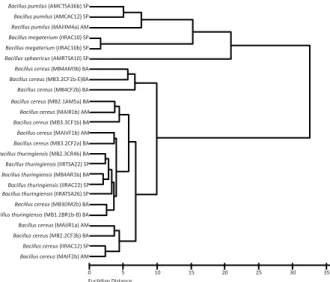
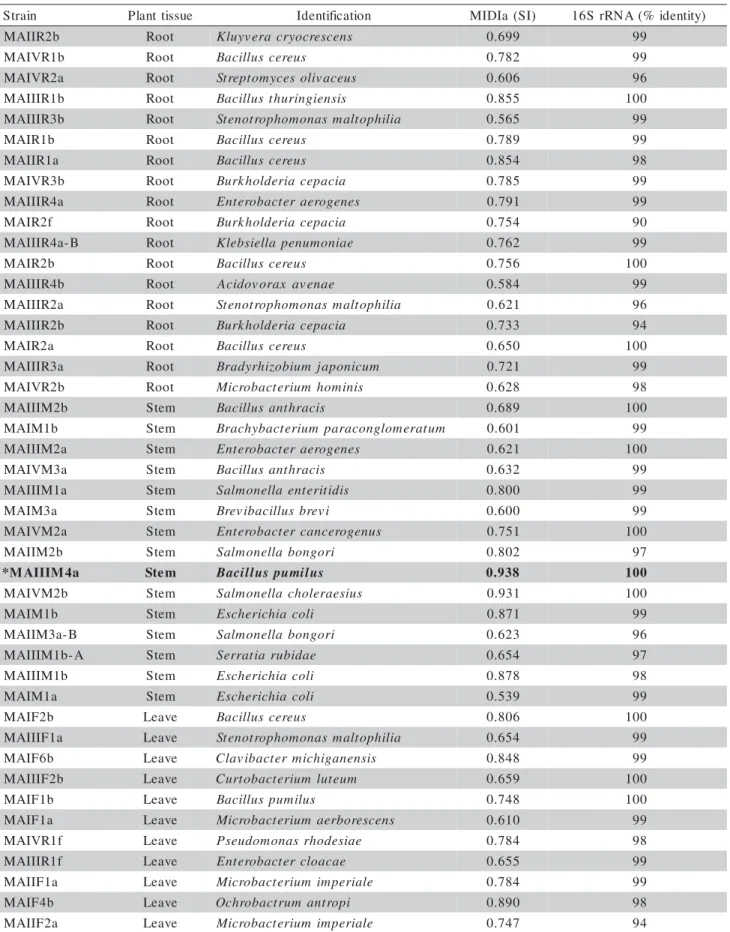
Sixty-seven strains of endophytic bacteria were iso-lated from six cassava plants. The taxonomic results revealed that out of 67 isolates subjected to 16S rRNA gene and FAME profile analysis, only 44 showed simi-larities comparing the two techniques. It was possible to identify 19 genera of endophytic bacteria in cassava plants (Table 1). More than 25% of the isolates were classified as Bacillus species. The bacteria identified in cassava were isolated from all colonizing parts of the plant. The percentage of colonization in roots was 41% of the total isolated bacteria, followed by the stem with 34% and leaves with 25%. Some of the strains were present in all tissues of the plant, while others colonized specific parts. The resulting profiles were identified with microbial identification software (MIDI) usingthe TSBA database, version 4.0 (MIDI, Newark, Del.) and a dendrogram can be visualized in Figure 1 (Teixeira, 2004). The 16S rRNA sequence of the B.
pumilus MAIIIM4a compared favourably (98%
iden-tity) with several sequences of B. pumilus (AF526907, AY289549, AY496869, AY167886) from public data-base by BLAST analysis. Phylogenetic relationships were investigated with other bacteria from Genebank using the joining method (Figure 2). All the B. pumilus
strains formed a monophyletic cluster in the tree. The bootstrap analysis based on 1,000 resamplings of the neighbour-joining data, used to test the robustness and stabilityof the branching, showed that the B. pumilus
strains sequences clustered with high bootstrap value (98%) separated from the other species of Bacillus.
Scanning electron microscopy of cassava tissues
Scanning electron microscopy analysis demon-strated that B. pumilus MAIIIM4a beneficially infected
Figure 1 - Dendrogram obtained by FAME profile analysis of
Table 1 - Identification of endophytic bacteria using FAME and 16S rRNA gene. The fat acids profiles were obtained comparing the data from the TSBA4.0 library (reference strains). Isolates with similarity index (SI) of 0.6 or higher were considered positively identified.
aFAME (Fatty acid methyl ester). aSource: Teixeira, 2004. *Strain selected for antimicrobial analysis
n i a r t
S Planttissue Identification MIDIa (SI) 16SrRNA(% identity)
b 2 R I I A
M Root Kluyvera cryocrescens 0.699 99 b 1 R V I A
M Root Bacilluscereus 0.782 99
a 2 R V I A
M Root Streptomycesolivaceus 0.606 96 b 1 R I I I A
M Root Bacillusthuringiensis 0.855 100 b 3 R I I I A
M Root Stenotrophomonas maltophilia 0.565 99 b
1 R I A
M Root Bacilluscereus 0.789 99
a 1 R I I A
M Root Bacilluscereus 0.854 98
b 3 R V I A
M Root Burkholderiacepacia 0.785 99 a 4 R I I I A
M Root Enterobacteraerogenes 0.791 99 f
2 R I A
M Root Burkholderiacepacia 0.754 90 B -a 4 R I I I A
M Root Klebsiellapenumoniae 0.762 99 b
2 R I A
M Root Bacilluscereus 0.756 100 b 4 R I I I A
M Root Acidovorax avenae 0.584 99 a 2 R I I I A
M Root Stenotrophomonas maltophilia 0.621 96 b 2 R I I I A
M Root Burkholderiacepacia 0.733 94 a
2 R I A
M Root Bacilluscereus 0.650 100 a 3 R I I I A
M Root Bradyrhizobium japonicum 0.721 99 b 2 R V I A
M Root Microbacterium hominis 0.628 98 b 2 M I I I A
M Stem Bacillusanthracis 0.689 100 b
1 M I A
M Stem Brachybacteriumparaconglomeratum 0.601 99 a 2 M I I I A
M Stem Enterobacteraerogenes 0.621 100 a 3 M V I A
M Stem Bacillusanthracis 0.632 99 a 1 M I I I A
M Stem Salmonellaenteritidis 0.800 99 a
3 M I A
M Stem Brevibacillusbrevi 0.600 99 a 2 M V I A
M Stem Enterobactercancerogenus 0.751 100 b 2 M I I A
M Stem Salmonellabongori 0.802 97 a 4 M I I I A M
* Stem Bacilluspumilus 0.938 100
b 2 M V I A
M Stem Salmonellacholeraesius 0.931 100 b
1 M I A
M Stem Escherichiacoli 0.871 99 B -a 3 M I I A
M Stem Salmonellabongori 0.623 96 A -b 1 M I I I A
M Stem Serratiarubidae 0.654 97 b 1 M I I I A
M Stem Escherichiacoli 0.878 98 a
1 M I A
M Stem Escherichiacoli 0.539 99 b
2 F I A
M Leave Bacilluscereus 0.806 100 a 1 F I I I A
M Leave Stenotrophomonas maltophilia 0.654 99 b
6 F I A
M Leave Clavibactermichiganensis 0.848 99 b 2 F I I I A
M Leave Curtobacterium luteum 0.659 100 b
1 F I A
M Leave Bacilluspumilus 0.748 100 a
1 F I A
M Leave Microbacterium aerborescens 0.610 99 f 1 R V I A
M Leave Pseudomonas rhodesiae 0.784 98 f 1 R I I I A
M Leave Enterobactercloacae 0.655 99 a 1 F I I A
M Leave Microbacterium imperiale 0.784 99 b
4 F I A
M Leave Ochrobactrumantropi 0.890 98 a 2 F I I A
the tissues, established and colonized leaves, stems and roots of cassava plants. However, the distribution of this species was variable according to the colonization microhabitats. Populations of B. pumilus MAIIIM4a in the stem and root of cassava are shown in Figure 3. The majority of cells of B. pumilus MAIIIM4a in-vaded and colonized intracellular and intercellular sites
of cassava tissues. It was not detected presence of any bacteria in tissues of the axenic plants.
Screening of bacteria for antagonism against P. aphanidermatum
A total of 67 bacterial isolates were screened for their antagonistic ability to suppress the mycelial growth of P. aphanidermatum in an in vitro assay. Ten isolates showed strong antagonistic activity against the phytopathogen. These antagonists were isolated from all parts of the plants, that is, 30% from roots, 20% from leaves and 50% from stem. The percentage of inhibition against P. aphanidermatum found is pre-sented as follow: Enterobacter cancerogenus MAIVM2a (33%), Bacillus anthracis MAIVM3a (26%), B.
anthracis MAIIIM2b (48%), Kluyvera cryocrescens
MAIIR2b (29%), Bradyrhizobium japonicum
MAIIIR3a (35%), B. pumilus MAIIIM4a (54%), B.
cereus MAIF4b (33%), Clavibacter michiganensis
isidiosum MAIF6b (34%), B. cereus MAIVM1b
(51%), Burkholderia cepacia MAIVR3b (35%). Due to the highest antifungal activity of B. pumilus
MAIIIM4a, this strain was chosen for further analy-sis.
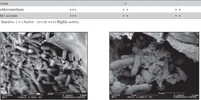
Antimicrobial activity
Dichloromethane and ethyl acetate extracts showed high bioactivity against the phytopathogens R. solani,
P. aphanidermatum and S. rolfsii, while the hexane
extract presented low bioactivity only against P.
aphanidermatum (Table 2).
Figure 3 - Scanning electron micrographs of B. pumilus MAIIIM4a colonizing cassava tissues. a) Stem colonization; b) Root colonization. s
t c a r t x
E R.solani P.aphanidermatum S. rolfsii
e n a x e
H - +
-e n a h t e m o r o l h c i
D +++ ++ ++
e t a t e c a l y h t
E +++ ++ ++
Table 2 - Bioactivity of different B. pumilus extracts against phytopathogenic fungi.
( - ) Inactive ( + ) Active (++ or +++) Highly active. Figure 2 - A phylogenetic tree of the B. pumilus MAIIIM4a 16S
TLC and bioautography
The choice of extraction conditions was very im-portant, since different solvents showed different frac-tions of compounds. Preliminary TLC analysis of B.
pumilus MAIIIM4a supernatant extracted into ethyl
acetate indicated that 10 fractions were obtained, while hexane extraction presented only two fractions (Table 3). The bioautography was performed only for the ethyl acetate extract. Clear inhibition zones at Rf of 0.62, 0.56 and 0.42 for B. pumilus MAIIIM4a extracts were observed against P. aphanidermatum, showing that the compounds responsible for the antifungal ac-tivity are strongly apolar.
GC-MS
The results obtained by GC-MS are presented in Table 4. Mass spectra data were compared with Nist 12, Nist 62 and Wiley 139 libraries. These data sug-gested that hexane extracts were constituted of long chain hydrocarbons. Dichloromethane extract presented phthalate derivative, composed of aromatic and differ-ent classes of oxygenated compositions (esters, alcohols, diols, ketones and others), while ethyl acetate extract had phthalate, fatty ester, long chains of un-saturated hydrocarbons and diols.
Mass Spectrometry
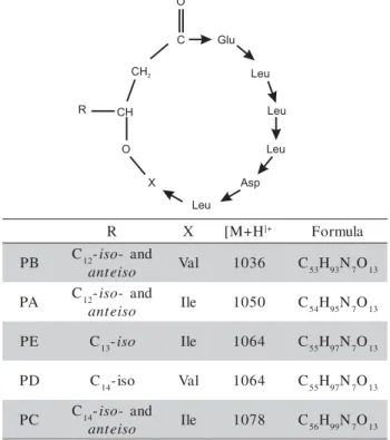
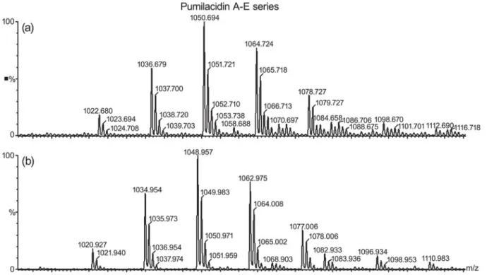
Mass peaks were assigned to lipopeptide species by comparison with reference data and by mass spectrometric in situ sequence analysis of selected compounds by ESI-MS/MS. These mass data cor-respond well to those determined by other authors indicating the presence of pumilacidin A-E series (Kalinovskaya et al., 2002; Pabel et al., 2003) (Fig-ure 4).
The predominant ions in ESI mass spectrum in the positive ion mode were that of m/z 1036.7, 1050.7, 1064.7 and 1078.7 (Figure 5a) and the more abundant one was that of m/z 1050.7, which corre-sponds to the protonated pumilacidin A with a C12 lipid carbon chain and Ile7. In a similar manner, ESI mass spectrum in the negative ion mode showed the pumilacidin A-E series [M-H]- (Figure 5b). Different
isoforms exist for each lipopeptide, which vary in
f o s t c a r t x
E B. pumilus *Rf
I II III IV V VI VII VIII IX X
l o r t n o
C 0.89 0.62 0.53 0.23 - - -
-e n a x e
H 0.33 0.24 - - -
-e n a h t e m o r o l h c i
D 0.89 0.80 0.65 0.60 0.53 0.23 - - -
-e t a t e c a l y h t
E 0.89 0.78 0.71 0.67 0.62 0.56 0.49 0.42 0.29 0.25
Table 3 - Rf values for different solvent extracts using UV light to visualize spots (254 and 366 nm).
*Rf is defined as the distance traveled by the compound divided by the distance traveled by the solvent
the chain length of their fatty acid components and amino acid residues in their peptide rings, respec-tively.
DISCUSSION
A total of 67 endophytic bacteria were isolated from cassava plants collected in the Brazilian Amazon region. The isolation methodology used probably underesti-mates the real bacterial population, since it has been isolated only bacteria that could grow in culture con-ditions. Several isolates were identified by FAME pro-file analysis or 16S ribosomal RNA gene sequencing. The selected isolate MAIIIM4a was confirmed to be-long to B. pumilus species and showed similarity with
B. pumilus strains AMCTSA36b and AMCAC12.
Ba-cillus species are found as the main endophytic
bac-teria in several plants, including Zea mays L. (Lalande et al., 1989), Gossypium hirsutum L. (Misaghi & Donndelinger, 1990), Beta vulgaris (Jacobs et al.,
Figure 4 - Structure and mass data of lipopeptides pumilacidins. (PA-PE indicates pumilacidins series A-E).
R X [M+H]+ Formula
B
P C12-iso- and
o s i e t n
a Val 1036 C53H93N7O13 A
P C12-iso- and
o s i e t n
a Ile 1050 C54H95N7O13 E
P C13-iso Ile 1064 C55H97N7O13
D
P C14-iso Val 1064 C55H97N7O13
C
P C14-iso- and
o s i e t n
Table 4 - Compounds identified in the hexane extract using GC/MS.
a = Compounds present in the culture medium. b = Not identified. t
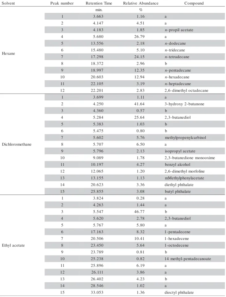
n e v l o
S Peak number RetentionTime RelativeAbundance Compound
. n i
m %
e n a x e H
1 3.663 1.16 a
2 4.147 4.51 a
3 4.183 1.85 n-propilacetate
4 5.680 26.79 a
5 13.556 2.18 n-dodecane 6 15.480 5.10 n-tridecane 7 17.298 24.15 n-tetradecane
8 18.372 2.96 b
9 18.997 12.35 n-pentadecane 0
1 20.603 12.94 n-hexadecane 1
1 22.105 3.19 n-heptadecane 2
1 22.201 2.83 2,6-dimethyloctadecane
e n a h t e m o r o l h c i D
1 3.699 1.11 a
2 4.250 41.64 3-hydroxy2-butanone
3 4.360 0.57 b
4 5.284 25.64 2,3-butanediol
5 5.383 1.03 b
6 5.475 0.80 b
7 5.602 5.76 methylpropenylcarbinol
8 5.707 6.50 a
9 5.796 2.13 isopropylacetate
0
1 9.089 1.78 2,3-butanedionemonooxime
1
1 10.197 4.27 benzylalcohol
2
1 12.065 1.20 2,6-dimethylmorfoline
3
1 13.155 1.13 mMethylphenylacetate
4
1 20.623 3.36 diethylphthalate
5
1 25.855 3.08 butylphthalate
e t a t e c a l y h t E
1 3.824 0.28 a
2 4.263 1.44 a
3 5.547 46.77 b
4 5.620 2.78 2,3-butanediol
5 5.767 5.80 a
6 17.163 8.32 1-pentadecene
7 20.506 10.41 1-hexadecene
8 23.450 5.64 1-octodecene
9 23.789 0.81 b
0
1 25.238 0.82 14 methyl-pentadecanoate
1
1 25.896 6.19 a
2
1 26.111 3.86 a
3
1 26.402 4.23 b
4
1 28.546 1.02 a
5
Figure 5 - ESI mass spectrum in the positive (a) and negative (b) ion mode of the pumilacidin A-E series.
1985), and Solanum tuberosum L. (Hollis, 1951), and the species that are often cited as endophytic are: B. cereus and B. lentus (Araújo et al., 2001), B. subtilis
(Bai et al., 2002), B.megaterium (Elvira-Recuenco & Vuurde, 2000; Araújo et al., 2001), B. insolitus and B.
brevis (Sturz et al., 1997) and B. pumilus (Araújo et
al., 2002). This genera has been reported as a potent biological control agent (Cook, 1986), demonstrating a great potential in agriculture.
Scanning electron microscopy was able to detect this strain in inner tissues in all parts of the plant.
Ba-cillus pumilus MAIIIM4a colonized roots and stem,
indicating that this isolate is efficient and a good can-didate as biological control agent.
With the increased occurrence of phytopathogens causing plant disease, the search for new antifungal bioagent has gained urgency. The production of bioactive compounds by Bacillus species is well es-tablished (Korzybski et al., 1978; Naruse et al., 1990; Munimbazi & Bullerman, 1998). These classes of bioactive compounds act as antifungal peptides, anti-fungal lipopeptides and antimicrobial polypeptides (Pabel et al., 2003). To characterize antimicrobial ac-tivity of B. pumilus MAIIIM4a, the isolate was tested against three phytopathogenic fungi: R. solani, P. aphanidermatum and S.rolfsii. The antifungal screen-ing revealed that B. pumilus MAIIIM4a produced a compound with high bioactivity against P.
aphanidermatum. The bioautography was considered
an efficient test to determine antimicrobial activity, since less than 2.5 μg of extract was enough to form
inhibition halo around the active fractions. LC-MS/MS was able to identify a compound assigned as pumilacidin by comparison with reference data. Pumilacidin is produced by B. pumilus and are cyclic acylheptapeptide composed of a beta-hydroxy fatty acid (Naruse et al., 1990). This lipopeptide has antimicro-bial (Pabel et al., 2003), antiviral and antiulcer activity (Naruse et al., 1990).
Mass spectrometric data corroborate the identifi-cation of one bioactive compound as a lipopeptide, pumilacidin, a characteristic of B. pumilus. This is the first time that an endophytic B. pumilus was charac-terized as pumilacidin producer.
ACKNOWLEDGEMENT
Financial support was provide by the Fundação de Amparo à Pesquisa do Estado de São Paulo (FAPESP 99/09177-1). F.M.P.M. was supported by a graduate scholarship from CAPES (Coordenação de Aperfeiçoamento de Pessoal de Nível Superior).
REFERENCES
ARAÚJO, W.L.; MACCHERONI JR, W.; AGUILLAR-VILDOSO, C.I.; BARROSO, P.A.V.; SARIDKIS, H.O.; AZEVEDO, J.L. Variability and interactions between endophytic bacteria and fungi isolated from leaf tissues of citrus rootstocks. Canadian Journal of Microbiology, v.47, p.229-236, 2001.
ARAÚJO, W.L.; MARCON, J.; MACCHERONI JR, W.; ELSAS, J.D.van; VUURDE, J.W.L.van; AZEVEDO, J.L. Diversity of endophytic bacterial populations and their interaction with
Xylella fastidiosa in citrus plants. Applied and Environmental
BAI, Y.; DÁOUST, F.; SMITH, D.L.; DRISCOLL, B.T. Isolation of plant-growth-promoting Bacillus strains from soybean root nodules. Canadian Journal of Microbiology, v.48, p.230-238, 2002.
COOK, R.J. Plant health and the sustainability of agriculture, with special reference to disease control by beneficial microorganisms.
Biological Agriculture and Horticulture, v.3, p.211-232, 1986.
ELVIRA-RECUENCO, M.; VUURDE, J.W.L.van. Natural incidence of endophytic bacteria in pea cultivars under field conditions.
Canadian Journal of Microbiology, v.46, p.1036–1041, 2000.
FAO. Championing the cause of cassava. 2000. Available in www.fao.org/NEWS/2000/000405-e.htm. Accessed 28 Sept. 2000. HALL, T. BioEdit: version 5.0.6. Raleigh: North Carolina State
University, Department of Microbiology, 2001. 192p. HALLMANN, J.; QUADT-HALLMANN, A.; MAHAFFEE, W.F.;
KLOEPPER, J.W. Bacterial endophytes in agricultural crops.
Canadian Journal of Microbiology, v.43, p.895-914, 1997. HEUER, H.; KRSEK, M.; BAKER, P.; SMALLA, K.; WELLINGTON, E.M. Analysis of actinomycete communities by specific amplification of genes encoding 16S rRNA and gel-electrophoretic separation in denaturing gradients. Applied and Environmental Microbiology, v.63, p.3233-3234, 1997. HOLLIS, J.P. Bacteria in healthy potato tissue. Phytopathology,
v.41, p.350-366, 1951.
JACOBS, M.J.; BUGDEE, W.M.; GABRIELSON, D.A. Enumeration, location, and characterization of endophytic bacteria within sugar beet roots. Canadian Journal of Botany, v.63, p.1262-1265, 1985.
KALINOVSKAYA, N.I.; KUZNETSOVA, T.A.; IVANOVA, E.P.; ROMANENKO, L.A.; VOINOV, V.G.; HUTH, F.; LAATSCH, H. Characterization of Surfactin-like Cyclic Depsipeptides Synthesized by Bacillus pumilus from Ascidian Halocynthia
aurantium. Marine Biotechnology, v.4, p.179–188, 2002.
KITAJIMA, E.W.; LEITE, B. Curso introdutório de microscopia eletrônica de varredura. 2. ed. Piracicaba: NAP/ MEPA-USP/ ESALQ, 1999.46p.
KORZYBSKI, T.; KOWSZYK-GIFINDER, Z.; KURYTOWICZ, W.
Antibiotics: origin, nature, and properties. Washington, D.C.: American Society for Microbiology, , 1978. p.501-534. KREBS, B.; HÖDING, B.; KÜBART, S.; ALEMAYEHU-WORKIE,
M.; JUNGE, H.; SCHMIEDEKNECHT, G.; GROSCH, R.; BOCHOW, H.; HEVESI, M. Use of Bacillus subtilis as biological control agent. I. Activities and characterization of Bacillus
subtilis strains. Journal of Plant Diseases and Protection,
v.105, p.181-197, 1998.
LALANDE, R.; BISSONNETTE, N.; COUTLEE, D.; ANTOUN, H. Identification of rhizobacteria from maize and determination of their plant-growth promoting potential. Plant and Soil, v.115, p.7-11, 1989.
LIMA, A.O.S.; QUECINE, M.C.; FUNGARO, M.H.P.; ANDREOTE, F.D.; MACCHERONI JR, W.; ARAÚJO, W.L.; SILVA FILHO, M.C.; PIZZIRANI-KLEINER, A.A.; AZEVEDO, J.L. Molecular characterization of a ß-1,4-endoglucanase from an endophytic
Bacillus pumilus strain. Applied Microbiology and
Biotechnology, v.68, p.57-65, 2005.
LOEFFLER, W.; TSCHEN, S.-M.; VANITTANAKOM, N.; KUGLER, M.; KNORPP, E.; HSIEH, T.-F.; WU, T.-G. Antifungal effects of bacilysin and fengymycin from Bacillus subtilis F29-3: a comparison with activities of other Bacillus antibiotics.
Journal of Phytopathology, v.115, p.204-213, 1986.
MISAGHI, I.J.; DONNDELINGER, C.R. Endophytic bacteria in symptom-free cotton plants. Phytopathology, v.80, p.808-811, 1990.
MUNIMBAZI, C.; BULLERMAN, L.B. Isolation and partial characterization of antifungal metabolites of Bacillus pumilus.
Journal of Applied Microbiology, v.84, p.959-968, 1998. MURASHIGE, T.; SKOOG, F. A revised medium for rapid growth
and bioassays with tobacco tissue cultures. Physiologia Plantarum, v.15, p.473-497, 1962.
NARUSE, N.; TENMYO, O.; KOBARU, S.; KAMEI, H.; MIYAKI, T.; KONISHI, M.; OKI, T. Pumilacidin, a complex of new antiviral antibiotics. Production, isolation, chemical properties, structure and biological activity. Journal of Antibiotics, v.43, p.267-280, 1990.
NÜBEL, U.; ENGELEN, B.; FELSKE, A.; SNAIDR, J.; WIESHUBER, A.; AMANN, R.I.; LUDWIG, W.; BACKHAUS, H. Sequence heterogeneities of genes encoding 16S rRNAs in
Paenibacillus polymyxa detected by temperature gradient gel
electrophoresis. Journal of Bacteriology, v.178, p.5636-5643, 1996.
PABEL, C.T.; VATER, J.; WILDE, C.; FRANKE, P.; HOFEMEISTER, J.; ADLER, B.; BRINGMANN, G.; HACKER, J.; HENTSCHEL, U. Antimicrobial activities and matrix-assisted laser desorption/ionization mass spectrometry of Bacillus
isolates from the marine sponge Aplysina aerophoba. Marine Biotechnology, v.5, p.424-434, 2003.
PLEBAN, S.; CHERNIN, L.; CHET, I. Chitinolytic activity of an endophytic strain of Bacillus cereus. Letters in Applied Microbiology, v.25, p.284-288, 1997.
SAITOU, N.; NEI, N. The neighbor-joining method: a new method for reconstructing phylogenetic trees. Molecular Biology and Evolution, v.4, p.406-25, 1987.
STIERLE, A.; STROBEL, G.; STIERLE, D. Taxol and taxane production by Taxomyces andreanae, an endophytic fungus of Pacific yew. Science, v.260, p.214-216, 1993.
STURZ, A.V.; CHRISTIE, B.R.; MATHESON, B.G.; NOWAK, J. Biodiversity of endophytic bacteria which colonize red clover nodules, roots, stems and foliage and their influence on host growth. Biology and Fertility of Soils, v.25, p.13-19, 1997. TEIXEIRA, M.A.; Diversidade de Bactérias endofíticas de mandioca (Manihotis esculenta Crantz) coletada de diferentes regiões do Brasil. São Paulo: USP/ICB, 2004. 102p. Tese (Doutorado). TEIXEIRA, M.A.; MELO, I.S.; VIEIRA, R.F.; COSTA, F.E.C.;
HARAKAVA, R. Microrganismos endofíticos de mandioca de áreas comerciais e etnovariedades em três Estados brasileiros. Pesquisa Agropecuária Brasileira, v.42, p.42-49, 2007. Available in http://www.scielo.br/pdf/pab/v42n1/06.pdf. Accessed 1 Jul. 2008.
THOMPSON, J.D.; HIGINS, D.G.; GIBSON, T.J. Clustal-W: improving the sensitivity of progressive multiple sequence alignment through sequence weighting, position-specific gap penalties and weight matrix choice. Nucleic Acids Research, v.22, p.4673-4680, 1994.
TRISCHMAN, J.A.; TAPIOLAS, D.M.; JENSEN, P.R.; DWIGHT, R.; FENICAL, W.; McKEE, T.C.; IRELAND, C.M.; STOUT, T.J.; CLARDY, J.C. Salinamide-A and salinamide-B: anti-inflammatory depsipeptides from a marine streptomycete.
American Chemical Society, v.116, p.757-758, 1994.