Importance of hyaluronan biosynthesis
and degradation
in cell differentiation
and tumor formation
Ludwig Institute for Cancer Research, Biomedical Center, Uppsala, Sweden P. Heldin
Abstract
Hyaluronan is an important connective tissue glycosaminoglycan. Elevated hyaluronan biosynthesis is a common feature during tissue remodeling under both physiological and pathological conditions. Through its interactions with hyaladherins, hyaluronan affects several cellular functions such as cell migration and differentiation. The activities of hyaluronan-synthesizing and -degrading enzymes have been shown to be regulated in response to growth factors. During tumor progression hyaluronan stimulates tumor cell growth and inva-siveness. Thus, elucidation of the molecular mechanisms which regu-late the activities of hyaluronan-synthesizing and -degrading enzymes during tumor progression is highly desired.
Correspondence
P. Heldin
Ludwig Institute for Cancer Research Biomedical Center
Box 595 S-751 24 Uppsala Sweden
Fax: +46-18-471-4244
E-mail: [email protected]
Presented at SIMEC 2002 (International Symposium on Extracellular Matrix), Angra dos Reis, RJ, Brazil, October 7-10, 2002.
Research supported by the Swedish Cancer Society, Konung Gustav V:s 80-års Fond and Q-Med AB (Uppsala).
Received December 13, 2002 Accepted February 3, 2003
Key words •Hyaluronan •Synthase •Hyaluronidase •Tumor invasion •Extracellular matrix •Pericellular matrix
Introduction
Hyaluronan (hyaluronic acid) is a linear glycosaminoglycan composed, on average, of 10,000 disaccharide units of glucuronic acid and N-acetylglucosamine, with a mo-lecular weight of 1-5 x 10⁶. It belongs to the
family of mucopolysaccharides that also in-cludes heparin, chondroitin sulfate, keratan sulfate and dermatan sulfate. Hyaluronan was first described in 1934 (1), but the hyaluro-nan-synthesizing and -catabolizing enzymes, as well as the cell surface interacting mol-ecules, were identified as late as in the 1990s. Hyaluronan is found in all vertebrate tissues and on the surface of certain Streptococcus
and Pasteurella bacterial pathogens. Despite its simple structure, hyaluronan is involved in several biological events; it influences the hydration and physical properties of tissues and is important in the organization of peri-cellular and extraperi-cellular matrices through its interactions with other extracellular
mac-romolecules such as aggrecan and versican. Furthermore, hyaluronan affects cell behav-ior, e.g., migration, proliferation and differ-entiation, via binding to specific cell surface receptors such as CD44. Absence of the CD44 gene virtually prevents osteosarcoma progression (2).
Alterations in the amount of hyaluronan have been documented in several diseases such as rheumatoid arthritis, inflammatory and vascular diseases, as well as cancer (3). This review will focus on the importance of hyaluronan for tumor cell proliferation and invasion.
Hyaluronan-synthesizing and -degrading enzymes
re-quired. The metabolism of hyaluronan in the mammalian body is rapid compared to other extracellular matrix components; for ex-ample, in the epidermis the half-life of hyaluronan is less than one day (4). The cells devote much energy to the synthesis of hyaluronan molecules consisting of thou-sands of sugars; therefore the metabolism of hyaluronan is carefully regulated in a tissue-specific manner.
The mammalian hyaluronan-synthesiz-ing (Has) enzymes were cloned and charac-terized in 1996. The first Has gene cloned was from the Streptococcus pyogenes group A bacteria in 1993 using transposon mu-tagenesis with Tn916. Mutant cells in which the insertion of the transposon element dis-rupted the locus of hyaluronan synthesis were selected by visual screening of small dry colonies versus large mucoid colonies (5-7). The availability of the bacterial Has sequence led to the identification of the eu-karyotic hyaluronan synthases, termed Has1, Has2, and Has3 (8-11). The mammalian hyaluronan synthase isoforms share 55-71% amino acid sequence similarity, whereas human and mouse homologous isoforms share about 97% similarity. Based on the sequence, the Has isoforms encode plasma membrane proteins with seven membrane-associated regions and a central cytoplas-matic domain which possesses consensus sequences for phosphorylation by protein kinase C (PKC; reviewed in Ref. 12). More recently a viral Has was discovered in the genome of the chlorella virus 1 which infects chlorella-like algae in fresh water world-wide. Using the transposon mutagenesis ap-proach, another bacterial Has has also been identified from Gram-negative Pasteurella
multocida bacteria which, importantly, is
about twice as large as the other Has iso-forms (reviewed in Ref. 13). The evolution of the vertebrate Has family involved gene duplication and divergence. The ancestral Has in animals was duplicated to form the Has1 and Has2 lineages. A subsequent pair
of duplication events resulted in the forma-tion of Has1 and a Has-related homologue, as well as in the formation of Has2 and Has3. Mice deficient in Has1 or Has3 activity are viable, whereas mice deficient in Has2 activity have severe developmental prob-lems such as cardiac defects. Thus, Has2 activity is required for embryonic develop-ment (14). In all vertebrates so far studied three Has isoforms have been found and are located on different chromosomes (12). The distinct expression patterns of the Has iso-forms during embryogenesis and adulthood suggest different functional roles (15,16). As a tool for a more direct assessment of possible differences in the enzymatic prop-erties of each Has isoform, our group and others have overexpressed Has genes in Chi-nese hamster ovary cells, COS-1 cells or rat 3Y1 fibroblasts (17,18). These studies dem-onstrated that each of the three Has isoforms is capable of hyaluronan polymerization; however, they exhibit different catalytic rates for hyaluronan synthesis and produce hyal-uronans of different size. In addition, the studies indicated that different cytoplasmic proteins interact specifically with each Has protein, and may have accessory or regula-tory roles in hyaluronan biosynthesis (17).
vitro, higher Hyal activities are found in media than in cell lysates (20). It remains to be elucidated whether hyaluronidases are lysosomal and/or secreted into the extracel-lular matrix. In addition to their role in hyal-uronan degradation, hyaluronidases may also play key roles in differentiation events; tu-mor cells exhibiting hyaluronidase activity induce angiogenesis of the cornea of mice (21), and more recently we have shown that PH-20-like hyaluronidase promotes differ-entiation of capillary endothelial cells grown on collagen gel in a CD44-dependent man-ner (22). The rapid turnover of hyaluronan, which varies considerably among different tissues in the human body, raises many ques-tions concerning the control mechanisms that modulate the expression levels and activities of hyaluronidases.
Growth factor regulation of hyaluronan metabolism
Increased levels of hyaluronan have been detected in several human cancers (for a review, see Ref. 23) and therefore we have investigated the molecular mechanisms in-volved in the perturbation of hyaluronan synthesis and catabolism. We have studied the regulation of Has and hyaluronidase gene expression in response to growth factors re-leased during tumor progression. We first investigated the effects of various growth factors on the synthesis of hyaluronan in sparse and dense fibroblast cultures. Plate-let-derived growth factor (PDGF-BB) had a potent stimulatory effect on hyaluronan syn-thesis whereas PDGF-AA was less active, suggesting that the stimulatory effect of PDGF-BB on hyaluronan synthesis is mainly mediated by its ß-receptor. No correlation with the mitogenic effects of the growth factors was found since epidermal growth factor (EGF) and basic fibroblast growth factor (bFGF), which are as potent mitogens as PDGF-BB, had much weaker effects than PDGF-BB on hyaluronan synthesis, and since
tumor growth factor beta (TGF-ß), which inhibits cell growth, stimulated hyaluronan synthesis (24). Studies by other groups also showed a growth factor-dependent stimula-tion of hyaluronan synthesis in cells of mes-enchymal origin (25,26). The effects of the external stimuli studied on hyaluronan syn-thesis were mediated partly by activation of PKC and protein kinase A (27,28). Further studies on the growth factor-dependent regu-lation of hyaluronan synthesis revealed that among the three Has isoforms, Has2 is most markedly up-regulated or suppressed in re-sponse to external stimuli in cultures of nor-mal human mesothelial cells (16).
Notably, the growth factor regulation of hyaluronan synthesis also affected the mi-croenvironment between the cells and the extracellular matrix. PDGF-BB and EGF, but not TGF-ß, stimulated the formation of a pericellular matrix around mesothelial cells (29). However, hyaluronan synthesis and pericellular matrix formation in chick em-bryo limb mesodermal cells were induced by bFGF and TGF-ß, but not by PDGF or EGF (30). Thus, growth factors appear to have different regulatory roles in hyaluronan syn-thesis and assembly in various cell types.
nonstimulated cells (20). Members of the inter-α-inhibitor family have also been shown
to inhibit serum hyaluronidase activity (31).
The importance of hyaluronan in tumor invasion
Several tumor types, both epithelial and connective tissue tumors, are often enriched in hyaluronan. Human mesotheliomas, nephroblastomas and, to a somewhat lesser extent, breast carcinomas, are considered to have the highest enrichment in hyaluronan. The mean level of hyaluronan in the serum of a middle-aged healthy person is about 40 µg/ml. Hyaluronan levels in serum increase in various diseases, and levels above 250 µg/ ml indicate progressive disease and can be of diagnostic value. In most tumors an increase in the proportion of hyaluronan over other glycosaminoglycans is commonly seen. Most of the hyaluronan is found within the tumor-associated stromal tissue at concentrations up to 0.1% of the wet weight (32). It should also be considered that elevation of the amounts of hyaluronan in tumors in vivo
may not result exclusively from increased hyaluronan synthesis. Rather, increased cel-lularity or decreased hyaluronidase activity may give rise to higher hyaluronan levels. Alternatively, local clearance of hyaluronan or clearance via the lymphatic tissue may be impaired in advanced tumors. However, in-creased hyaluronan deposition is also found during embryonic development, wound heal-ing and regeneration. This suggests that the accumulation of hyaluronan is a common feature during tissue reconstruction. It is possible that hyaluronan, as a consequence of its physicochemical properties, creates permeable matrices within which cells can easily change shape and migrate. The func-tion of hyaluronan accumulafunc-tion in tumor tissues remains to be determined.
Immunohistochemical staining of tumor tissues for hyaluronan using a hyaluronan-binding protein isolated from bovine
carti-lage proteoglycan followed by biotinylation (33) has indicated that most human solid tumors have increased amounts of hyaluro-nan in the stromal tissue. However, in vitro
studies of tumor cell lines have revealed that not all tumor cells synthesize hyaluronan by themselves. Whereas some tumor cells such as fibrosarcoma (34) and glioma (35) pro-duce large amounts of hyaluronan, others, such as mesothelioma and lung carcinoma cell lines, do not synthesize hyaluronan; rather these tumors secrete factors that stimu-late hyaluronan synthesis by normal connec-tive tissue cells such as mesothelial cells and fibroblasts (36,37). Co-culture of fibroblasts and tumor cells also leads to stimulation of hyaluronan synthesis. Thus, most likely the host connective tissue responds to the pres-ence of actively proliferating and migrating tumor cells.
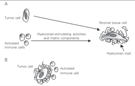
hyaluronan-con-taining pericellular matrices around meso-thelial cells (40). It is also possible that during tumor invasion the host immune sys-tem may also play a role in the assembly of pericellular matrices through induction of hyaluronan synthesis by tumor-adjacent fi-broblasts by secretion of inflammatory cyto-kines (41). Thus, these types of pericellular matrices may assemble and function as a protective shield around connective cells in the stromal tissue, in vivo, as a response to the release of both matrix components and growth factors by tumor cells (Figure 1A).
Several recent studies have shown that hyaluronan overexpression by various tu-mor cells after transfection of tutu-mor cells with different Has isoforms enhances their malignant phenotype (42-45). In one of our experimental models we have investi-gated the effect of hyaluronan production on the malignant properties of mesothelioma cells.
About 70% of malignant pleura mesothe-lioma cases are associated with elevated amounts of hyaluronan which have been directly correlated with tumor burden. How-ever, our in vitro studies revealed that not all mesothelioma cells obtained from various patients produce hyaluronan (36). In an ef-fort to explore the relation between hyaluro-nan-producing mesotheliomas and their ag-gressiveness, we have compared the biologi-cal properties of the non-hyaluronan-pro-ducing mesothelioma cell line Mero-25 with those of the same cells made to produce hyaluronan by transfection with Has2 cDNA. Our data revealed that hyaluronan-produc-ing mesotheliomas are more aggressive and invasive than the non-hyaluronan-produc-ing mesotheliomas (44). A possible explana-tion could be that hyaluronan-synthesizing tumor cells are surrounded by protective pericellular matrices which may help tumor cells evade cellular immune attack, as has been shown for glioma cells (46) (Figure 1B). Another possibility is that the hyaluro-nan synthesized by tumor cells contributes
to the expansion of extracellular matrix and thereby facilitates tumor cell migration.
Recently, several contradictory findings on the functional significance of hyalu-ronidases in terms of tumor development have been published. In some of the studies hyaluronidase expression and activity was detected in metastatic human melanoma, glio-blastoma and colon carcinoma, whereas in other studies hyaluronidase activity sup-pressed tumor development (47,48). Based on the knowledge that Has overexpression increases tumorigenicity in the systems stud-ied whereas Hyal overexpression increases tumor development in some experimental models and suppresses it in others, we de-cided to investigate the impact of Hyal1 and Has2 expression in a colon carcinoma model both in vitro and in vivo. These studies re-vealed that Has2 overexpression promotes tumorigenicity, whereas Hyal1 overexpres-sion delays tumor development in a colon carcinoma model (49). It would therefore be interesting to study the mechanisms that regu-late the activities of hyaluronan-synthesiz-ing and -degradhyaluronan-synthesiz-ing enzymes durhyaluronan-synthesiz-ing tumor progression in different tissues and organs.
Figure 1. Hyaluronan synthesis during tumor progression. A host tumor stromal cell is surrounded by a hyaluronan-containing coat in response to hyaluronan-stimulating activities and matrix components secreted by a tumor cell and/or activated immune cells (A). A tumor cell surrounded by a hyaluronan-containing coat excludes host immune cells (B).
Tumor cell
Activated immune cells
Hyaluronan-stimulating activities and matrix components
Stromal tissue cell
Hyaluronan coat
Tumor cell
Activated immune cell
A
Future perspectives
The cloning and characterization of eu-karyotic hyaluronan-synthesizing and -de-grading enzymes, as well as hyaluronan re-ceptors, were major breakthroughs in hyal-uronan research. These reagents will make it possible to raise monoclonal antibodies and construct probes suitable for immunohis-tochemistry and in situ hybridization,
re-spectively, which can be used to increase our understanding of their roles in normal and pathological situations. For example, it will be interesting to further elucidate the roles of hyaluronan in pericellular coats and in ma-trices and its interactions with other mol-ecules. Such studies should shed light on the hyaluronan-induced enhancement of tumor invasion.
References
1. Meyer K & Palmer JW (1934). The polysaccharide of the vitreous humor. Journal of Biological Chemistry, 107: 629-634.
2. Weber GF, Bronson RT, Ilagan J, Cantor H, Schmits R & Mak TW (2002). Absence of the CD44 gene prevents sarcoma metastasis.
Cancer Research, 62: 2281-2286.
3. Laurent TC & Fraser JRE (1992). Hyaluronan. FASEB Journal, 6: 2397-2404.
4. Tammi MI, Day AJ & Turley EA (2002). Hyaluronan and homeostasis: a balancing act. Journal of Biological Chemistry, 277: 4581-4584. 5. DeAngelis PL, Papaconstantinou J & Weigel PH (1993). Molecular
cloning, identification, and sequence of the hyaluronan synthase gene from group A Streptococcus pyogenes. Journal of Biological Chemistry, 268: 19181-19184.
6. DeAngelis PL, Papaconstantinou J & Weigel PH (1993). Isolation of a
Streptococcus pyogenes gene locus that directs hyaluronan biosyn-thesis in acapsular mutants and in heterologous bacteria. Journal of Biological Chemistry, 268: 14568-14571.
7. Dougherty BA & van de Rijn I (1994). Molecular characterization of
hasA from an operon required for hyaluronic acid synthesis in group A Streptococci. Journal of Biological Chemistry, 269: 169-175. 8. Itano N & Kimata K (1996). Expression cloning and molecular
charac-terization of HAS protein, a eukaryotic hyaluronan synthase. Journal of Biological Chemistry, 271: 9875-9878.
9. Shyjan AM, Heldin P, Butcher EC, Yoshino T & Briskin MJ (1996). Functional cloning of the cDNA for a human hyaluronan synthase.
Journal of Biological Chemistry, 271: 23395-23399.
10. Spicer AP, Augustine ML & McDonald JA (1996). Molecular cloning and characterization of a putative mouse hyaluronan synthase. Jour-nal of Biological Chemistry, 271: 23400-23406.
11. Spicer AP, Olson JS & McDonald JA (1997). Molecular cloning and characterization of a cDNA encoding the third putative mammalian hyaluronan synthase. Journal of Biological Chemistry, 272: 8957-8961.
12. Weigel PH, Hascall VC & Tammi M (1997). Hyaluronan synthases.
Journal of Biological Chemistry, 272: 13997-14000.
13. DeAngelis PL (1999). Hyaluronan synthases: fascinating glycosyl-transferases from vertebrates, bacterial pathogens, and algar vi-ruses. Cellular and Molecular Life Sciences, 56: 670-682.
14. Camenisch TD, Spicer AP, Brehm-Gibson T, Biesterfeldt J, August-ine ML, Calabro A, Kubalak S, Klewer SE & McDonald JA (2000). Distruption of hyaluronan synthase-2 abrogates normal cardiac mor-phogenesis and hyaluronan-mediated transformation of epithelium to mesenchyme. Journal of Clinical Investigation, 106: 349-360.
15. Spicer AP & McDonald JA (1998). Characterization and molecular evolution of a vertebrate hyaluronan synthase gene family. Journal of Biological Chemistry, 273: 1923-1932.
16. Jacobson A, Brinck J, Briskin MJ, Spicer AP & Heldin P (2000). Expression of human hyaluronan synthases in response to external stimuli. Biochemical Journal, 348: 29-35.
17. Brinck J & Heldin P (1999). Expression of recombinant hyaluronan synthase (HAS) isoforms in CHO cells reduces cell migration and cell surface CD44. Experimental Cell Research, 252: 342-351. 18. Itano N, Sawai T, Yoshida M, Lenas P, Yamada Y, Imagawa M,
Shinomura T, Hamaguchi M & Yoshida Y (1999). Three isoforms of mammalian hyaluronan synthases have distinct enzymatic proper-ties. Journal of Biological Chemistry, 274: 25085-25092.
19. Stern R & Csoka AB (2000). Mammalian hyaluronidases. www.glycoforum.gr.jp/science/hyaluronan/HA15/HA15E.html. 20. Li Y, Rahmanian M, Widström C, Lepperdinger G, Frost GI & Heldin
P (2000). Irradiation-induced expression of hyaluronan (HA) synthase 2 and hyaluronidase 2 genes in rat lung tissue accompanies active turnover of HA and induction of types I and III collagen gene expres-sion. American Journal of Respiratory Cell and Molecular Biology, 23: 411-418.
21. Liu D, Pearlman E, Diaconu E, Guo K, Mori H, Haqqi T, Markowitz S, Willson J & Man-Sun S (1996). Expression of hyaluronidase by tumor cells induces angiogenesis in vivo. Proceedings of the Na-tional Academy of Sciences, USA, 93: 7832-7837.
22. Rahmanian M & Heldin P (2002). Testicular hyaluronidase induces tubular structures of endothelial cells grown in three-dimensional collagen gel through a CD44-mediated mechanism. International Journal of Cancer, 97: 601-607.
23. Toole BP, Wight TN & Tammi MI (2002). Hyaluronan-cell interac-tions in cancer and vascular disease. Journal of Biological Chemistry, 277: 4593-4596.
24. Heldin P, Laurent TC & Heldin C-H (1989). Effect of growth factors on hyaluronan synthesis in cultured human fibroblasts. Biochemical Journal, 258: 919-922.
25. Tirone ED, Alessandris C, Hascall VC, Siracusa G & Salustri A (1997). Hyaluronan synthesis by mouse cumulus cells is regulated by inter-actions between follicle-stimulating hormone (or epidermal growth factor) and a soluble oocyte factor (or transforming growth factor ß1). Journal of Biological Chemistry, 272: 4787-4794.
inhibitor genistein, in cultured mesothelial cells from rabbit pericar-dial cavity. Journal of Cell Science, 98: 91-98.
27. Klewes L & Prehm P (1994). Intracellular signal transduction for serum activation of the hyaluronan synthase in eukaryotic cell lines.
Journal of Cellular Physiology, 160: 539-544.
28. Suzuki M, Asplund T, Yamashita H, Heldin C-H & Heldin P (1995). Stimulation of hyaluronan biosynthesis by platelet-derived growth factor-BB and transforming growth factor-beta1 involves activation of protein kinase C. Biochemical Journal, 307: 817-821.
29. Heldin P & Pertoft H (1993). Synthesis and assembly of the hyaluro-nan-containing coats around normal human mesothelial cells. Exper-imental Cell Research, 208: 422-429.
30. Munaim SI, Klagsbrun M & Toole BP (1991). Hyaluronan-dependent pericellular coats of chick embryo limb mesodermal cells: Induction by basic fibroblast growth factor. Developmental Biology, 143: 297-302.
31. Mio K, Carrette O, Maibach HI & Stern R (2000). Evidence that the serum inhibitor of hyaluronidase may be a member of the Inter-α
-inhibitor family. Journal of Biological Chemistry, 275: 32413-32421. 32. Laurent TC, Laurent UB & Fraser JR (1996). Serum hyaluronan as a
disease marker. Annual Medicine, 28: 241-253.
33. Hellström S, Tengblad A, Johansson C, Hedlund U & Axelsson E (1990). An improved technique for hyaluronan histochemistry using microwave irradiation. Histochemical Journal, 22: 677-682. 34. Hopwood J, Fitch FW & Dorfman A (1974). Hyaluronic acid
synthe-sis in a cell-free system from rat fibrosarcoma. Biochemical and Biophysical Research Communications, 61: 583-590.
35. Philipson LH & Schwartz NB (1984). Subcellular localization of hyalu-ronate synthetase in oligodendroglioma cells. Journal of Biological Chemistry,259: 5017-5023.
36. Asplund T, Versnel MA, Laurent TC & Heldin P (1993). Human mesothelioma cells produce factors that stimulate the production of hyaluronan by mesothelial cells and fibroblasts. Cancer Research, 53: 388-392.
37. Teder P, Bergh J & Heldin P (1995). Functional hyaluronan receptors are expressed on squamous cell lung carcinoma cell line but not on other lung carcinoma cell lines. Cancer Research,55: 3908-3914. 38. Knudson W & Knudson CB (1991). Assembly of a chondrocyte-like
pericellular matrix on non-chondrogenic cells. Role of the cell sur-face hyaluronan receptors in the assembly of a pericellular matrix.
Journal of Cell Science, 99: 227-235.
39. Lee GM, Johnstone B, Jacobson K & Caterson B (1993). The dynam-ic structure of the perdynam-icellular matrix of living cells. Journal of Cell Biology, 123: 1899-1907.
40. Heldin P, Suzuki M, Teder P & Pertoft H (1995). Chondroitin sulfate proteoglycan modulates the permeability of hyaluronan-containing coats around normal human mesothelial cells. Journal of Cellular Physiology,165: 54-61.
41. Pure E & Cuff CA (2001). A crucial role for CD44 in inflammation.
Trends in Molecular Medicine,7: 213-221.
42. Itano N, Sawai T, Miyaishi O & Kimata K (1999). Relationship be-tween hyaluronan production and metastatic potential of mouse mammary carcinoma cells. Cancer Research,59: 2499-2504. 43. Kosaki R, Watanabe K & Yamaguchi Y (1999). Overproduction of
hyaluronan by expression of the hyaluronan synthase Has2 en-hances anchorage-independent growth and tumorigenicity. Cancer Research,59: 1141-1145.
44. Li Y & Heldin P (2001). Hyaluronan production increases the malig-nant properties of mesothelioma cells. British Journal of Cancer, 85: 600-607.
45. Liu N, Gao F, Han Z, Xu X, Underhill CB & Zhang L (2001). Hyaluronan synthase 3 overexpression promotes the growth of TSU prostate cancer cells. Cancer Research, 61: 5207-5214.
46. Gately CL, Muul LM, Greenwood MA, Papazoglou S, Dick SJ, Kornblith PL, Smith BH & Gately MK (1984). In vitro studies on the cell-mediated immune response to human brain tumors. II. Leuko-cyte-induced coats of glycosaminoglycan increase the resistance of glioma cells to cellular immune attack. Journal of Immunology,133: 3387-3395.
47. Jackson DG, Schenker T, Waibel R, Bell JI & Stahel RA (1994). Expression of alternatively spliced forms of the CD44 extracellular-matrix receptor on human lung carcinomas. International Journal of Cancer, 8 (Suppl): 110-115.
48. Lokeshwar VB, Young MJ, Goudarzi G, Iida N, Yudin AI, Cherr GN & Selzer MG (1999). Identification of bladder tumor-derived hyalu-ronidase: its similarity to HYAL1. Cancer Research, 59: 4464-4470. 49. Jacobson A, Rahmanian M, Rubin K & Heldin P (2002). Expression of