Article
J. Braz. Chem. Soc., Vol. 26, No. 4, 804-809, 2015. Printed in Brazil - ©2015 Sociedade Brasileira de Química 0103 - 5053 $6.00+0.00
A
*e-mail: [email protected]
Caesalpinioflavone, a New Cytotoxic Biflavonoid Isolated from
Caesalpinia pluviosa
var
. peltophoroides
João L. B. Zanin,a Murilo Massoni,a Marcelo H. dos Santos,a Giovana C. de Freitas,b Evandro L. O. Niero,c Renata R. Schefer,b João H. G. Lago,d Marisa Iontae and
Marisi G. Soares*,a
aInstituto de Química and eInstituto de Ciências Biomédicas, Universidade Federal de Alfenas, 37130-000 Alfenas-MG, Brazil
bInstituto de Química and cInstituto de Ciências Biomédicas, Universidade de São Paulo, 05508-000 São Paulo-SP, Brazil
dInstituto de Ciências Ambientais, Químicas e Farmacêuticas, Universidade Federal de São Paulo, 09920-000 São Paulo-SP, Brazil
The present study aimed to investigate the presence of compounds with antitumor activity in the plant Caesalpinia pluviosa var. peltophoroides. From bioactivity guided studies it was possible to isolate a new biflavonoid, named caesalpinioflavone, whose chemical structure was determined by spectroscopic (1H and 13C nucler magnetic ressonace, homonuclear correlation spectroscopy and heteronuclear multiple-bond correlation spectroscopy) and spectrometric (high resolution electrospray ionization mass) methods. According to in vitro assays, caesalpinioflavone was effective in reducing the cell viability of tumor cell lines A549, MCF7, Hst578T and HTC. This effect was consequent of cell cycle arrest in G1/S transition (A549 and MCF7) and cytotoxic activity (Hs578T and HTC). Taken together, these data indicate that caesalpinioflavone has a promising antitumor activity.
Keywords:Caesalpinia pluviosa var. peltophoroides, biflavonoid, antiproliferative activity, cancer
Introduction
Caesalpinia L. is a genus of Fabaceae plants belonging to the Caesalpinioideae sub-family, which is found in tropical and subtropical zones, and includes about 500 species, most of which have not been investigated in relation to their chemical compositions and biological properties. Caesalpinia pulcherrima presents emmenagogue and
abortifacient effects,1 Caesalpinia bonduc (L.) Roxb.
displays anthelmintic, anticancer and antimalarial
properties,2 and Caesalpinia sappan has been used as
an anti-inflammatory agent.2-4 In addition, it has been
described that sappanchalcone, extracted from C. sappan,
suppresses oral cancer cell growth and induces apoptosis
in oral squamous cell carcinoma.5 Several classes of natural
compounds have been isolated from the Caesalpinia
genus, including flavonoids, diterpenes, steroids, organic
acids and sugars.6 Additionally, hydrolysable tannins
were isolated from Caesalpinia pluviosa (synonym
Poincianella pluviosa).7
Considering that Caesalpinia represents a valuable
source for identifying new chemical compounds with therapeutic potential, in this meaning, this study aimed the identification of cytotoxic compounds from the
stem bark methanol extract of Caesalpinia pluviosa var.
peltophoroides using a bioguided fractionation procedure.
Experimental
General
All solvents and reagents used were analytically pure. Silica gel (Merck, 230-400 mesh) and sephadex LH-20 (Sigma-Aldrich) were used for column chromatographic (CC) separations while silica gel plates (0.25 mm)
chromatography (TLC). Plates were revealed using
iodine vapor, vanillin-H2SO4 (3%), 1% FeCl3 in ethanol
or using ultraviolet radiation (λ = 254 and 356 nm).
Ultraviolet (UV) measurements were performed using an UV-spectrophotometer model UVvis2550 (Shimadzu). Melting point was determined using a PFM II Aaker apparatus. Infrared (IR) spectrum was obtained on Shimadzu IR-Prestig-21 and manipulated with the software
IR-Solution. 1H (500 MHz) and 13C (125 MHz) nuclear
magnetic resonance (NMR) spectra were recorded on a Bruker 500 MHz apparatus DRX spectrometer (Bruker
BioSpin, Germany) using CD3OD as solvent. High
resolution electrospray ionization mass (HRESIMS) was obtained using a Bruker MicroTOF II spectrometer (Bruker Daltonics, Germany) in negative mode. Optical rotation measurement was recorded at 25 °C on a Perkin Elmer model 343 polarimeter. High pressure liquid chromatography (HPLC) chromatograms were obtained on a UFLC Shimadzu 20 A, with a diode array detector (DAD) and a VP-ODS (Shimadzu) C-18 column (150 × 4.6 mm, 5 mm particle size).
Plant material
Stem bark of Caesalpinia pluviosa var. peltophoroides
was collected on the campus of the federal university of Alfenas-UNIFAL/MG (Latitude: 21° 25’ 45’’ south and longitude: 45° 56’ 50’’ west). The botanical identification was carried out at the federal university of Alfenas by professor Dr. Marcelo Polo. A voucher specimen is deposited at the herbarium of federal university of Alfenas under number of UALF-1634.
Extraction and isolation
Fresh stem barks from Caesalpinia pluviosa var.
peltophoroides were air-dried at 45 °C for 72 h, and the ground powder (3.0 kg) was extracted by maceration with ethanol (EtOH) at room temperature four times (4 × 7 L). The resulting solution was concentrated under reduced pressure to yield 100 g of crude EtOH extract, which
was ressuspended in EtOH/H2O 3:1 and successively
partitioned using n-hexane and ethyl acetate (EtOAc) to afford 62 g of n-hexane and 20 g of EtOAc phases. Cytotoxic assay on these both phases indicated that the bioactivity was concentrated on EtOAc phase. Part of this material (8 g) was subjected to CC on silica gel (135.5 g), using increasing amounts of EtOAc in n-hexane as eluent, affording twenty five fractions (250 mL each) which were pooled in five groups (A-E). Bioactive group C (2.4 g) was purified by CC over silica gel (80 g) using mixtures
of n-hexane/EtOAc as eluent to give 105 fractions (50 mL each), which were pooled into 12 groups (C1-C12) being bioactivity concentered at group C6. This group (120 mg) was subjected to sephadex LH-20 (30 × 2 cm) column chromatography using methanol (MeOH) as eluent to afford 80 fractions (12 mL each) which were pooled in seven groups (C6/1-C6/7). Bioactive group C6/3 (40 mg) was composed by caesalpinioflavone as a dark yellow solid.
Caesalpinioflavone: [α]D
25 +135 (c 1.0, MeOH);
mp 227 °C; λmax/nm 264, 311; IR (KBr) νmax/cm-1 3415,
2952, 1653, 1615, 1512, 1445, 1273, 1242, 839; HRESIMS
m/z 525.1185 [M-H]− (calcd. to C
30H21O9 525.1186). 1H and
13C NMR spectra (see Table 1).
Cytotoxic assays
Four tumor cell lines were used in this study: human lung carcinoma (A549), human breast carcinomas (MCF-7 and Hs578T) and rat hepatocellular carcinoma (HTC). The cell cultures were maintained in Dulbecco’s modified Eagle’s medium (DMEM, Sigma, CA, USA) supplemented with 10% fetal bovine serum (Vitrocell,
Campinas, Brazil). Cells were grown in a 37 °C humidified
incubator containing 5% CO2 and seeded into 96 wells
plates at 5 × 10³ (HTC and A549) or 1 × 10⁴ (MCF7 and
Hs578T) cells per well. After attachment (24 h), the cells
were treated for 48 h with caesalpinioflavone at different
concentrations (5 - 160 µmol L-1). The Promega
non-radioactive cell proliferation assay was used to determinate the cell viability. This assay measures the amount of formazan produced from [3-(4,5-dimethylthiazol-2-yl)- 5-(3-carboxymethoxyphenyl)-2-(4-sulfophenyl-2H-tetrazolium, inner salt, MTS)] by the dehydrogenase enzymes of metabolically active cells. Thus, the quantity of formazan produced (as measured by the absorbance at 490 nm) is directly proportional to the number of living cells. Absorbance values of the treated cells were compared with the absorbance values of the untreated cells. The experiments were conducted in triplicate wells and repeated twice. The data are presented as the mean ± standard deviation (SD). Deoxyribonucleic acid (DNA) content was evaluated by flow cytometry (Attune, life technology) after 1 h of staining [2-phenylbenzimidazole-5-sulfonic acid (PBSA) containing
propidium iodide (30 µg mL-1) and RNAase (3 mg mL-1)].
The data shown are representative of three independent experiments. The results presented in this study correspond to the average of three replicates (n = 3) ± standard deviation. The results were considered significantly different if they
had values of p < 0.05, using an ANOVA followed by the
Results and Discussion
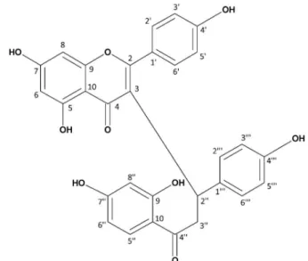
Caesalpinioflavone (Figure 1) was isolated as a
yellow crystalline solid with an optical activity [α]D
25 of
+135° (c 0.1, MeOH) and a melting point of 227 °C. The
molecular formula was established as C30H22O9 based on
the negative mode HRESIMS (Figure S1), which exhibited
the deprotonated molecule [M-H]− at m/z 525.1185 (calcd.
525.1186). The IR spectrum (Figure S2) showed strong
absorptions at 3415 cm-1 (OH), 2952 cm-1 (CH=CH),
and 1653/1615 cm-1. The UV spectrum displayed
absorptions with λmax at 264 and 311 nm, characteristic
of flavonoids. The 13C NMR spectrum (Table 1) showed
carbonyl resonances at δC 203.5 (C-4”) and 182.6 (C-4),
oxygenated carbons at δC 166.3 (C-7), 165.9 (C-5), 165.8
(C-2), 163.3 (C-9”), 163.0 (C-7”), 161.1 (C4’), 159.3
(C-9), 156.9 (C-4”’), the p-substitutedaromatic carbons
at δC 131.5 (C-3’ and C-5’), 131.4 (C-3”’ and C-5”’),
116.3 (C-2’ and C-6’), 116.1 (C-2”’ and C-6”’), as well
as saturated carbons at δC 49.2 (C-2”) and δC 36.0 (C-3”).
The presence of characteristic carbon signals of flavones and chalcones associated to HRESIMS, the occurrence of
a biflavonoid derivative was defined.91H NMR spectrum of
caesalpinioflavone (Table 1) showed signals ranging from
δH 7.00 to 6.50 (8H) which referred to the hydrogen atoms
of two p-substituted aromatic rings. These data, associated
to the presence of doublets at δH 6.24 (d, J 2.5 Hz, H-8) and
δH 6.19 (d, J 2.5 Hz, H-6), indicated a subunit of apigenin.
10
Other signals were observed atδH 7.26 (d, J 8.7 Hz, H-5”),
6.19 (d, J 2.5 Hz, H-8”) and 6.15 (dd, J 8.7 and 2.5 Hz,
H-6”), indicating the presence of a 1,2,4-trisubstituted
aromatic ring. Signals at δH 4.69 (dd, J 7.5 and 6.5 Hz,
H-2”), 3.47 (dd, J 14.0 and 6.5 Hz, H-3a”) and 3.09
(dd, J 14.0 and 7.5 Hz, H-3b”) indicated the occurrence
of a 4,2’,4’-trihydroxychalcone unit. The homonuclear
correlation spectroscopy (COSY) couplings between
the signals at δH 6.19 (d, J 2.5 Hz, H-6) with δH 6.24 (d,
J 2.5 Hz, H-8), δH 6.15 (dd, J 8.7 and 2.5 Hz, H-6”) with
δH 7.26 (d, J 8.7 Hz, H-5”) and δH 6.19 (d, J 2.5 Hz, H-8”),
and δH 4.69 (H-2”) with δH 3.47 (H-3”a) and δH 3.09
(H-3”b) confirmed the proposed substructures. Finally, the heteronuclear multiple-bond correlation spectroscopy (HMBC) spectrum showed the correlation of the signal at
δH 4.69 (H-2”) with those at δC 182.6 (C-4), 36.0 (C-3”),
165.8 (C-2) and 203.5 (C-4”), and the correlations of
the signals at δH 3.47/3.09 (H-3”) with those at δC 36.0
(C-3”) and 203.5 (C-4”), which confirmed the connection between C-2” and C-3 in the biflavonoid structure (Figure 2 and Figures S3-S7). All the observed correlations are shown in Table 1. The configuration at C-2” was not assigned.
The cytotoxic potential of caesalpinioflavone was determined by MTS assay, and the results showed a drastic reduction of the cell viability in cells treated with
caesalpinioflavone in concentrations upward of 40 µmol L-1
(Figure 3). Among the cell lines studied, the hepatoma cell line (HTC) was the most responsive to treatment
(IC50 value = 48.00 ± 1.60 µmol L-1). When HTC cells were
treated under the same conditions with cisplatin, a powerful
cytotoxic anticancer agent, the IC50 value was found to be
38.53 ± 1.49 µmol L-1. IC
50 values found for the other cell
lines were 121 ± 9.7 µmol L-1 (MCF7), 108 ± 7.6 µmol L-1
(A549) and 97.65 ± 3.2 µmol L-1 (Hs578T). These results
indicate that caesalpinioflavone displays antiproliferative activity against HTC cells, which is in agreement with recent studies reporting that different flavonoids can
Figure 1. Chemical structure of caesalpinioflavone, a novel biflavonoid isolated from the stem bark of Caesalpinia pluviosa var. peltophoroides.
exert growth inhibition and/or cytotoxic activities on
hepatocellular carcinoma cell lines.9-12
The images obtained by phase contrast microscopy (Figure 4) show the morphological features of the cell
lines studied. The images clearly show lower cell densities in the treated cultures compared to the control cultures. To investigate whether the cytotoxic effects were a consequence of cell death induction or cell cycle arrest, we performed the cell cycle analysis by DNA quantification. Thus, the cultures were treated with caesalpinioflavone
at 40 µmol L-1 and 80 µmol L-1, and the samples were
analyzed by flow cytometry. The results showed that the effects of caesalpinioflavone on the cell cycle progression were concentration-dependent, and they varied depending on the cell type (Figure 5).
In MCF7, we observed cell cycle arrest in the G1/S
transition (at 40 µmol L-1) and the G2/M arrest (at
80 µmol L-1). In A549, both concentrations caused G1/S
transition inhibition. It has been reported that phenolic compounds present pro-oxidant activity when used at
high concentrations,13-15 and may contribute for activating
proteins associated to cell cycle control such as p53, p21
and GADD45.16,17 In the present work, the cell lines with
wild p53 displayed inhibition of the G1/S transition as a consequence of treatment, suggesting that the p53 pathway could be activated by caesalpinioflavone.
In Hs578T and HTC cells, an increased subG1 population was observed in addition to cell cycle arrest.
When concentrations around the IC50 were used (80 µmol L-1
and 40 µmol L-1 for Hs578T and HTC, respectively) cell
cycle arrest in G2/M was observed. In these cell lines, at the same concentrations, the subG1 populations were 2.5-fold (Hs578T) and 5-fold (HTC) higher than in control cultures.
Table 1. NMR data for caesalpinioflavone in CD3OD
Position δC δH (m, J / Hz) HMBC
2 165.8 - H2”
3 120.9 - H2”/H3”
4 182.6 - H2”
5 165.9 - H6
6 100.1 6.19 (d, 2.5) H8
7 166.3 - H8
8 94.6 6.24 (d, 2.5) H6
9 159.2 - H8
10 104.9 - H8
1’ 124.6 -
-2’; 6’ 116.3 6.82 (d, 8.7)
-3’; 5’ 131.5 7.02 (d, 8.7)
-4’ 161.1 - H3’/H5’
2” 49.2 4.69 (dd, 7.5; 6.5) H3”
3a” 36.0 3.46 (dd, 14.0; 6.5) H3b”/H2”
3b” 36.0 3.09 (dd, 14.0; 7.5) H3a”/H2”
4” 203.5 - H2”/H3”
5” 132.1 7.25 (d, 9.0) H6”
6” 108.5 6.15 (dd, 9.0; 2.5) H5”/H8”
7” 163.0 - H8”
8” 103.9 6.19 (d, 2.5) H6”
9” 163.3 - H8”
10” 113.7 - H5
1”’ 132.1 - H2”/H2”’
2”’; 6”’ 116.1 6.64 (d, 8.5)
-3”’; 5”’ 131.4 6.95 (d, 8.5)
-4”’ 156.8 - H3”’/H5”’
0 20 40 60 80 100 120 140
5 10 20 40 80 160
R
ela
ve cell via
bi
ty
A549 HTC MCF7 Hs578T
Concentraon / ( mol L )µ -1
Figure 3. Relative cell viability of caesalpinioflavone against tumor cell lines A549, HCT, MCF7 and Hs578T, obtained by the MTS assay.
You et al.18 demonstrated that the increase in the subG1
population in human PC-3 (prostate cancer) cell cultures after treatment with ginkgetin (a biflavonoid) for 24 h was
due to apoptosis induction.Phenolic compounds, such as
caffeic acid phenethyl ester, induced apoptosis in glioma cells by the activation of the p53 pathway whereas cinnamic acid was effective in inducing apoptosis in melanoma cells
by different pathways.12,19,20 The results obtained in the
present work showed that caesalpinioflavone inhibits cell proliferation of MCF7 and A549 cells, and has cytotoxic activity against Hs578T and HTC cell lines.
Conclusions
A novel biflavonoid, named as caesalpinioflavone was
isolated from the stem bark of Caesalpinia pluviosa var.
peltophoroides. Caesalpinioflavone reduced cell viability of tumor cell lines A549, MCF7, Hst578T and HTC, as consequence of cell cycle arrest in G1/S transition (A549 and MCF7) and cytotoxic activity (Hs578T and HTC). Taken together, these data indicate that caesalpinioflavone has a promising antitumor activity.
Supplementary Information
Supplementary information, including 1H NMR,
13C NMR, COSY, HSQC, and HMBC spectra, as well
as mass and IR spectra, are available free of charge at http://jbcs.org.br as a PDF file.
Acknowledgements
The authors acknowledge Dr. Glaucia Maria Machado Santelli for providing the cell lines used in this study, and aid received from FAPEMIG, CAPES, CNPq and FINEP through grants and subsidies.
References
1. Srinivas, K. V. N. S.; Rao, Y. K.; Das, I. M. B.; Krishna, K. V. S. R.; Kishore, K. H.; Murty U. S. N.; Phytochemistry2003, 63, 789.
2. Udenigwe, C. C; Ata, A.; Samarasekera, R.; Chem. Pharm. Bull. 2007, 55, 442.
3. Nagumo, S.; Whasiyama, M.; Sasaki, Y.; Hosokawa, T.; Biol. Pharm. Bull.2009, 32, 941.
4. Choi, B. M.; Lee, J.; Gao, S. S.; Eun, S. Y.; Kim, Y.; Ryu, S.; Choi, Y.; Park, R.; Kwon, D. Y.; Kim, B.; Biofactors2007, 30, 149. 5. Lee, Y. M.; Kim, Y. C.; Choi, B. J.; Lee, D. W.; Yoon. J. H.;
Kim, E. C.; Toxicol. in vitro2011, 25, 1782.
6. Zanin, J. L. B.; de Carvalho, B. A.; Martineli, P. S.; dos Santos, M. H.; Lago, J. H. G.; Sartorelli, P.; Viegas. C. Jr.; Soares, M. G.;
Molecules2012, 17, 7887.
7. Bueno, F. G.; Panizzon, G. P.; Mello, E. V. S. L.; Lechtenberg, M.; Petereit, F.; de Mello, J. C. P.; Hensel, A.; Fitoterapia2014, 99, 252.
8. Ferreira, D. F.; Ciênc. Agrotec. 2011, 35, 1039.
9. Yang, J.; Yang, Y.; Tian, L.; Sheng, X. F.; Liu, F.; Cao. J. G.;
World J. Gastroenterol.2011, 14, 4298. 0% 50% 100% 150% control control control control 40 40 40 40 80 80 80 80 Cell p o pula on Cell p o pula on Cell p o pula on Cell p o pula on
Concentraon / ( mol L )µ -1
Concentraon / ( mol L )µ -1
Concentraon / ( mol L )µ -1
Concentraon / ( mol L )µ -1
A549 MCF7 Hs578T 0% 20% 40% 60% 80% 100% 120% G2/M S G1 SubG1 0% 20% 40% 60% 80% 100% 120% G2/M S G1 SubG1 G2/M S G1 SubG1 0% 20% 40% 60% 80% 100% 120% G2/M S G1 SubG1 HTC
10. Kim, B. R.; Jeon, Y. K.; Nam, M. J.; Food Chem. Toxicol.2011,
49, 1626.
11. Liang, R. R.; Zhang, S.; Qi, J. A.; Wang, Z. D.; Li, J.; Liu, P. J.; Huang, C.; Le, X. F.; Yang, J.; Li, Z. F.; Int. J. Oncol.2012, 41: 969.
12. Lee, Y. J.; Kuo, H. C.; Chu, C. Y.; Wang, C. J.; Lin, W. C.; Tseng, T. H.; Biochem. Pharmacol.2003, 66, 2281.
13. Banskota, A. H.; Nagaoka, T.; Sumioka, I. Y.; Tezuka, Y.; Awale, S.; Midorikawa, K.; Matsushige, K.; Kadota, S.;
J. Ethnopharmacol.2002, 80, 67.
14. Hsu, Y. L.; Chen, C. Y.; Hou, M. F.; Tsai, E. M.; Jong, Y. J.; Hung, C. H.; Kuo, P. L.; Mol. Nutr. Food Res.2010, 54, 1307. 15. Thayyullathil, F.; Chathoth, S.; Hago, A.; Patel, M.; Galadari, S.;
Free Rad. Biol. Med.2008, 45, 1403.
16. El-Deiry, W. S.; Tokino, T.; Velculescu, V. E.; Levy, D. B.; Parsons, R.; Trent, J. M.; Lin, D.; Mercer, W. E.; Kinzler, K. W.; Vogelstein, B.; Cell1993, 75, 817.
17. Kastan, M. B.; Zhan, Q.; El-Deiry, W. S.; Carrier, F.; Jacks, T.; Walsh, W. V.; Plunkett, B. S.; Vogelstein, B.; Fornace, A. J.;
Cell1992, 71, 587.
18. You, O. H.; Kim, S. H.; Kim, B.; Sohn, E. J.; Lee, H. J.; Shim, B. S.; Yun, M.; Kwon, B. M.; Kim, S. H.; Bioorg. Med. Chem. Lett.2013, 23, 2692.
19. Sje, Q. B.; Bode, A. M.; Ma, W. Y.; Chen, N. Y.; Dong, Z.;
Cancer Res.2001, 61, 1604.
20. Niero, E. L.; Machado-Santelli, G. M.; J. Exp. Clin. Cancer Res.2013, 32, 31.
Submitted: October 14, 2014
Published online: February 24, 2015