ORIGINAL
ARTICLE
Hyaluronic acid in the evaluation of liver fibrosis
in patients with hepatitis C on haemodialysis
Authors
Renata Eliane de Ávila1 Ricardo Andrade Carmo2 Kátia de Paula Farah3 Antônio Lúcio Teixeira3 Lucas Viana Coimbra3 Carlos Maurício de Figueiredo Antunes4 José Roberto Lambertucci1
1Infectious Diseases Branch, Department of Internal Medicine, Faculdade de Medicina, Universidade Federal de Minas Gerais; Núcleo de Ações e Pesquisa em Apoio - NUPAD/ FM / UFMG.
2Fundação Hemominas. 3Infectious Diseases Branch, Department of Internal Medicine, Faculdade de Medicina, Universidade Federal de Minas Gerais. 4Department of Parasitology, Instituto de Ciências Biológicas – UFMG.
Submitted on: 08/12/2009 Approved on: 11/18/2009
Correspondence to:
Dr. José Roberto Lambertucci
Faculdade de Medicina da UFMG
Departamento de Clínica Médica
Avenida Alfredo Balena, 190. Belo Horizonte - Minas Gerais - Brazil CEP: 30130-100 Phone: +55-31-34099820 E-mail:
This work was partially supported by Fundação de Amparo à Pesquisa do Estado de Minas Gerais (FAPEMIG) and Conselho Nacional de Desenvolvimento Científi co e Tecnológico (CNPq).
ABSTRACT
Background: This study evaluated the role of HA as a marker of liver fi brosis in patients with hepa-titis C on haemodialysis. Methods: This is a cross-sectional study in which 52 patients were divided into two groups: Group 1: patients with hepatitis C and end-stage renal disease (ESRD) undergoing haemodialysis (n = 23); and Group 2: patients with hepatitis C without ESRD (n = 29). Plasma levels of HA were associated with histological data of the samples obtained by liver biopsy and classifi ed by METAVIR group scoring system. Results: Higher plasma levels were signifi cantly correlated to sig-nifi cant liver fi brosis (METAVIR ≥ F2). In Group 1, the HA cutoff to discriminate sigsig-nifi cant fi brosis was 984.8 ng/mL, with accuracy, sensitivity and specifi city of 80.8%, 83.0%, and 70.0%, respectively. In Group 2, the HA cutoff was 222.3 ng/mL, with accuracy, sensitivity and specifi city of 74.5%, 70.0%, and 94.0%, respectively. Conclusion: HA was an accurate noninvasive marker in predicting signifi cant fi brosis in patients with hepatitis C on haemodialysis.
Keywords: liver fi brosis, hyaluronic acid, end-stage renal disease, haemodialysis, hepatitis C. [Braz J Infect Dis 2010;14(4):335-341]©Elsevier Editora Ltda.
INTRODUCTION
Chronic hepatitis C virus (HCV) infection is common in patients on haemodialysis (HD) and has been associated with chronic liver disease in this setting. The prevalence of HCV infection in HD units ranges, ac-cording to geographical area, from 9% to 80% worldwide.1 In Brazil, several dialysis
centers2-4 reported rates from 11% to 52%.
The transfusion-associated transmissions have been reduced due to the implementa-tion of blood donor screening for hepatitis C in the early 1990s and the use of recom-binant erythropoietin. Currently, nosoco-mial transmission is the main cause of HCV infection in this population.5,6
The mortality rate of HCV infected HD patients is higher than non-infected subjects.7,8 Candidates for renal
transplanta-tion have to be treated for hepatitis C before surgery, since HCV infection has a negative impact on graft and patient survival.9 The
degree of fibrosis evaluation is paramount for treatment decision.
Liver biopsy remains the gold stand-ard procedure for diagnosing liver fibrosis.
However, it is an invasive method associated with sampling error, interobserver variabil-ity and potential complications.10 In HD
pa-tients, the rate of complications is elevated due to higher risk of bleeding secondary to haemostatic disorders.11,12 In a recent study,
severe complications of liver biopsies were demonstrated in 13.2% of HD patients.13
Therefore, there is a need to assess the util-ity of noninvasive markers of fibrosis in this population.
Hyaluronic acid (HA) has been used to predict liver fibrosis in patients with chronic hepatitis C.14-17 APRI
(aspartate-aminotrans-ferase to platelet ratio index) is another promising liver fibrosis marker in hepatitis C.18-20 In a recent study, APRI identified
sig-nificant liver fibrosis (METAVIR ≥ F2) with accuracy of 80.1% in patients with hepatitis C on haemodialysis.21
PATIENTS AND METHODS
This is a cross-sectional study of patients attending an Infectious Diseases Outpatient Clinic and Haemodialy-sis Unit at the Hospital of the Universidade Federal de Minas Gerais-HC-UFMG (Belo Horizonte, Brazil).
Patients with chronic hepatitis C and end-stage renal disease (ESRD) on haemodialysis are referred to our out-patient clinic to be evaluated and treated for hepatitis C. Our current practice is to treat hepatitis C according to the degree of fibrosis found in liver fragments obtained by needle liver biopsy. From May 2000 to October 2007, 69 patients with chronic hepatitis C and ESRD on HD who underwent liver biopsy in a previous study13 were
registered under our care. Among those subjects, all in-dividuals who had stored plasma sample (used in HCV-RNA detection) and liver fragments collected within one year by liver biopsy were considered for enrollment. These patients were selected for group 1 (hepatitis C and ESRD on HD). The other 67 consecutive patients with chronic hepatitis C without ESRD who had undergone liver biopsy for treatment evaluation and collection of plasma sample were also considered for enrollment (group 2).
The study was approved by the Ethics Committee of Universidade Federal de Minas Gerais (Belo Horizonte, MG, Brazil). A written informed consent was obtained from each participant prior to commencement of the study.
Criteria for selection of subjects
The inclusion criteria for subjects of groups 1 and 2 were availability of liver fragments obtained by liver biopsy and a plasma sample collected within one year; age between 18 and 70 years old; presence of chronic HCV infection (defined as a reactive anti-HCV antibod-ies for more than six months and a positive HCV-RNA by PCR); absence of other chronic liver diseases such as hepatitis B (defined as negative reaction to HBV sur-face antigen and HBV core antibody), auto-immune hepatitis (negative reaction to antinuclear, anti-smooth muscle, anti-mitochondrial and anti-liver-kidney mi-crosomal antibodies), schistosomiasis mansoni (no pre-vious history and negative stool examination), negative reaction to anti-HIV-1/2 (EIA); no previous history of regular use of hepatotoxic drugs or alcohol abuse (> 40 g of alcohol/day). Patients previously treated for hepati-tis C were excluded.
Socio-demographic, epidemiological and clinical data
Baseline data were collected at the time of liver biopsy (in Groups 1 and 2) including: age, gender and length of time under haemodialysis (in group 1).
Laboratory data
Levels of aminotransferases (alanina-aminotransferase-ALT and aspartate-aminotransferase-AST), hemoglob-in, platelets, and albumin were collected at the time of liver biopsy in groups 1 and 2. Parathormone (PTH) was measured in group 1.
Assay for anti-HCV antibodies was determined us-ing enzyme immunoassay EIA III (Abbot Laboratories, North Chicago, IL, USA). For RNA extraction, blood HCV-RNA was detected by transcription followed by nested-RT-PCR using primers derived from the 5’-UTR non-coding region of the HCV. The genotype was deter-mined through Restriction Fragment Length Polymor-phism (RFLP) analysis of the PCR product.
Histological analyses
Liver samples were obtained by percutaneous ultrasound-directed hepatic biopsy. Two experienced pathologists ex-amined (reviewed) liver fragments in a blinded manner (they knew the patients had hepatitis C but did not have any information about their clinical statuses). After disclo-sure of the diagnosis, the discordant cases were re-analyzed and a consensus was reached. The histological samples were analyzed using the METAVIR algorithm:22 F0 = no fi brosis,
F1 = portal fi brosis without septa, F2 = few septa, F3 = nu-merous septa without cirrhosis, and F4 = cirrhosis. Stages of fi brosis were categorized as signifi cant fi brosis (META-VIR ≥ F2) and absent or mild fi brosis (METAVIR < F2).
Plasma hyaluronic acid assay
HA plasma levels were assessed by a quantitative en-zyme-linked antibody (HA-ELISA) and colorimetric detection with a commercially available test Kit (Ech-elon Biosciences®, Salt Lake City, USA). For this test, HA normal value was < 50 ng/mL.
Statistical analyses
RESULTS
Socio-demographic, clinical and laboratory characteristics
Socio-demographic, clinical and laboratory features of the patients are shown in Table 1.
A total of 136 patients were considered for enrollment in groups 1 and 2. However, the plasma samples were not col-lected within one year of liver biopsy in 20 and 12 in groups 1 and 2 patients, respectively. Other 22 patients of group 1 were not included because of the following reasons: age < 18 or > 70 years (n = 2), schistosomiasis (n = 7), hepatitis B (n = 3), use of hepatotoxic drugs (n = 6), and auto-immune hepatitis (n = 1). In group 2, 26 patients were not included because of the following fi ndings: age < 18 or > 70 years (n = 2), schistosomiasis (n = 4), alcohol abuse (n = 8), use of hepatotoxic drugs (n = 4), hepatitis B (n = 4), previously treated for hepatitis C (n = 4).
Baseline characteristics (age, gender and time on haemo-dialysis in group 1) of patients who were excluded due to unavailable plasma sample collected at least one year of liver biopsy were compared to the characteristics of the included participants in groups 1 and 2. In group 1, excluded patients were signifi cantly older than those included (p = 0.01), whereas in group 2 no difference was observed between in-cluded and exin-cluded patients.
A total of 52 patients have been selected for this study: group 1 (Hepatitis C and ESRD, n = 23) and group 2 (Hepa-titis C and no ESRD, n = 29).
The two groups were similar regarding age and gender. None of the included patients had clinical manifestations of liver failure. There was a predominance of genotype 1, be-ing of 95.7% in group 1 and of 79.0% in group 2. ALT and AST levels were signifi cantly lower in patients with hepatitis C and ESRD , when compared to patients without ESRD. These patients also had a lower level of albumin and hemo-globin (Table 1).
Table 1. Distribution of socio-demographic, clinical and laboratory features of 52 patients included in groups 1 (hepatitis C and end-stage renal disease) and 2 (hepatitis C alone)
Characteristics Group 1 Group 2
(Hepatitis C and ESRD) (Hepatits C alone) p-value*
n = 23 n = 29
Age (mean ± SD years) 43.47 (± 12.37) 46.07 (± 12.39) 0.74
Gender
Male n (%) 11 (48.1) 18 (62.1) 0.45
Female n (%) 12 (51.9) 11 (37.9)
-Skin color
White n (%) 07 (30.4) 13 (44.8)
-Black n (%) 05 (21.7) 01 (3.4) 0.67
Others n (%) 11 (47.8) 15 (51.7)
-Time of haemodialysis 9.67 ± 5.00 -
-(years, mean ± SD)
ALT** (U/L median) 37.0 74.0 0.00
AST*** (U/L median) 29.0 53.3 0.00
Haemoglobin (g/dL median) 11.5 15.6 0.00
Platelets (/mm3 median) 194,000 212,000 0.51
Albumin (g/dL median) 3.7 4.3 0.00
PTH**** (pg/L mean) 381.04 -
-*Student t test, Fishers exact test, Chi-square test, as appropriate comparisons of groups. **ALT: alanine-aminotransferase.
***AST: aspartate-aminotransferase.
Histological data
The mean number of portal tracts in the biopsies was 9 in both groups. Steatosis was observed in 55.2% of group 2 patients, and hepatic siderosis in 56.5% of group 1 sub-jects. Histological examination revealed signifi cant fi brosis in 56.5% in patients of group 1 and in 44.3% in group 2. However, the frequency of advanced fi brosis and cirrhosis (METAVIR ≥ F3) was low in both groups (Figure 1).
Table 2. Hyaluronic acid levels in groups 1 (hepatitis C and end-stage renal disease) and 2 (hepatitis C alone)
Hyaluronic acid (ng/mL)
Study groups n Mean Median Minimal Maximum SD p-value*
1 (ESRD**/hepatitis C) 23 5002.8 1546.3 302.5 3922.3 9455.9 0.00
2 (Hepatitis C) 29 268.3 160.8 80.9 1029.3 255.3
-*Kruskal-Wallis.
** ESRD: end-stage renal disease. 50 45 40 35 30 25 20 15 10 5 0 %
0 1 2 3 4
Fibrosis
1 - ESRD/hepatitis C
2 - Hepatitis C
Figure 1: Frequency distribution of histological fibrosis stage according to the METAVIR score in 23 patients with end-stage renal disease (ESRD) and hepatitis C (group 1) and 29 patients with hepatitis C alone (group 2).
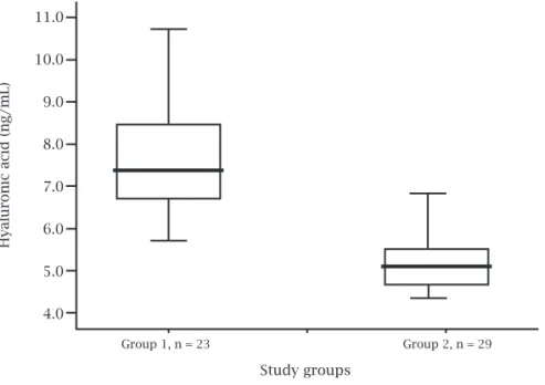
Figure 2: Box plot of logarithmic hyaluronic acid (HA) level in relation to study groups. There was significant difference between HA levels in groups 1 and 2 (p = 0.00).
Study groups
Hyaluro
nic acid (ng/mL)
Group 1, n = 23 Group 2, n = 29
11.0
10.0
9.0
8.0
7.0
6.0
5.0
4.0
Variables associated with signifi cant fi brosis
was 141.0 ng/mL in patients without signifi cant fi brosis and 277.6 ng/mL in patients with signifi cant fi brosis (p = 0.02).
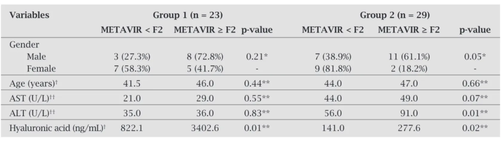
In group 1, only HA levels were correlated with the de-gree of liver fi brosis by METAVIR algorithm. There was no signifi cant association between age, gender, AST and ALT levels with degree of fi brosis. In group 2, HA and ALT lev-els were signifi cantly associated with degree of liver fi brosis (Table 3).
In Group 1, the HA cutoff applied to discriminate sig-nifi cant fi brosis was 984.8 ng/mL, with accuracy (AUROC), sensitivity and specifi city of 80.8%, 83.0%, and 70.0%, re-spectively. In group 2, the HA cutoff was 222.3 ng/mL, with
Table 3. Variables associated with the degree of liver fibrosis (absent or mild: METAVIR < F2 or significant: METAVIR ≥ F2, in groups 1 (hepatitis C and end-stage renal disease) and 2 (hepatitis C alone)
Variables Group 1 (n = 23) Group 2 (n = 29)
METAVIR < F2 METAVIR ≥ F2 p-value METAVIR < F2 METAVIR ≥ F2 p-value
Gender
Male 3 (27.3%) 8 (72.8%) 0.21* 7 (38.9%) 11 (61.1%) 0.05*
Female 7 (58.3%) 5 (41.7%) - 9 (81.8%) 2 (18.2%)
-Age (years)† 41.5 46.0 0.44** 44.0 47.0 0.66**
AST (U/L)†† 21.0 29.0 0.55** 44.0 49.0 0.07**
ALT (U/L)†† 35.0 36.0 0.83** 56.0 91.0 0.01**
Hyaluronic acid (ng/mL)† 822.1 3402.6 0.01** 141.0 277.6 0.02**
† Median ; †† Mean.
* Fisher’s exact test; ** Mann-Whitney test.
accuracy, sensitivity and specifi city of 74.5%, 70.0%, and 94.0%, respectively (Figure 3).
Variables associated with plasma levels of hyaluronic acid in patients on hemodialysis
In group 1, only HA levels were correlated with the degree of liver fi brosis by METAVIR algorithm (p = 0.01). There was no signifi cant correlation among age, gender and time of haemodialysis in this group. Hyperparathyroidism (PTH > 69 pg/L) was found in 95.7% of group 1 patients, but there was no signifi cant correlation between serum parathormone (PTH) and HA levels.
Figure 3: Receiver operating characteristic curves of hyaluronic acid for the prediction of significant fibrosis (METAVIR ≥ F2). A: group 1 (patients with end-stage renal disease with hepatitis C). B: group 2 (Hepatitis C alone).
1.0
0.8
0.6
0.4
0.2
0.0
1.0
0.8
0.6
0.4
0.2
0.0
Sensitivity
A
B
1 - Specificity
*AUC: Area under the curve. **CI: Confidence interval.
AUC* = 0.808 CI** = 0.662-0.995
AUC* = 0.745 CI** = 0.55-0.935
1 - Specificity
Sensitivity
DISCUSSION
In this study, plasma hyaluronic acid (HA) level was a good noninvasive marker of signifi cant fi brosis in patients with hepatitis C on haemodialysis. For hepatitis C without ESRD, our fi ndings are comparable to those reported previously.14-17
For patients with ESRD, this is much more important than for patients with hepatitis C without co-morbidities. The frequency and severity of complications of liver biopsy in patients with renal failure has been documented.11-13
There-fore, HA levels may be used to defi ne when treatment for hepatitis C should be considered, especially in subjects with coagulation dysfunction caused by renal failure. Besides, it may be used as a screening test before a decision in favor of liver biopsy is to be presented to the patient.
Our fi ndings contrast with the observations of a recent study.22 In the latter, the diagnostic value of hyaluronic acid
(HA) as a noninvasive marker of liver fi brosis was evaluated in 185 ESRD HCV-infected patients. For the prediction of signifi cant fi brosis, the AUROC (0.798) of the regression model, including AST, platelet count and HA, was signifi -cantly higher than the AUROC of HA (0.650).
The specifi city of HA in detecting signifi cant liver fi bro-sis was lower in group 1 than in group 2. It may result from other factors that increase HA levels in ESRD patients. High plasma levels of HA have already been described in ESRD.24-26
The increase of plasmatic HA in ESRD has been attributed to an “infl ammatory state” caused by haemodialysis itself.24
Notwithstanding, as observed for patients in group 1, plas-ma HA levels reached even higher levels in the presence of hepatitis C infection, validating the test in the identifi cation of patients with signifi cant liver fi brosis.
Patients on haemodialysis usually develop second-ary hyperparathyroidism and it was found in most group 1 patients (Table 1). Parathyroid hormone (PTH) is a key hormone regulating bone mineral homeostasis and it also stimulates the production of HA in cultures of bone cells24, 25 Nevertheless, in comparing PTH levels with HA levels in
group 1 (13 patients with fi brosis and 10 without) no statis-tical correlation was found (i.e. high HA levels were associ-ated with liver fi brosis, but not with PTH values). Certainly, a larger number of patients should be evaluated to confi rm whether PTH, due to its action in bone metabolism, has any infl uence in plasma HA levels. If HA is to be used alone as a marker of fi brosis, it may give a false positive test for fi brosis that may result in unnecessary and unwanted treatment for hepatitis C.
In the present study, women with ESRD had lower levels of plasma HA. It has been shown that estrogen decreases os-teoclast number, decreasing therefore bone resorption.26
Es-trogen seems to regulate osteoclastogenesis by modulating cytokine production (interleukins 1 and 6 and tumor necro-sis factor), and expression of adhesion molecules on bone marrow stromal cells. Sex steroids also act directly upon the
parathyroid gland to increase PTH mRNA at physiological relevant doses.26 The decrease of bone metabolism in
es-trogen producing women may explain the decrease in HA liberation by bone cells and their lower levels in patients with ESRD.
The longer the patient stays on haemodialysis, probably the stronger is the infl ammatory process associated with chronic haemodialysis. It has been previously reported that the timespan on haemodialysis and certain markers of chronic infl ammation, such as dialysis-related amyloid, correlate to HA serum levels.16 Cytokine production may
be stimulated by contact of the blood with bioincompat-ible dialysis membranes, and cytokines have been shown to stimulate the synthesis of HA. The length of time on haemodialysis did not correlate to HA levels in our study, and the reason for that is uncertain. However, two factors should be considered: (1) 12 out of our 23 patients (52.2%) on haemodialysis were women and estrogen decreases HA serum levels; and (2) the mean time on haemodialysis in the present study was of 9.7 years and a signifi cant differ-ence in HA levels can take more than 10 years to be de-tected.
Older people tend to have a series of disadvantages on haemodialysis. As survival has increased with dialysis, they are usually those who have endured haemodialysis for long-er plong-eriods (and consequently they have spent more time in an environment with persistent chronic infl ammation), and the chronic infl ammatory process caused by dialysis may result in decreased albumin synthesis by the liver as a result of cytokines stimulation of acute-phase reactants.21
In addition, as sex steroids decrease with aging, they may increase osteoporosis and the levels of plasma HA.
However, no statistical correlation was found between age, gender and time on haemodialysis in group 1. This fi nding indicates that liver fi brosis was the main factor as-sociated with the increase of HA serum levels in group 1 patients. In addition, aminotransferase serum levels were not associated with liver fi brosis on haemodialysis patients with hepatitis C, as shown by others.25
The number of patients studied and the low frequency of advanced fi brosis and cirrhosis (METAVIR ≥ F3) were the main limitations of this study. As a matter of fact, pa-tients with decompensated liver cirrhosis are not referred to our outpatient clinic for therapeutic decision because such patients are not candidates for hepatitis C treatment before kidney transplantation.
In conclusion, plasma HA levels can predict the pres-ence of signifi cant fi brosis in patients with hepatitis C on haemodialysis with good sensitivity and specifi city. It is desirable to start treatment for hepatitis C in this group of patients and to follow the behavior of hyaluronic acid levels after successful treatment to establish its value as a marker of fi brosis regression.
ACKNOWLEDGEMENTS
This work was partially supported by Fundação de Amparo à Pesquisa do Estado de Minas Gerais (FAPEMIG) and Conselho Nacional de Desenvolvimento Científi co e Tec-nológico (CNPq).
REFERENCES
1. Fabrizi F, Poordad FF, Martin P. Hepatitis C infection and the patient with end-stage renal disease. Hepatology 2002; 36:3-10.
2. Carneiro MA, Martins RM, Teles AS et al. Hepatitis C preva-lence and risk factors in hemodialysis patients in Brazil: a sur-vey by polimerase chain reaction and serological. Mem Inst Oswaldo Cruz 2001; 96:765-9.
3. Santana GO, Cotrim HP, Mota E et al. Anti-HCV in patients undergoing hemodialysis in Salvador, BA, Brazil. Arq Gastro-enterol 2001; 38:24-31.
4. Medeiros MTG, Coelho-Filho JM. Prevalence and associated factors to hepatitis C in hemodialisys patients in Brazil. Rev Saúde Pública 2004; 38:187-93.
5. Fabrizi F, Martin P, Lunghi G, Ponticelli C. Nosocomial trans-mission of hepatitis C virus infection in hemodialysis patients: Clinical perspectives. Int J Artif Organs 2000; 23:805-16. 6. Jadoul M. Epidemiology and mechanisms of transmission of
the hepatitis C virus in hemodialysis. Nephrol Dial Trans 2000; 15:S39-41.
7. Nakayama E, Akiba T, Marumo F et al. Prognosis of anti-hep-atitis C virus antibody-positive patients on regular hemodialy-sis therapy. J Am Soc Nephrol 2000; 11:1896-902.
8. Yokosuka O, Okuda K. Natural history of chronic hepatitis C in patients on hemodialysis: case-control study with 4-23 years of follow-up. World J Gastroenterol 2004; 10:2209-12. 9. Gonzáles-Roncero F, Gentil MA, Valdivia MA. Outcome of
kidney transplant in chronic hepatitis C virus patients: ef-fect of pretransplantation interferon-alpha 2b monotherapy. Transplant Proceed 2003; 35:1745-7.
10. Fontana RJ, Lok ASF. Noninvasive monitoring of patients with chronic hepatitis C. Hepatology 2002; 36:557-63.
11. Cotler SJ, Diaz G, Gundlapalli S et al. Characteristics of hepati-tis C in renal transplantation candidates. J Clin Gastroenterol 2002; 35:191-5.
12. Albuquerque W, Arantes V, de Paula Farah K et al. Acute pan-creatitis and acute cholecystitis caused by hemobilia after per-cutaneous ultrasound-guided liver biopsy. Endoscopy 2005; 37:1159-60.
13. de Paula Farah K, Carmo RA, de Figueiredo Antunes CM, Se-rufo JC et al. Hepatitis C, HCV genotypes and hepatic siderosis in patients with chronic renal failure on haemodialysis in Bra-zil. Nephrol Dial Transplant 2007, 2027-31.
14. Guechot J, Laudat A, Loria A et al. Diagnostic accuracy of hyaluronan and type III procollagen amino-terminal pep-tide serum assays as markers of liver fi brosis in chronic vi-ral hepatitis C evaluated by ROC curve analysis. Clin Chem 1996; 42:558-63.
15. Wong VS, Hughes V, Trull A. Serum hyaluronic acid is a use-ful marker of liver fi brosis in chronic hepatitis C virus infec-tion. J Viral Hepatitis 1998; 5:187-92.
16. Plevris JN, Haydon GH, Simpson KJ. Serum hyaluronan: a non-invasive test for diagnosing liver cirrhosis. Eur J Gas-troenterol Hepatol 2000; 12:1121-7.
17. Halfon P, Bourliere M, Penaranda G et al. Accuracy of hy-aluronic acid level for predicting liver fi brosis stages in pa-tients with hepatitis C virus. Comp Hepatol 2005; 11:4-6. 18. Wai CT, Greensson JK, Fontana RJ et al. A simple
noninva-sive index can predict both signifi cant fi brosis and cirrho-sis in patients with chronic hepatitis C. Hepatology 2003; 38:518-26.
19. Imbert-Bismut F, Ratziu V, Pieroni L et al. Biochemical markers of liver fi brosis in patients with hepatitis C virus infection: a prospective study. Lancet 2001; 357:1069-75. 20. Chrysanthos NV, Papatheodoridis GV, Savvas S et al.
As-partate aminotransferase to platelet ratio index for fi brosis evaluation in chronic viral hepatitis. Eur J Gastroenterol Hepatol 2006; 18:389-96.
21. Schiavon LL, Carvalho Filho RJ, Narcizo Schiavon JL et al. Simple blood test as noninvasive markers of liver fi brosis in hemodialysis patients with chronic hepatitis C virus infec-tion. Hepatology 2007; 46:307-14.
22. Schiavon LL, Narciso-Schiavon JL, Carvalho Filho RJ et al. Serum levels of YKL-40 and hyaluronic acid as nonin-vasive markers of liver fi brosis in haemodialysis patients with chronic hepatitis C virus infection. J Viral Hepat 2008; 15:666-74.
23. Bedossa P, Poynard T. An algorithm for the grading of activ-ity in chronic hepatits C. Hepatology 1996; 24:289-93. 24. Turney J H, Davison AM, Forbes MA et al. Hyaluronic acid
in end-stage renal failure treated by hemodialysis: Clinical correlates and implications. Nephrol Dial Transplant 1991; 6:566-70.
25. Furusyo N, Nayashi J, Kanamoto-Tanaka Y et al. Liver dam-age in hemodialysis patients with hepatitis C virus viremia: A prospective 10-year study. Dig Dis Sci 2000; 45:2221-8. 26. de Medina M, Hill M, Sullivan HO, Leclerq B et al. Detection
of anti-hepatitis C virus antibodies in patients undergoing dialysis utilizing a hepatitis C virus 3.0 assay: correlation with hepatitis C virus RNA. J Lab Clin Med 1998; 132:73-5. 27. Wong GL. Paracrine interactions in bone-secreted products
of osteoblasts permit osteoclasts to respond to parathyroid hormone. J Biol Chem 1984; 259:4019-22.
28. Midura RJ, Su X, Morcuende JA et al. Parathyroid hormone rapidly stimulates hyaluronan synthesis by periosteal oste-oblasts in the tibial diaphysis of the growing rat. J Biol Chem 2003; 278:51462-8.
29. Hughes D, Dai A, Tiffee JC, Li HH et al. Estrogen promotes apoptosis of murine osteoclasts mediated by TGF-beta. Nat Med 1996; 2:1132-6.
30. Suda T, Udagawa N, Nakamura I et al. Modulation of osteo-clast differentiation by local factors. Bone 1995; 28:87-91. 31. Trocme C, Leroy V, Sturn N et al. Longitudinal evaluation of