AR
TIGO ORIGINAL / ORIGINAL AR
TICLE
ANALYSIS OF THE SUSTAINED
VIROLOGICAL RESPONSE IN PATIENTS
WITH CHRONIC HEPATITIS C AND
LIVER STEATOSIS
Leonora De Zorzi
PICCOLI
1, Angelo Alves de
MATTOS
1, Gabriela Perdomo
CORAL
1,
Ângelo Zambam de
MATTOS
1and Diogo Edele dos
SANTOS
2ABSTRACT – Context - Chronic hepatitis C as well as non-alcoholic fatty liver disease are recognized as the main cause of liver disease
in Western countries. It is common to see the concomitance of the diseases and the inluence of steatosis in the sustained virological response of patients with hepatitis C virus. Objective - Assess the sustained virological response in chronic hepatitis C patients according to the presence of liver steatosis. Methods - One hundred sixty patients with chronic hepatitis C were retrospectively evaluated. Demographic data such as gender, age, body mass index, presence of diabetes mellitus and systemic arterial hypertension, virus genotype and use of pegylated interferon were analyzed, as was the staging of ibrosis and the presence of steatosis at histology.
Results - Most patients were male (57.5%), with a mean age of 48 ± 9.7 years. The most frequent genotype observed was 3 (56.9%) and, in the histological evaluation, steatosis was observed in 65% of the patients (104/160). Sustained virological response in patients with steatosis occurred in 38.5%, and in 32.1% in patients without steatosis (P = 0.54). When we analyzed possible factors associated with the presence of steatosis, only body mass index and systemic arterial hypertension revealed a signiicant association. When the factors that inluenced sustained virological response were evaluated in a logistic regression, genotype and use of pegylated interferon proved to be independent factors associated to the response. Conclusion - In the evaluated patients the presence of liver steatosis did not inluence the sustained virological response of patients with chronic hepatitis C treated with interferon and ribavirin.
HEADINGS - Hepatitis C, chronic. Fatty liver. Antiviral agents.
INTRODUCTION
Non-alcoholic fatty liver disease (NAFLD) is a pathological clinical syndrome that ranges from isolated liver steatosis to non-alcoholic steatohepatitis (NASH),
which can progress to advanced ibrosis and cirrhosis(7).
Estimates based on imaging studies and autopsies suggest that approximately 20%-30% of American adults and those from Western countries have an accumulation
of liver fat(3, 6, 7, 15, 46), and approximately 10% of these
individuals have NASH. This is frequently associated with adult age, female gender, obesity, diabetes mellitus (DM) type 2, hypertriglyceridemia, dyslipidemia and other conditions characterized by resistance to insulin
and hyperinsulinemia(7, 11, 52). A study conducted by our
group(60) demonstrated that NASH is also a common
disease among obese, non-diabetic patients, with altered levels of aminotransferases, occurring in 3.18% and,
Study conducted at the Gastroenterology and Hepatology Unit of Universidade Federal de Ciências da Saúde de Porto Alegre (UFCSPA), Porto Alegre, RS, Brasil. 1 Post-Graduate Course in Hepatology of UFCSPA; 2 Hospital Santa Casa de Misericórdia de Porto Alegre, RS, Brasil.
Correspondence: Prof. Angelo Alves de Mattos - Rua Cel. Aurélio Bitencourt, 35/201 - 90430-080 - Porto Alegre, RS, Brasil. E-mail: [email protected]
as a rule, presenting itself as a mild disease, although more intense cases may also be observed.
Liver steatosis is one of the most frequent histological indings in chronic hepatitis C (CHC) patients, observed
in approximately 55% of those patients(1, 8, 14, 19, 26, 28, 29, 37,
40, 42, 43, 45, 49, 59). In a recent study, Coral et al.(17) revealed
steatosis in 55 % of the patients infected by hepatitis C virus (HCV), correlating with genotype 3, but not with the degree of liver ibrosis.
The mechanisms through which the HCV causes liver damage associated with steatosis are poorly understood. It seems that the accumulation of fat in the hepatocytes of patients with CHC occurs mainly due to an effect of the virus itself strengthened by metabolic
factors related to the host(19, 42, 50, 53). The virus interferes
necrosis factor), leading to insulin resistance: TNF probably affects insulin receptor substrate (IRS-1) phosphorylation, decreasing glucose captivation and promoting compensatory hyperinsulinemia. Besides this, it is possible that liver siderosis and alterations in the levels of circulating adipocytokines
might as well have a role in insulin sensitivity in CHC(39).
Nevertheless, Mello et al.(35) failed to ind an enhancement in
insulin resistance, evaluated by HOMA (homeostatic model assessment), with the treatment of HCV.
There is agreement among several authors that the physiopathology of liver steatosis associated with HCV is
different in patients infected by genotype 1 or 3(4, 12, 23, 31, 47, 49).
Thus, the association between CHC and steatosis in patients infected by HCV genotype non-3, especially in those with genotype 1, seems to be related to pre-existing factors of the host, and is called “metabolic steatosis”. In this group of patients, the intensity of fat accumulation is proportional to BMI and the degree of visceral obesity, and there is no improvement after negativation of the virus through antiviral
treatment(13, 33). On the other hand, in patients with HCV
genotype 3, steatosis is related to higher viral loads and usually decreases after HVC treatment; this virus probably leads to hypobetalipoproteinemia, affecting liver release of triglycerides in bloodstream as VLDL (very low density
lipoprotein)(39).
The presence and intensity of liver steatosis is considered a marker of liver disease progression in patients with CHC, and it also seems to have an impact on sustained virological response (SVR) in these patients when submitted to antiviral
therapy(48, 53). It is possible that hyperinsulinemia could
block the inhibition of HCV replication by interferon, since insulin and interferon share signal transduction factors, such as p38 MAP kinase; PI3 kinase and IRF-1 also might
be associated(18, 39).
The main objective of this study was to assess SVR in patients with CHC and to relate it to the presence of hepatic steatosis.
METHODS
This is a retrospective study in which medical records of patients with CHC were assessed after they had undergone liver biopsy between January 2001 and December 2006, at Hospital Irmandade Santa Casa de Misericórdia de Porto Alegre, RS, Brasil.
The criteria for inclusion were: be over the age of 18, be infected by HCV, have undergone a liver biopsy, have concluded treatment with conventional or pegylated interferon associated with ribavirin at least 6 months prior to data collection with the intent of evaluating sustained virological response. Currently, in Brazil, the Public Health System provides treatment for naïve CHC patients with pegylated interferon if HCV genotype-1 and with conventional interferon if other genotypes of HCV.
In order to avoid possible confusion factors, daily alcohol consumption of more than or equal to 40 g/day for men and 20 g/day for women and other causes of liver disease such
as infection by hepatitis B virus, auto-immune hepatitis, hemochromatosis, Wilson’s disease, alpha 1-antitrypsin deiciency and use of hepatotoxic medications were considered criteria for exclusion. Patients with decompensated liver disease, patients infected with HIV, patients with renal insuficiency and patients with any sort of neoplasia were also excluded. Other criteria for exclusion were liver biopsies with histological representation of less than seven portal spaces and incomplete medical records.
All patients had their diagnosis of HCV infection conirmed
by PCR(56) and genotyping was conducted by restriction
fragment length polymorphism technique(55).
Age, gender, body mass index (BMI – using the weight/height²
formula)(16), presence of DM (deined as fasting glycemia greater
than or equal to 126 mg/dL in at least two measurements)(5),
presence of SAH – systemic arterial hypertension (deined as systolic blood pressure - BPs – greater than or equal to 140 mm Hg or diastolic blood pressure - BPd – greater than or
equal to 90 mm Hg)(51) and the serum levels of the following
exams were evaluated: aminotransferases, total cholesterol,
HDL (high density lipoproteins), triglycerides and γ-GT.
With regard to BMI, it was considered normal a BMI less than or equal to 25 kg/m²; overweight, BMI between 25.1 and 29.9 kg/m²; and obesity, BMI greater than or equal to 30 kg/m². Two groups were considered for statistical analysis:
BMI ≤25 kg/m² and BMI >25 kg/m².
The type of treatment conducted, interferon α and
ribavirin or pegylated interferon and ribavirin, was also considered. In order to analyze response to treatment, two groups were considered: those who achieved SVR when the qualitative RNA-HCV was negative in the 6th month after conclusion of treatment and the non-responders, patients who did not have a 2 log drop in quantitative RNA-HCV in week 12, patients who did not negativate qualitative RNA-HCV at the end of treatment or who had a positive qualitative RNA-HCV in the 6th month after conclusion of the same (recurrents).
Liver fragments obtained in liver biopsies were colored with H-E, picrosirius and Prussian blue dyes to assess steatosis and inlammatory activity, liver ibrosis and iron load, respectively.
The Metavir(30) classiication was used for chronic hepatitis
C and the Brunt(11) classiication for NAFLD. For statistical
analysis, the patients were divided into two groups according to degree of ibrosis: absence of ibrosis to moderate ibrosis (F0-F1-F2) and intense ibrosis (F3-F4).
NASH was deined according to the American Association
for the Study of Liver Diseases classiication(11).
All biopsies were reviewed by a single pathologist who did not know the patients’ clinical and laboratory data.
The study was submitted to and approved by the Ethics Committee of the Hospital Irmandade Santa Casa de Misericórdia de Porto Alegre.
Age, weight, height and body mass index were analyzed as means and standard deviation. Laboratory exams such
as serum glycemia, aminotransferases and γ-GT were also
virological response was calculated by frequency and percent, generating an SVR rate. Other categorical variables were also expressed in frequency and percent. The association of the
variables and SVR was evaluated by OR - odds ratio, with
a95% conidence interval. Chi-square or Fisher’s exact test
were used when necessary. Potential confusion effects were removed in a logistics regression model and a 5% index of
signiicance was adopted (α = 0.05). The statistical analysis
was done using the SPSS 12.0 package.
RESULTS
Of a total of 396 patients with CHC, 120 did not meet the criteria for inclusion: 24 for being in treatment during the data collection period; 2 because they suspended treatment before completing it due to intolerance; 28 for being HIV-positive; 11 for having concomitant hepatitis B virus; 4 for undergoing lung and kidney transplants during the study period; 8 for having several types of neoplasia (including a case of hepatocellular carcinoma); 3 patients with decompensated cirrhosis; 4 alcohol abusing patients; 18 patients with chronic renal insuficiency who were undergoing treatment with interferon in monotherapy; and 18 patients with chronic renal insuficiency whose liver biopsy was considered insuficient.
Ninety-six patients were also excluded from the protocol for not having any indication of treatment during this period and 20 patients for incomplete data because they had missed follow-ups at the clinic.
Thus, 160 patients met the previously established criteria of inclusion and were evaluated for this study.
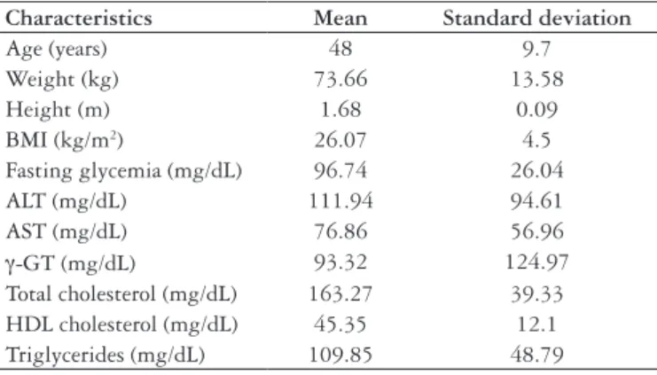
The majority of patients was male (92/160 - 57.7%) and mean age was 48 years, with a standard deviation of 9.7. The mean weight of the patients was 73.66 kg, with a standard deviation of 13.58 kg. Mean height was 1.68 m, with a standard deviation of 0.09 m. Mean body weight index was 26.07 kg/m², with a standard deviation of 4.5 kg/m². These data and those that refer to the laboratory exams can be appreciated in Table 1.
Of the 160 patients studied, genotype 3 was the most prevalent, being found in 91 patients (56.9%), while genotype 1 was found in 60 patients (37.5%), and genotype 2, in 9 patients (5.6%).
In the analysis of liver biopsy, grade 0 ibrosis was found in 3 patients (1.9%); grade 1 ibrosis, in 31 (19.4%); grade 2 ibrosis, in 68 patients (42.5%); grade 3 ibrosis, in 34 (21.3%); and grade 4 ibrosis, in 24 (15%). Most patients (102 – 63.8%) belonged to the absent ibrosis to moderate ibrosis group.
Analyzing the degree of steatosis found in liver biopsies, steatosis was observed to be present in 104/160 patients (65%). Among these, 76 (47.5%) presented steatosis in less than 33% of the hepatocytes, that is, grade 1 steatosis; 21 patients (13.1%) had between 33% and 66% of the hepatocytes with steatosis, that is, grade 2; and 7 patients (4.4%) presented more than 66% of their hepatocytes with steatosis, characterizing grade 3 steatosis. Among the patients with steatosis, in 98/104, it was macrovesicular, which represents 94.2%. Mixed steatosis was observed in 6/104, representing 5.8% of the sample.
The presence of NASH was only observed in eight patients studied (5%) and, for that reason, this data was not submitted to statistical analysis.
Among patients with genotype 3, steatosis was observed in 64/91 patients (61.5%). In the other genotypes, the presence of steatosis was observed in 40/69 (38.5%) patients.
DM was found in 19 patients, which represents 11.9% of the sample studied. Twenty-four patients (15%) were diagnosed with arterial hypertension.
When the antiviral treatment used by the patients was analyzed, only 48 (30%) patients had undergone treatment with pegylated interferon associated with ribavirin. Conventional interferon associated with ribavirin was the treatment of choice for 70% of the patients (112/160).
In analysis of the virological response, SVR was found in 58 patients, representing 36.3% of the sample, while non-responders represented 73 patients (45.6%), and recurrents, 29 patients (18.1%).
The results found, in accordance with group characteristics, taking into account the presence of steatosis, are shown in Table 2.
Characteristics Mean Standard deviation
Age (years) 48 9.7
Weight (kg) 73.66 13.58
Height (m) 1.68 0.09
BMI (kg/m2) 26.07 4.5
Fasting glycemia (mg/dL) 96.74 26.04
ALT (mg/dL) 111.94 94.61
AST (mg/dL) 76.86 56.96
γ-GT (mg/dL) 93.32 124.97
Total cholesterol (mg/dL) 163.27 39.33 HDL cholesterol (mg/dL) 45.35 12.1 Triglycerides (mg/dL) 109.85 48.79
TABLE 1. Demographic characteristics and laboratory data of evaluated
patients
BMI = body mass index
Characteristics Steatosis
n = 104 (65%)
Without steatosis
n = 56 (35%) P
Age >40 years 89 (85.6%) 41 (73.2%) 0.09 BMI >25 kg/m² 65 (62.5%) 18 (32.1%) <0.01
Genotype 0.08
1 37 (35.6%) 23 (41.1%)
2 3 (2.9%) 6 (10.7%)
3 64 (61.5%) 27 (48.2%)
Fibrosis 0.45
0.1 and 2 69 (66.3%) 33 (58.9%) 3 and 4 35 (33.7%) 23 (41.1%)
Diabetes 11 (10.6%) 8 (14.3%) 0.66
SAH 22 (21.2%) 2 (3.6%) 0.01
TABLE 2. Characteristics analyzed in relation to the presence of
steatosis
BMI = body mass index
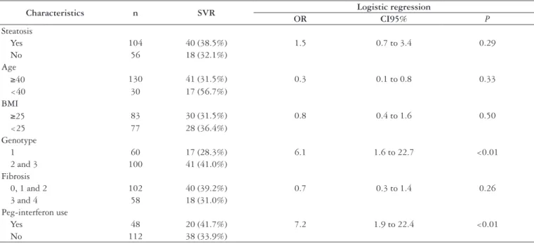
Analyzing liver steatosis and its relation to SVR, it was observed that from 104 patients who had liver steatosis in the biopsy, 40 (38.5%) had SVR, whereas in the group of patients without steatosis this number was 18/56 (32.1%), with no statistical difference. These data and those that refer to the relation between SVR and age, BMI, genotype, grade of ibrosis and type of interferon used can be analyzed in Table 3.
This study revealed that, in genotype 1, the presence of steatosis was associated with SVR in 14/37 (11.2%) versus
2/23 (2.98%) without steatosis (P = 0.06). In patients with
genotypes 2 and 3 who achieved SVR, steatosis was present in 33% (26/67) versus 34.2% (15/33) of those without steatosis
(P = 0.99).
DISCUSSION
There is a study that suggests that steatosis can induce a
mechanism of resistance to antiviral therapy(42). This mechanism
is still unknown, but it seems to be independent of other known response factors, such as genotype 1, high viral load and extensive ibrosis as well as metabolic factors. This mechanism also seems to be speciic of the so-called “metabolic steatosis”, since the “viral steatosis” observed in genotype 3 is not associated with a low rate of sustained virological response.
The negative effect of steatosis on the response to antiviral treatment may be caused by the reduction in bioavailability of antiviral drugs and/or host factors, such as more advanced
disease and/or coexistence of other associated diseases(55).
Another possible explanation for steatosis’ interference in the
response to antiviral treatment is associated with resistance to insulin, considering the “two stage” hypothesis suggested by
Day et al.(20), in 1998. It is possible that hyperinsulinemia blocks
inhibition of HCV mediated by interferon, by interacting with
interferon’s regulator factor, impeding its activation(31).
Liver steatosis is present in around 55% of patients infected
by HCV, varying between 40% and 86% of the cases(36, 43). In
the present casuistics, steatosis occurred in 65% of the patients,
which is in agreement with the data found in literature(42).
In a general sense, steatosis present in patients infected by
HCV is grade 1, affecting less than 33% of the hepatocytes(8, 12, 17,
45), which was also found in this study (47.5% of the biopsies).
This study did not show any signiicant association between age and the presence of steatosis, but did with BMI, which was
also shown by other authors(25, 26, 27, 29, 37, 42, 47, 52).
There is a tendency to associate the hepatitis C genotype
3 virus with the presence of steatosis(2, 8, 12, 28, 36, 37, 40, 42, 43, 45, 48, 57),
also found in this study, in which 61.5% of HCV genotype 3 patients had steatosis in the biopsy. The presence of steatosis was observed in fewer patients with genotypes other than 3. However, despite the higher prevalence of steatosis in genotype 3, there was no statistical difference among groups. Unfortunately, it was not possible to evaluate the existence of a relation between HCV genotype 3 viral load and steatosis, since Brazilian Public Health System does not provide viral load measurements for this genotype.
Some authors(1, 36, 43), when seeing an association between
liver steatosis and genotype 3, suggested that a speciic viral sequence of this genotype could be responsible for this “steatotic phenotype”. It must be pointed out that improvement in steatosis
Characteristics n SVR Logistic regression
OR CI95% P
Steatosis
Yes 104 40 (38.5%) 1.5 0.7 to 3.4 0.29
No 56 18 (32.1%)
Age
≥40 130 41 (31.5%) 0.3 0.1 to 0.8 0.33
<40 30 17 (56.7%)
BMI
≥25 83 30 (31.5%) 0.8 0.4 to 1.6 0.50
<25 77 28 (36.4%)
Genotype
1 60 17 (28.3%) 6.1 1.6 to 22.7 <0.01
2 and 3 100 41 (41.0%)
Fibrosis
0, 1 and 2 102 40 (39.2%) 0.7 0.3 to 1.4 0.26
3 and 4 58 18 (31.0%)
Peg-interferon use
Yes 48 20 (41.7%) 7.2 1.9 to 22.4 <0.01
No 112 38 (33.9%)
TABLE 3. Evaluated factors and their relation with sustained virological response
SVR = sustained virological response OR = odds ratio
is associated with the negativation of the HCV genome in the hepatocyte.
Rubbia-Brandt et al.(44) conirmed, in a multivariate analysis,
that the association between the presence and severity of liver steatosis in patients infected by genotype 3 is not fortuitous, since HCV genotype 3 patients had a tenfold greater chance of presenting liver steatosis in biopsy, regardless of other factors such as alcohol use, overweight/obesity and use of injectable drugs.
With regard to the relationship between steatosis and liver
ibrosis, several studies have demonstrated a positive association(19,
26, 29). Studies that evaluated sequential liver biopsies proved this
association(12, 22, 57). Thus steatosis has been considered a co-factor
for liver ibrosis. In the casuistics under study, and as referred by
other authors(8, 32), an association was not demonstrated between
the presence of steatosis and ibrosis.
The relationship between the metabolic syndrome and the presence of liver steatosis is well-known. Furthermore, common indings of this syndrome, such as obesity, diabetes, arterial hypertension and hypertriglyceridemia, are strongly associated
with the presence of NAFLD in CHC patients(47).
In our study, the presence of DM was only observed in 19 patients, without any relationship between this inding and the presence of steatosis. This fact is believed to result from the small number of cases with diabetes. However, it was possible to demonstrate that the presence of systemic arterial hypertension was related to the inding of liver steatosis.
In this study, 36.3% of the patients achieved SVR, which is in agreement with the values found in a previous study conducted
by our group(21).
In relation to SVR, of the diverse parameters evaluated, only genotype and pegylated interferon use in antiviral treatment achieved statistical signiicance in a multivariate analysis. BMI, age, ibrosis and steatosis did not correlate with SVR in this analysis.
The mechanisms through which obesity can affect response to antiviral treatment are not totally understood. BMI has been related to the degree of steatosis observed in chronic hepatitis C(10, 26). Bressler et al.(10) demonstrated that obesity, deined as
BMI>30 kg/m², is a risk factor for non-response to antiviral treatment, regardless of genotype and the presence of liver cirrhosis. In this study, BMI was not associated with SVR, as
in other studies(40, 42).
Most of the patients in this study were over 40 years of age, which could probably be one of the factors to explain the
population’s low SVR. In younger patients, SVR was found in 56.7% corroborating the fact that age is related to response to
treatment in patients infected by HCV(9, 24, 33, 34, 40, 61). However,
it must be underscored that no association was found between age and SVR in this study.
When analyzing liver ibrosis, no statistically signiicant correlation was found between the grade of ibrosis and SVR, although patients with lower grades of ibrosis obtained a
better SVR(38, 41).
In the casuistics in question, the presence of steatosis did not exert any inluence on SVR, different from what was found
in other studies(40, 42). It is possible that it could relate to a
type-II error, since our study has a smaller sample than others, but, recently, it has been published some data corroborating our
indings. Cross et al.(18), for instance, found that only HCV
genotype and pretreatment weight were associated to SVR in a multivariate analysis; neither statosis, nor steatohepatitis were considered independently associated with this outcome. It is possible, then, that pretreatment weight and not steatosis would affect the treatment results; nevertheless, we did not ind association between BMI and SVR either.
As in literature(24, 31, 34, 42, 53), in a multivariate analysis, this study
demonstrated that patients with HCV genotypes 2 and 3 had a better SVR rate when compared to those with genotype 1.
Westin et al.(58), in a European multicentric study, demonstrated
that 88% of the genotype 3 patients obtained SVR, regardless of the high prevalence of steatosis and pretreatment viral load found in this group. In genotypes other than 3, a 56% SVR rate was found, and, in patients with steatosis, this rate was of 46%, whereas, in those without steatosis this rate was of 65%, demonstrating the impact of steatosis on SVR in these patients. This group suggests that weight loss should be the objective of an intervention in the patients’ life habits with the intent of creating better conditions for antiviral therapy success.
The improvement in SVR rate has been demonstrated by several authors when the antiviral treatment of choice is pegylated
interferon associated with ribavirin(24, 34, 38, 61). This study did not
present any different results, despite the small number of patients treated with pegylated interferon. When a logistics regression analysis was made, a statistically signiicant association was found between type of interferon used and SVR.
Piccoli LZ, Mattos AA, Coral GP, Mattos AZ, Santos DE. Análise da resposta virológica sustentada em pacientes com hepatite crônica pelo vírus da hepatite C e esteatose hepática. Arq Gastroenterol. 2011;48(3):179-85.
RESUMO – Contexto - Tanto a hepatite crônica pelo vírus C quanto a doença hepática gordurosa não-alcoólica são reconhecidas como causas frequentes de doença hepática nos países ocidentais. É comum observar a concomitância das duas doenças e a inluência da esteatose na resposta virológica sustentada dos pacientes com hepatite crônica pelo vírus C. Objetivo - Avaliar a resposta virológica sustentada nos pacientes com hepatite crônica
pelo vírus C de acordo com a presença de esteatose. Métodos - Foram avaliados, retrospectivamente, 160 pacientes com hepatite crônica C. Dados demográicos, como sexo, idade, índice de massa corpórea, presença de diabetes mellitus e hipertensão arterial sistêmica, genótipo do vírus e uso de interferon peguilado foram analisados, bem como o estadiamento e a presença de esteatose, quando da histologia. Resultados - A maioria dos
pacientes era masculina (57,5%), com média de idade de 48 anos ± 9,7. O genótipo mais frequente foi o 3 (56,9%) e, na avaliação histológica, foi observada esteatose em 65% dos pacientes (104/160). A resposta virológica sustentada nos pacientes com esteatose foi de 38,5%, sendo de 32,1% nos pacientes sem esteatose (P = 0,54). Quando se analisaram possíveis fatores associados à presença de esteatose, somente índice de massa corpórea e hipertensão arterial sistêmica estiveram associados de forma signiicativa. Quando se avaliaram em regressão logística os fatores que inluenciaram a resposta virológica sustentada, genótipo e uso de interferon peguilado mostraram-se fatores independentes associados à resposta. Conclusão - A
presença de esteatose hepática não inluenciou a resposta virológica sustentada de pacientes com hepatite crônica pelo vírus C tratados com interferon e ribavirina.
DESCRITORES - Hepatite C crônica. Fígado gorduroso. Antivirais.
REFERENCES
1. Adinoli LE, Gambardella M, Andreana A, Tripodi MF, Utili R, Riggiero G. Steatosis accelerates the progression of liver damage of chronic hepatitis C patients and correlates with speciic HCV genotype and visceral obesity. Hepatology. 2001;33:1358-64.
2. Adinoli LE, Utili R, Andreana A, Tripodi MF, Utili R, Ruggiero G. Serum HCV RNA levels correlate with histological liver damage and concur with steatosis in progression of chronic hepatitis C. Dig Dis Sci. 2001;46:1677-83.
3. Adinoli LE, Durante-Mangoni E, Zampino R, Ruggiero G. Hepatitis C virus-associated steatosis – pathogenic mechanisms and clinical implications [review article]. Aliment Pharmacol Ther. 2005;22:52-5.
4. Almeida PRL, Mattos AA; Peixoto MF, Both CT. Estudo clínico, laboratorial e histológico em doadores de sangue anti-HCV positivos. GED Gastroenterol Endosc Dig. 1999;18:85-90.
5. American Diabetes Association. Clinical practice recommendations 2000. Diabetes Care. 2000;23(suppl 1):1-116.
6. Angulo P, Keach JC, Batts KP, Lindor KD. Independent predictors of liver ibrosis patients with nonalcoholic steatohepatitis. Hepatology. 1999;30:1356-62. 7. Angulo P. Nonalcoholic fatty liver disease. N Engl J Med. 2002;346:1221-31. 8. Asselah T, Boyer N, Guimont MC, Cazals-Hatem D, Tubach F, Nahon K, Daïkha
H. Vidaud D, Martinot M, Vidaud M, Degott C, Valla D, Marcellin P. Liver ibrosis is not associated with steatosis but with necroinlammation in French patients with chronic hepatitis C. Gut. 2003;52:1638-43.
9. Bach N, Thung SN, Schaffner F. The histological features of chronic hepatitis C and autoimmune chronic hepatitis a comparative analysis. Hepatology. 1992;15:572-7.
10. Bressler BL, Guindi M, Tomlinson G, Heathcote J. High body mass index is an independent risk factor for o response to antiviral treatment in chronic hepatitis C. Hepatology. 2003;38:639-44.
11. Brunt EM. Nonalcoholic steatohepatitis: deinition and pathology. Semin Liver Dis. 2001;21:3-16.
12. Castéra L, Hézode C, Roudot-Thoraval F, Bastie A, Zafrani ES, Pawlotsky JM, Dhumeaux D. Worsening of steatosis in an independent factor of ibrosis progression in untreated patients with chronic hepatitis C and paired liver biopsies. Gut. 2003;52:288-92.
13. Castéra L, Hézode C, Roudot-Thoraval F, Lonjon I, Zafrani ES, Pawlotsky JM, Dhumeaux D. Effect of antiviral treatment on evolution of liver steatosis in patients with chronic hepatitis C: indirect evidence of a role of hepatitis C virus genotype 3 in steatosis. Gut. 2004;53:420-4.
14. Castéra L, Chouteau P, Hezode C, Zafrani ES, Dhumeaux D, Pawlotsky JM. Hepatitis C virus-induced hepatocellular steatosis. Am J Gastroenterol. 2005;100:711-5.
15. Clark JM, Brancati FL, Diehl AM. Nonalcoholic fatty liver disease. Gastroenterology. 2002;122:1649-57.
16. Clinical guidelines on the identiication, evaluation, and treatment of overweight and obesity in adults: executive summary. Expert panel on the identiication, evaluation, and treatment of overweight in adults. Am J Clin Nutr. 1998;68:899-917. 17. Coral G, de Mattos AA, de Mattos AZ, dos Santos DE. [Steatosis and
non-alcoholic steatohepatitis in patients with chronic hepatitis due to hepatitis C virus infection]. Arq Gastroenterol. 2006;43:265-8.
18. Cross TJ, Quaglia A, Nolan J, Hughes S, Harrison PM. Do steatosis and steatohepatitis impact on sustained virological response (SVR) rates in patients receiving pegylated interferon and ribavirin for chronic hepatitis C infection? J Med Virol. 2010;82:958-64.
19. Czaja AJ, Carpenter HA, Santrach PJ, Moore SB. Host and disease speciic factors affecting steatosis in chronic hepatitis C. J Hepatol. 1998;29:198-206. 20. Day CP, James OF. Hepatitic steatosis: innocent bystander of guilty party?
Hepatology. 1998;27:1463-66.
21. de Almeida PR, de Mattos AA, Amaral KM, Feltrin AA, Zamin P, Tovo CV, Picon PD. Treatment of hepatitis C with peginterferon and ribavirin in a public health program. Hepatogastroenterology. 2009;56:223-6.
22. Fartoux L, Chazouillères O, Wendum D, Poupon R, Serfaty L. Impact of steatosis on progression of ibrosis in patients with mild hepatitis C. Hepatology. 2005;41:82-7.
23. Fartoux L, Poujol-Robert A, Guéchot J, Wendum D, Poupon R, Serfaty L. Insulin resistance is a cause of steatosis and ibrosis progression in chronic hepatitis C. Gut. 2005;54:1003-8.
24. Fried MW, Shiffman ML, Reddy KR, Smith C, Marinos G, Gonçalves FL Jr, Häussinger D, Diago M, Carosi G, Dhumeaux D, Craxi A, Lin A, Hoffman J, Yu J. Peginterferon alfa-2a plus ribavirina for chronic hepatitis C virus infection. N Engl J Med. 2002;347:975-82.
25. Friedenberg F, Pungpapong S, Zaeri N, Braitman L. The impact of diabetes and obesity on liver histology in patients with hepatitis C. Diabetes Obes Metab. 2003;5:150-5.
26. Hourigan LF, MacDonald GA, Purdie D, Whitehall VH, Shorthouse C, Clouston A, Powell EE. Fibrosis in chronic hepatitis C correlates signiicantly with body mass index and steatosis. Hepatology. 1999;29:1215-9.
27. Hu KQ, Currie SL, Shen H, Cheung RC, Ho SB, Bini EJ, McCracken JD, Morgan T, Bräu N, Schmidt WN, Jeffers L, Wright T. Clinic implications of hepatic steatosis in patients with chronic hepatitis C: a multicenter study of U.S. Veterans. Dig Dis Sci. 2007;52:570-8.
28. Hui JM, Kench J, Farrell GC, Lin R, Samarasinghe D, Liddle C, Byth K, George J. Genotype-speciic mechanisms for hepatic steatosis in chronic hepatitis C infection. J Gastroenterol Hepatol. 2002;17:873-81.
29. Hwang SJ, Luo JC, Chu CW, Lai CR, Lu CL, Tsay SH, Wu JC, Chang FY, Lee SD. Hepatic steatosis in chronic hepatitis C virus infection: prevalence and clinical correlation. J Gastroenterol Hepatol. 2001;16:190-5.
31. Jian Wu Y, Shu Chen L, Gui-Qiang W. Effects of fatty liver and related factors on eficacy of combination antiviral therapy in patients with chronic hepatitis C. Liver Int. 2006;26:166-72.
32. Leandro G, Mangia A, Hui J, Fabris P, Rubbia-Brandt L, Colloredo G, Adinoli LE, Asselah T, Jonsson JR, Smedile A, Terrault N, Pazienza V, Giordani MT, Giostra E, Sonzogni A, Ruggiero G, Marcellin P, Powell EE, George J, Negro F. Relationship between steatosis, inlammation, and ibrosis in chronic hepatitis C: a meta-analysis of individual patients data. Gastroenterology. 2006;130:1636-42. 33. Lonardo A, Loria P, Adinoli LE, Carulli N, Ruggiero G. Hepatitis C and steatosis:
a reappraisal. J Viral Hepat. 2006;13:73-80.
34. Manns MP, Mchutchison JG, Gordon SC, Rustgi VK, Schiffman M, Reindollar R, Goodman ZD, Koury K, Ling M, Albrecht JK. Peginterferon alfa-2b plus ribavirin compared with interferon alfa-2b plus ribavirin for initial treatment of chronic hepatitis C: a randomized trial. Lancet. 2001;358:958-65.
35. Mello V, Cruz T, Nuñez G, Simões MT, Ney-Oliveira F, Braga H, Araújo C, Cunha S, Schinoni MI, Cruz M, Parana R. Peripheral insulin resistance during treatment of chronic hepatitis C with peguilated interferon plus ribavirin. J Med Virol. 2006;78:1406-10.
36. Mihm S, Fayyazi A, Hartmann H, Ramadori G. Analysis of histopathological manifestations of chronic hepatitis C virus infection with respect to virus genotype. Hepatology. 1997;25:735-9.
37. Monto A, Alonzo J, Watson JJ, Grunfeld C, Wright TL. Steatosis in chronic hepatitis C: relative contributions of obesity, diabetes mellitus and alcohol. Hepatology. 2002;36:729-36.
38. National Institute of Health Consensus Development Conference Statement. Management of hepatitis C: 2002 – June 10-12, 2002. Hepatology. 2003;36: s3-20.
39. Parise ER, Oliveira AC. [Insulin resistance in chronic hepatitis C]. Arq Gastroenterol. 2007;44:178-84.
40. Patton HM, Patel K, Behling C, Bylund D, Blatt LM, Vallée M, Heaton S, Conrad A, Pockros PJ, McHutchison JG. The impact of steatosis on disease progression and early sustained treatment response in chronic hepatitis C patients. J Hepatol. 2004;40:484-90.
41. Poynard T, Bedossa P, Opolon P. Natural history of liver ibrosis progression in patients with chronic hepatitis C. The OBSVIRC, METAVIR, CLINIVIR, and DOSVIRC group. Lancet. 1997;349:825-32.
42. Poynard T, Ratziu V, McHutchison J, Manns M, Goodman Z, Zeuzem S, Younossi Z, Albrecht J. Effect of treatment with peginterferon or interferon alfa-2b and ribavirina on steatosis in patients infected with hepatitis C. Hepatology. 2003;38:75-85.
43. Rubbia-Brandt L, Quadri R, Abid K, Giostra E, Malé PJ, Mentha G, Spahr L, Zarski JP, Borisch B, Hadengue A, Negro F. Hepatocyte steatosis is a cytopathic effect of hepatitis C virus genotype 3. J Hepatol. 2000;33:106-15.
44. Rubbia-Brandt L, Leandro G, Spahr L, Giostra E, Quadri R, Male PJ, Negro F. Liver steatosis in chronic hepatitis C: a morphological sign suggesting infection with HCV genotype 3. Histopathology. 2001;39:119-24.
45. Rubbia-Brandt L, Fabris P, Pagnanin S, Leandro G, Male PJ, Giostra E, Carlotto A, Bozzola L, Smedile A, Negro F. Steatosis affects chronic hepatitis C progression in a genotype speciic way. Gut. 2004;53:406-12.
46. Sanyal AJ, Contos MJ, Sterling RK, Luketic VA, Shiffman ML, Stravitz RT, Mills AS. Nonalcoholic fatty liver disease in patients with hepatitis C is associated with features of the metabolic syndrome. Am J Gastroenterol. 2003;98:2064-71.
47. Sanyal AJ. Nonalcoholic fatty liver disease and hepatitis C – risk factors and clinical implications [review article]. Aliment Pharmacol Ther. 2005;22: 48-51.
48. Serfaty L, Andreani T, Giral P, Carbonell N, Chazouillères O, Poupon R. Hepatitis C virus induced hypobetalipoproteinemia: a possible mechanism for steatosis in chronic hepatitis C. J Hepatol. 2001;34:428-34.
49. Serfaty L, Poujol-Robert A, Carbonell N, Chazouillères O, Poupon RE, Poupon R. Effect of the interaction between steatosis and alcohol intake on liver ibrosis progression in chronic hepatitis C. Am J Gastroenterol. 2002;97:1807-12. 50. Simmonds P. Variability of hepatitis C virus. Hepatology. 1995;21:570-83. 51. The sixth report of the Joint National Committee on prevention, detection,
evaluation, and treatment of high blood pressure. Arch Intern Med. 1997;157:2413-46.
52. Solis-Herruzo JA, Pérez-Carreras M, Rivas E, Fernández-Vásquez I, Garia C, Bernardos E, Castellano G, Colina F. Factors associated with the presence of nonalcoholic steatohepatitis in patients with chronic hepatitis C. Am J Gastroenterol. 2005;100:1091-8.
53. Soresi M, Tripi S, Franco V, Giannitrapani L, Alessandri A, Rappa F, Vuturo O, Montalto G. Impact of liver steatosis on the antiviral response in the hepatitis C virus-associated chronic hepatitis. Liver Int. 2006;26:1119-25.
54. Stuyver L, Rossau R, Wyseur A, Duhamel IM, Vanderborgth B, van Heuverswyn H, Maertens G. Typing of hepatitis C virus isolates and characterisation of new subtypes using a line probe assay. J Gen Virol.1993;74:1093-102.
55. Thomopoulos KC, Theocharis GJ, Tsamantas AC, Siagris D, Dimitropoulou D, Gogos C, Labropoulou-Karatza C. Liver steatosis is an independent risk factor for treatment failure in patients with chronic hepatitis C. Eur J Gastroenterol Hepatol. 2005;17:149-53.
56. Tong MJ, Hwang SJ, Lefkowitz M, Lee SD, Co RL, Conrad A, Schmid P, Lo KJ. Correlation of serum HCV-RNA and alanine aminotransferases levels in chronic hepatitis C patients during treatment with ribavirin. J Gastroenterol Hepatol. 1994;9:587-91.
57. Westin J, Nordlinder H, Lagging M, Norkrans G, Wejstal R. Steatosis accelerates ibrosis development over time in hepatitis C virus genotype 3 infected patients. J Hepatol. 2002;37:837-42.
58. Westin J, Lagging M, Dhillon AP, Norkrans G, Romero AI, Pawlotsky JM, Zeuzem S, Schalm SW, Verheij-Hart E, Negro F, Missale G, Neumann AU, Hellstrand K for the DITTO-HCV Group. Impact of hepatic steatosis on viral kinetics and treatment outcome during antiviral treatment of chronic HCV infection. J Viral Hepat. 2007;14:29-35.
59. Younossi ZM, Mccullough A, Ong PJ, Barnes DS, Post A, Tavill A, Bringman D, Martin LM, Assmann J, Gramlich T, Mullen KD, O’Shea R, Carey WD, Ferguson R. Obesity and non-alcoholic fatty liver disease in chronic hepatitis C. J Clin Gastroenterol. 2004;38:705-9.
60. Zamin I Jr, de Mattos AA, Zettler CG. Nonalcoholic steatohepatitis in nondiabetic obese patients. Can J Gastroenterol. 2002;16:303-7.
61. Zeuzem S, Hultcrantz R, Bourliere M, Goeser T, Marcellin P, Sanchez-Tapias J, Sarrazin C, Harvey J, Brass C, Albrecht J. Peginterferon alfa-2b plus ribavirina for treatment of chronic hepatitis C in previously untreated patients infected with HCV genotype 2 or 3. J Hepatol. 2004;40:993-9.