Motility to Alter Host Colonization
Hiroshi Asakura1*, Noritaka Hashii2, Masashi Uema1, Nana Kawasaki2, Yoshiko Sugita-Konishi3, Shizunobu Igimi1, Shigeki Yamamoto1¤
1Division of Biomedical Food Research, National Institute of Health Sciences, Setagaya-ku, Tokyo, Japan,2Division of Biological Chemistry and Biologicals, National Institute of Health Sciences, Setagaya-ku, Tokyo, Japan,3Department of Food and Life Sciences, Azabu University, Fuchinobe Chuo-ku, Sagamihara, Kanagawa, Japan
Abstract
Vitamin B6 (pyridoxal-5’-phosphate, PLP) is linked to a variety of biological functions in prokaryotes. Here, we report that the pdxA(putative 4-hydroxy-L-threonine phosphate dehydrogenase) gene plays a pivotal role in the PLP-dependent regulation of flagellar motility, thereby altering host colonization in a leading foodborne pathogen,Campylobacter jejuni. AC. jejuni pdxAmutant failed to produce PLP and exhibited a coincident loss of flagellar motility. Mass spectrometric analyses showed a 3-fold reduction in the main flagellar glycan pseudaminic acid (Pse) associated with the disruption ofpdxA. ThepdxA mutant also exhibited reduced growth rates compared with the WT strain. Comparative metabolomic analyses revealed differences in respiratory/energy metabolism between WTC. jejuniand thepdxAmutant, providing a possible explanation for the differential growth fitness between the two strains. Consistent with the lack of flagellar motility, thepdxAmutant showed impaired motility-mediated responses (bacterial adhesion, ERK1/2 activation, and IL-8 production) in INT407 cells and reduced colonization of chickens compared with the WT strain. Overall, this study demonstrated that thepdxAgene affects the PLP-mediated flagellar motility function, mainly through alteration of Pse modification, and the disruption of this gene also alters the respiratory/energy metabolisms to potentially affect host colonization. Our data therefore present novel implications regarding the utility of PLP and its dependent enzymes as potent target(s) for the control of this pathogen in the poultry host.
Citation:Asakura H, Hashii N, Uema M, Kawasaki N, Sugita-Konishi Y, et al. (2013)Campylobacter jejuni pdxAAffects Flagellum-Mediated Motility to Alter Host Colonization. PLoS ONE 8(8): e70418. doi:10.1371/journal.pone.0070418
Editor:Stefan Bereswill, Charite´-University Medicine Berlin, Germany
ReceivedMarch 30, 2013;AcceptedJune 24, 2013;PublishedAugust 6, 2013
Copyright:ß2013 Asakura et al. This is an open-access article distributed under the terms of the Creative Commons Attribution License, which permits unrestricted use, distribution, and reproduction in any medium, provided the original author and source are credited.
Funding:This work was supported in part through funding from a Grant-in-Aid for Scientific Research from the Japan Society for the Promotion of Science (JSPS) (22780275), and a grant from Ministry of Health, Labour, and Welfare, Japan (H24-shokuhin-ippan-009). The funders had no role in study design, data collection and analysis, decision to publish, or preparation of the manuscript.
Competing Interests:The authors have declared that no competing interests exist.
* E-mail: [email protected]
¤ Current address: Department of Fisheries, Tokai University, Orido, Shimizu-shi, Shizuoka, Japan
Introduction
Campylobacter jejuni is a Gram-negative, spiral-shaped, micro-aerophilic bacterium that causes foodborne diarrheal illness worldwide [1,2]. Recent epidemiological and biochemical studies have shown that Campylobacter infection is also implicated in neuropathies, including Guillain-Barre´ syndrome (GBS), through the production of autoantibodies induced by bacterial lipooligo-saccharides [3,4]. The chicken is the predominant natural host for this pathogen, through whichCampylobactercan be transmitted to humans [5,6,7]. Thus, control of this pathogen in poultry habitats is associated with the global public health benefit of preventing human campylobacteriosis. However, how to controlCampylobacter remains unresolved, mainly due to our lack of understanding regarding how this pathogen colonizes chickens and establishes persistent infections and how it is involved in human virulence [7]. An increased understanding of the molecular biology of Campylo-bacter would therefore provide valuable information for the development of therapeutic strategies and vaccines targeting this pathogen.
The differential expression of metabolic gene products in relation to pathogenesis has largely been left unexplored.
fromC. jejunigenerates UDP-4-amino-4,6-dideoxy-alNAc with the catalytic support of PLP as a co-factor under in vitroconditions [17], which led us to the assumption that PLP biosynthesis also affects flagellation and certain types of metabolism in this pathogen, thereby altering bacterial fitness andin vivocolonization, for which flagellar motility is a prerequisite [18].
Given this background, we studied the PLP biosynthesis pathway in C. jejuni throughin silico prediction and mutagenesis analyses. Biochemical and phenotypic analyses showed that the lack of thepdxAgene abolished PLP production and impaired the ability ofC. jejunito form flagella. We then focused on this mutant to characterize its biological effect(s) on host colonization through biochemical, metabolomic, and host infection approaches.
Materials and Methods
Bacterial strains and media
The bacterial strains and plasmids used in this study are listed in Table 1. C. jejuni strain 81–176 [19] was grown using routine methods in Mueller-Hinton (MH) broth or on MH agar (Becton-Dickinson, Franklin Lakes, NJ, USA) at 37uC for 24 h in a humidified CO2 AnaeroPack-Microaero gas system (Mitsubishi Gas Chemicals, Tokyo, Japan). The media were supplemented with chloramphenicol (Cm) (20mg ml21) or kanamycin (Km) (30mg ml21), as appropriate. TheE. coliDH5astrain, which was used as the host for subcloning and routine DNA manipulation, was grown in LB agar or LB broth unless otherwise indicated.
Construction ofC. jejunimutants and complementation of thepdxAmutant
The 81–176 mutant, in which most of thepdxA orflaAgenes were replaced with a cat cassette (encoding a Cm-resistance protein), was constructed as described previously [20]. To construct a pdxAmutant, a 500-bp fragment upstream of the 59 end and a 500-bp fragment downstream of the 39end of thepdxA locus were amplified from the wild-type (WT) strainviaPCR using either pdxA-s and pdxA-as-BI or pdxA-s-BI and pdxA-as primers (Table S1). AfterBamHI digestion, the two fragments were ligated and cloned into pGEM-T vector (Promega, Madison, WI, USA). Acatgene from the plasmid pRY109 [21] was then inserted into theBamHI site in the pGEM-T plasmid, and this allelic exchange plasmid (pGEM-pdxA-Cm, Table 1) was introduced into the genome of strain 81–176 through natural transformation [22].
Successful transformants were selected on MH agar supplemented with 5% horse blood and Cm (20mg ml21). Allelic replacement was confirmedvianucleotide sequencing. Disruption of the flaA gene was performed in the same manner (the oligonucleotide primers used in these procedures are listed in Table S1). The pdxAJ locus and the upstream region predicted by the Neural Network Promoter Prediction program (http://www.fruitfly.org/ seq_tools/promoter.html) to contain 235 and 210 promoter binding sites were amplified via PCR using the pdxA-CF and pdxA-CR primers (Table S1). The resultant PCR fragments were cloned into the XbaI/EcoRI sites of the pRY108 plasmid [21], yielding pRY-pdxA-Km (Table 1). This plasmid was introduced into thepdxAmutant strain through natural transformation, and the transformants were recovered on MH agar containing Km (10mg ml21) and Cm (20mg ml21). The construction of this pdxA2/+mutant strain was confirmedviaPCR using the pdxA-conF and pdxA-conR primers (Table S1).
Quantification of PLP
Bacteria were grown microaerobically in 10 ml of MH broth to mid-logarithmic phase (an OD600of 0.6), and crude homogenates were prepared in 20 mM Tris-HCl (pH 7.4)via bead crushing. After centrifugation for 10 min at 7,000 rpm at 4uC, the PLP contents in 50mg of protein of the lysate and serial dilutions of the lysate were measured using a vitamin B6 ELISA kit (Uscn Life Science, Houston, TX, USA) according to the manufacturer’s instructions. Fresh MH broth was also tested for the measurement of PLP.
Protein fractionation, SDS-PAGE, and immunoblotting Membrane and cytoplasmic proteins fromC. jejuniwere isolated as described previously [23]. These protein samples were then separated on a 7.5% sodium dodecyl sulfate-polyacrylamide gel (SDS-PAGE) and stained with CBB (Coomassie Brilliant Blue) to visualize the protein profiles. The proteins on the gel were simultaneously transferred to a PVDF membrane (Merck-Milli-pore, Billerica, MA, USA), and the FlaA protein was detected using a rabbit polyclonal antibody generated against C. jejuni flagellin [24] and an HRP-conjugated goat anti-rabbit secondary antibody (GE Healthcare, Little Chalfont, UK). The blots were developed using the ECL detection system (GE Healthcare).
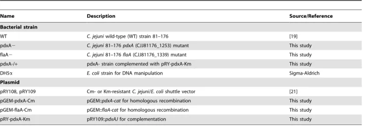
Table 1.Bacterial strains and plasmids used in this study.
Name Description Source/Reference
Bacterial strain
WT C. jejuniwild-type (WT) strain 81–176 [19]
pdxA2 C. jejuni81–176pdxA(CJJ81176_1253) mutant This study
flaA2 C. jejuni81–176flaA(CJJ81176_1339) mutant This study
pdxA-/+ pdxA- strain complemented with pRY-pdxA-Km This study
DH5a E. colistrain for DNA manipulation Sigma-Aldrich
Plasmid
pRY108, pRY109 Cm- or Km-resistantC. jejuni/E. colishuttle vector [21]
pGEM-pdxA-Cm pGEM::pdxA-catfor homologous recombination This study
pGEM-flaA-Cm pGEM::flaA-catfor homologous recombination This study
pRY-pdxA-Km pRY109::pdxAJfor complementation This study
Motility and growth assays
The motility of the WT,pdxA mutant, and complementedC. jejuni strains was assayed on 0.4% soft MH agar plates as previously described [25]. To measure bacterial growth, 1.4– 1.96106cells ofC. jejunithat were microaerobically grown in MH broth to an OD600of 1.2–1.3 at 37uC, were incubated in 10 ml of fresh MH broth supplemented with or without PLP (10 mg l21) with agitation (120 rpm) for 0, 12, 24, 36, 48, 72 h. At each time points, turbidity of the medium was measured at 600 nm.
Detection of pseudaminic acid (Pse)
(i) Derivatization of Pse. Pse was released from the 30mg of cytosolic protein fractions from the WT andpdxAmutant strains grown in MH broth to OD600 of 0.55–0.60, using the GlycoProfileTM b-elimination kit (Sigma-Aldrich) according to the manufacturer’s instructions. The released Pse was labeled with 1,2-diamino-4,5- methylenedioxybenzene (DMB) using a sialic acid fluorescence-labeling kit (TaKaRa, Shiga, Japan), and the reaction mixture was applied to a solid-phase extraction cartridge (Envi-Carb C, Supelco, Bellefonte, PA, USA). After washing with 2.5 ml of 5 mM ammonium acetate (pH 9.6), the labeled Pse was eluted using 3 ml of 45% acetonitrile/5 mM ammonium acetate (pH 9.6) and freeze-dried. Fresh MH broth was also subjected to the above sample preparation to observe the effect of background growth medium.
(ii) Liquid chromatography/mass spectrometry (LC/ MS). Chromatographic separation of DMB-labeled Pse was performed using the Paradigm MS4 HPLC system (Michrom BioResource, Auburn, CA, USA). The separated DMB-labeled Pse was applied to a C18 trap column (L-column Micro, CERI) and eluted using 0.1% formic acid/2% acetonitrile (buffer A) and 0.1% formic acid/90% acetonitrile (buffer B) with a linear gradient of 10–90% buffer B over 30 min at a flow rate of 300ml min21. Mass spectrometric analysis of DMB-labeled Pse was performed using a Fourier transformation ion cyclotron resonance (FT-ICR)/ion trap (IT)-type mass spectrometer (LTQ-FT) (Thermo Electron, San Jose, CA, USA) equipped with a nanoelectrospray ion source (AMR, Tokyo, Japan). The presence of DMB-Pse was determined via sequential scans consisting of selected ion monitoring (SIM, m/z441–461) using FT-ICR-MS and data-dependent MS/MS-MS/MS/MS/MS (MSn) with IT-MS.
Detection of metabolic compounds
(i) Sample preparation. A total of 3.2–3.46108C. jejuniWT orpdxAmutant cells grown in MH broth to an OD600of 0.6 were trapped on a 0.4-mm filter membrane (Merck-Millipore), washed twice with 10 ml of water, and immersed in 2 ml of methanol containing 10mM internal standard solution 1 (Human Metabo-lome Technologies (HMT), Yamagata, Japan). After sonication for 30 s, 1.6 ml of each suspension was mixed with 640mL of water and 1.6 ml of chloroform, followed by centrifugation for 5 min at 2,3006g. The 750ml of upper aqueous layer was filtered through a 5 KDa-cutoff filter (Millipore), lyophilized, and resuspended in 25ml of water.
(ii) Capillary Electrophoresis-Time of Flight/mass spectrometry (CE-TOF/MS). Cationic metabolites were ana-lyzed using a fused silica capillary tube (50mm680 cm) and Cation Buffer Solution (HMT) in a capillary electrophoresis system equipped with a Time-of-Flight mass spectrometer (CE-TOF/MS) and a CE-ESI-MS sprayer (Agilent Technologies, Santa Clara, CA, USA). Electrospray ionization-mass spectrom-etry (ESI-MS) was conducted in positive ion mode at 4,000 V. Anionic metabolites were analyzed using a fused silica capillary
and Anion Buffer Solution (HMT). ESI-MS was conducted in negative ion mode at 3,500 V. In both modes, the spectrometer was scanned from m/z 50 to 1,000. The other conditions were followed the cation analysis methodology of Soga and Heiger [26]. (iii) Data analysis. Raw data were processed using the MasterHands program [27]. Signal peaks corresponding to the isotopomers of 108 compounds (including the intermediates of the glycolytic system, the intermediates of the TCA cycle, and amino acids; see Table S2 for more details) were extracted. Each obtained migration time (MT) was normalized using the values of the internal standards. The resultant relative area values were further normalized based on the sample amounts. We used duplicate sets of samples from two independent experiments.
ATP assay
To determine the intracellular ATP concentration of bacterial samples, the BacTiter-Glo Microbial Cell Viability assay kit (Promega) was used. After growing the bacteria in MH broth at 37uC under microaerobic conditions to an OD595of 0.55–0.60, serial dilutions of all samples were prepared according to the manufacturer’s instructions. Following incubation of the samples at room temperature in a 96-well plate, luminescence was measured together with an ATP standard using GloMax Multi system (Promega), according to the manufacturer’s instructions. Simultaneously, we measured the bacterial numbers in the originally cultured MH broth by plate count.
Cell adhesion assay, IL-8 measurements, and immunoblotting
INT407 cells were seeded into 24-well culture plates (TPP) (3.06105cells well21) and incubated in RPMI1640 medium (Life Technologies, Carlsbad, CA, USA) for 20 h at 37uC in a humidified CO2 incubator. The cells were then rinsed and inoculated withC. jejuniat a multiplicity of infection (m.o.i.) of 100. At 60 min post-infection, the cells were washed three times with PBS to remove non-adherent bacteria, followed by cell detach-ment using 0.1% saponin in PBS. Serial dilutions of the suspensions were plated onto MH agar to determine the numbers of viable, cell-associated bacteria. To measure IL-8 secretion from the INT407 cells after infection, INT407 cells were infected with C. jejuni at an m.o.i. of 100 for 0, 4, and 16 h, and the culture supernatants were used to measure IL-8 levels with a human IL-8 ELISA kit (Becton-Dickinson), according to the manufacturer’s instructions. ERK activation was examined viawestern blotting using tyrosine-phosphorylated and total ERK1/2 monoclonal antibodies (Cell Signaling Technology, Danvers, MA, USA).
Chicken colonization assay
Specific pathogen-free, 14-day-old white leghorn chickens (obtained from Nisseiken Co., Ltd., Japan) were orally challenged with 500ml of MH broth containing approximately 3.06107WT or pdxA C. jejuni cells. The animals were euthanized at 7 and 28 days post-infection, and post-mortem cecal samples were collected after aseptic removal of the ceca.C. jejuni colonization of the cecum was examined by counting viable cells on mCCDA agar plates (Oxoid, Hampshire, UK). A control group was confirmed to be negative for Campylobacter. The above animal experiments were approved by the Committee for Animal Care and Use of the National Institute of Health Sciences, Japan.
Statistics and web tool
in various prokaryotes based on their genomic sequences, was used to illustrate the putative PLP and Pse biosynthesis pathways in the C. jejuni81–176 strain. The results from the motility, growth, ATP activity, cell adhesion, IL-8 production, and chicken colonization assays were expressed as the mean6 standard deviations of at least three independent observations. The significant differences between the measurements obtained from the WT and mutant strains were determined using Student’st-test.Pvalues,0.05 were considered statistically significant.
Results
Disruption of thepdxAgene abolishes PLP production in
C. jejuni
To predict the PLP biosynthesis pathway inC. jejuni81–176, we used the in silico pathway tool PATRIC (http://www.patricbrc. org/portal/portal/patric/Home). The result of this prediction illustrated that at least five genes might constitute two independent pathways for PLP biosynthesis in this pathogen (right box, Fig. 1A). Among these genes, pdxA (CJJ81176_1253) and pdxJ (CJJ81176_1252) are known to be involved in thede novosynthesis of PLP in E. coli[12] and are, indeed, conserved in the C. jejuni genome [28]. Recently, Stahl and Stintzi [29] reported that the pdxAgene (Cj1239 in the NCTC11168 strain) may be essential for microbial growth. We therefore decided to use thepdxA gene to study the role of PLP biosynthesis in the biology of this pathogen and constructed an insertionalpdxAmutant inC. jejunistrain 81– 176. Biochemical assays collectively detected very less amounts of PLP (0.1560.10mg 10 ml21) in the pdxA mutant than the WT strain (34.5567.61mg 10 ml21), and complementation of thepdxA gene in thepdxAmutant restored PLP production (33.8567.45mg 10 ml21) (Fig. 1B). Thus, we could demonstrate that thepdxAgene is truly a prerequisite for the PLP biosynthetic metabolism of this pathogen.
ThepdxAmutant impairs Pse production, flagellin glycosylation, and flagellation
Campylobacter flagellins are decorated with O-linked glycans, which are derivatives of Pse synthesized through sequential enzymatic reactions (i.e., transamination, decarboxylation, and racemization) [30], and this type of glycomodification is a prerequisite for the biogenesis, transport, and assembly of functional flagellar filaments [31,32]. Among components of the Pse biosynthesis process, thepseC(Cj1294/CJJ81176_1311) gene product, UDP-4-keto-deoxy-GlcNAc transaminase, is reported to require PLP to generate UDP-4-amino-4,6-dideoxy-GalNAc, a spectrometric analintermediate in the synthesis of Pse (left box, Fig. 1A) [17]. Immunoblot analyses showed the less glycosylation but expression of the flagellin A (FlaA) in cytoplasmic fraction of thepdxAmutant compared with that of WT strain (Fig. 2A), and the complementation of thepdxAgene restored the glycosylation of FlaA in thepdxAmutant (Fig. 2A). Having less detection of FlaA in the membrane fraction of thepdxAmutant than that of the WT strain (Fig. 2A), it could be considered that the less glycosylated FlaA was not transported to the surface of the pdxA mutant. In agreement, mass spectrometric analyses clearly showed that the pdxAmutant produced approximately 3-fold less Pse than the WT strain (Fig. 2B, Fig. S1, S2, S3), providing a link between pdxA, PLP, and Pse biosynthesis in this pathogen. In consistent with the fact that the glycosylation and surface expression of flagellar filaments are prerequisite for bacterial motility [31,32], phenotypic assays clearly demonstrated that thepdxAmutant was not motile, and the complementation of the pdxA gene restored motility (Fig. 2C). Furthermore, microscopic analyses consistently showed
that thepdxAmutant did not generate any flagellar filaments and that complementation of thepdxAgene restored flagellation, likely to the same level as in the isogenic WT strain (Fig. 2D). Addition of PLP did not restore the flagellar production of thepdxAmutant (Fig. 2D). Together, we were able to demonstrate that disruption of thepdxA gene impaired the glycosylation of flagellin, thereby reducing bacterial motility.
ThepdxAmutant exhibits altered Pse biosynthetic metabolism
Considering that PLP mediates a variety of enzymatic processes [33], a comparative metabolomic analysis was performed to characterize/confirm the types of metabolism that might be related to PLP activity. CE-TOF/MS (capillary electrophoresis time-of-flight/mass spectrometry) analysis detected 99 metabolic compounds extracted from the WT and pdxA mutant strains, among which the levels of 10 and 6 compounds were either increased or reduced .2-fold, respectively, in the pdxA mutant compared with the WT strain (Table 2 and more detailed information in Table S2). In support of the link between the presence of thepdxA gene and Pse production, thepdxAmutant exhibited greater amounts of UDP-glucose (a Pse precursor, Fig. 1A left panel) and a PLP precursor, pyridoxamine-5’-phosphate (PNP, Fig. 1A right panel), which showed concentra-tions that were at 3.6-fold and 2.5-fold higher than were exhibited by the WT strain, respectively (Table 2). Thus, these data clearly indicated an essential role of thepdxAgene in Pse biosynthesis inC. jejuni.
TheC. jejuni pdxAmutant exhibits altered respiratory/ energy metabolism
As an additional characteristic, we notified that theC. jejuni pdxA mutant exhibited growth defect compared with the WT strain, but this mutant showed successive growth in the absence of PLP, indicating that thepdxAgene was not essential for the growth of this pathogen (Fig. 3A, left panel). Different fromH. pylori pdxAmutant [16], the addition of PLP did not restore growth of theC. jejuni pdxA mutant (Fig. 3A). To investigate the metabolisms associated with the altered growth kinetics of theC. jejuni pdxAmutant, we thus focused indicators of significant alterations in energy/respiratory metabo-lisms because of the pivotal role of these metabometabo-lisms in the growth in this pathogen [34]. The metabolomic data showed that thepdxA mutant indeed produced greater amounts of ATP and GDP (by 2.7-fold each) and, hence, decreased amounts of NADP+ (0.5-fold) compared with the levels in the WT strain (Table 2). In agreement with these findings, thepdxA mutant exhibited an approximately 2.14-fold greater amount of ATP compared with the WT strain (when 1.66108cells were assayed; Fig. 3B). Energy metabolism is well known to depend on the respiratory cycle. Corroborating this fact, thepdxAmutant showed alterations in the concentrations of TCA cycle intermediates including cis-aconitic acid (2.7-fold), isocitrate (2.1-fold), succinate (2.1-fold), malate (1.5-fold), citrate (0.6-fold), and serine (0.5-fold, a major carbon source for the respiratory cycle in this pathogen [35]) compared with the WT strain (Fig. 3C, Table 2, Table S2). Thus, we were able to show that theC. jejuni pdxAmutant exhibited altered growth and respiratory/energy metabolism.
ThepdxAmutant exhibits impairedin vitro cell adhesion and chicken colonization
mutant, WT, andflaAmutant strains, the cell adhesion score of the pdxAmutant was found to be almost identical to that of the flaA mutant, exhibiting a 3-fold reduction compared with the WT strain at 1 h post infection (p.i.) (Fig. 4A). Following adherence,C. jejuni activates ERK1/2 MAPK signaling, which stimulates the
production of interleukin (IL)-8 in INT407 cells [39]. In agreement with the above cell adhesion scores, thepdxAmutant caused delayed phosphorylation of ERK1/2 MAPK compared with the WT strain (Fig. 4B), which was similar to theflaAmutant [39]. Additionally, similar to theflaAmutant [39], thepdxAmutant
Figure 1. Inactivation of thepdxAgene impairs the biosynthesis of vitamin B6 (PLP) inC. jejuni.(A) A scheme for the PLP production pathway (right box) inC. jejuniin relation to Pse biosynthesis (left box) is illustrated based onin silicopathway analysis performed using PATRIC (http://patricbrc.vbi.vt.edu/portal/portal/patric/Home). (B) ThepdxAmutant produced no PLP. The C. jejuni81–176 WT, pdxAmutant, and the complemented strains were grown in 10ml of MH broth to an OD600of 0.60. The suspensions were then homogenized, serially diluted, and subjected to ELISA to quantify the amounts of PLP (mg 10 ml21
induced significantly less IL-8 production in the INT407 cells compared with the WT strain at 4 h p.i. (p,0.05, Fig. 4C). A prolonged period after infection (18 hp.i.) resulted in
normaliza-tion of the IL-8 secrenormaliza-tion observed in response to all of the bacterial strains tested (Fig. 4C). These data indicated that the pdxA gene is a prerequisite for cell adhesion, with the mutant
Figure 2. ThepdxAmutant shows impaired pseudaminic acid (Pse) production and flagellar motility.(A) ThepdxAmutant shows less glycosylation of FlaA. SDS-PAGE and western blotting were conducted to detect theC. jejuniFlaA protein. Crude extracts and subcellular (cytoplasmic and membrane) fractions were extracted fromC. jejuniand visualized using CBB staining in an SDS-polyacrylamide gel (left panel). Western blot analyses were simultaneously performed to detect the FlaA protein (arrow, right panel). (B) ThepdxAmutant shows reduced Pse production. The left panel shows an extracted ion chromatogram atm/z441.0–461.0 obtained through SIM of DMB-labeled Pse from the WT andpdxAmutant strains (arrowed). The extracted ion chromatogram of blank sample (fresh MH broth) was simultaneously subjected to confirm the absence of Pse. AA, peak area in arbitrary units. Each ion signal is expressed as a relative percentage of the WT-derived sample (set to 100%) from two independent tests (right panel). MSndata were shown in Fig. S1, S2, S3. (C) The disruption of thepdxAgene impairs motility ofC. jejuni. The WT,pdxAmutant,pdxA -complemented (pdxA2/+), andflaAmutant (flaA-) strains were spotted and incubated onto 0.4% soft agar. Scale bars represent 3 mm. The motility of pdxAmutant was also assayed in the supplementation of 10 mg l21of PLP (pdxA
2+PLP). (D) ThepdxAmutant is aflagellated. Electron micrographs of theC. jejuniWT,pdxAmutant with or without supplementation of PLP (10 mg l21
exhibiting delayed activation of ERK1/2 signaling and impaired IL-8 production in intestinal epithelial cells. We also tested the colonization ability of the pdxA mutant in chickens because this host represents the most important reservoir of the pathogen for human infection [5] as well as the fact that the flagellin mutants exhibited less colonization in chicken [38]. At 7 and 28 days after infection, the pdxA mutant exhibited approximately 100-fold (2.1462.926104CFU/g at day 7) and 4.66104-fold (6.0067.486103CFU/g at day 28) decreases in the colonization of chicken cecum tissues compared with the parental strain (2.1462.126106CFU/g at day 7 and 2.7462.556108CFU/g at day 28, both of which were significantly different (p,0.05) from the pdxA mutant-infected animals) (Fig. 4D). Together, we were able to demonstrate that disruption of thepdxAgene impaired the colonization of chicken intestine byC. jejuni.
Discussion
Here, we examined the role of the PLP synthetic pathway in the biology ofC. jejuni.Disruption of thepdxAgene clearly impaired PLP production. Mass spectrometric and biochemical analyses revealed a reduced production and glycosylation of flagellins in the pdxA mutant, which is likely to impair bacterial motility. Having the altered growth by disruption of thepdxAgene in this pathogen, we then performed comparative metabolomic approaches, further revealing the association of pdxA gene to energy/respiratory metabolisms. We finally showed that The pdxAmutant exhibited decreased cell adhesion-dependent responsesin vitroandin vivohost colonization.
Based on thein silicopathway prediction for the PLP biogenesis, we selected the pdxA as a putative essential gene for the PLP production in this pathogen. A mutation of thepdxAgene impaired production of PLP inC. jejuni81–176, supporting our prediction. The reduced Pse production in the pdxA mutant was also
supportable to a previous study demonstrating the essentiality of PLP in the Pse production in C. jejuni [17]. The reduced production, but not complete loss of Pse in the pdxA mutant might be explained by the fact that small amounts of PLP (0.1460.07mg 10ml21) were also detected from basal MH broth (Fig. 1B). Perhaps, the residual Pse peak in thepdxAmutant might be stem from residual PLP in the medium.
Since flagellin glycosylation is prerequisite for the biogenesis, transport, and assembly of flagellar filaments in this pathogen [31,32] and which thereby alters the motility and host colonization of this pathogen [37,38], it was plausible that the decreased Pse levels in thepdxAmutant, affected flagellar glycosylation, thereby altering transport of flagellin to the bacterial surface. Phenotypic and infection studies indeed showed impaired motility and host colonization ofC. jejuniby disruption of thepdxAgene, supporting the idea that the reduced motility of thepdxAmutant was mainly due to the altered network between PLP and Pse.
We identified a link between PLP and the Pse modification system in C. jejuni 81–176, in agreement with the previously reported essential role of thepdxAgene in flagellar glycosylation in a closely related pathogen, H. pylori [16]. Moreover, the less glycosylation of FlaA protein in the cytoplasm ofpdxAmutant was in agreement with the previous report demonstrating that theC. jejuni pseCmutant expressed unglycosylated FlaA in the cytoplasm [40]. Unlike H. pylori, however, the C. jejuni pdxA mutant could grow without supplementation of PLP, and the addition of PLP did not restore the motility and growth ofC. jejuni pdxA mutant. These suggest the distinct metabolic impacts of PLP to the growth and/or viability betweenH. pyloriandC. jejuni.A protein-protein network prediction tool, STRING database (http://string.embl. de/) indeed shows differential networks of thepdxAgene between the two pathogens (Fig. S4).
Campylobacter exhibits unique nutritional requirements, and it has been thought to only utilize amino acids and TCA cycle
Table 2.Representative metabolites that are altered between theC. jejuniWT andpdxAmutant strains.
Compound name m/z*1 MT*2 Relative Area Ratio*3(pdxA2/WT)
WT pdxA2
Increased
UDP-glucose/galactose 565.05 8.22 2.25E-04 7.99E-04 3.55
Azelaic acid 187.10 11.56 2.84E-04 9.31E-04 3.28
2-Amino-2-(hydroxymethyl)-1,3-propanediol 122.08 7.83 7.14E-04 2.01E-03 2.82
cis-Aconitic acid 173.01 26.16 3.36E-03 9.17E-03 2.73
ATP 505.99 11.16 4.37E-04 1.16E-03 2.66
GDP 442.02 10.11 1.91E-04 5.09E-04 2.66
Pyridoxamine-5’-phosphate (PNP) 249.06 9.78 2.25E-04 5.55E-04 2.46
b-Alanine 90.06 6.96 6.85E-04 1.65E-03 2.40
Glycine 76.04 7.87 4.58E-03 1.04E-02 2.27
Isocitrate 191.02 26.91 3.00E-03 6.39E-03 2.13
Decreased
NADP+
742.07 8.92 5.3E-03 2.6E-03 0.49
ppGpp_divalent 300.47 13.69 4.3E-03 2.0E-03 0.48
Asparagine 133.06 9.80 1.3E-03 5.0E-04 0.40
Agmatine 131.13 4.94 5.5E-03 1.1E-03 0.19
The detected metabolites exhibiting.2.0-fold differences between the WT and pdxA- strains are shown. Each mean represents average from two independent tests. Candidate compounds are identified based on the detection peak (m/z)*1and migration time (MT)*2through HTM database.*3Relative mean of the pdxA-/WT ratio. Full lists are shown in Table S2.
intermediates as carbon sources for energy production [41]. The TCA cycle is a sequential process involving enzymatic reactions in which a two-carbon acetyl unit is oxidized to CO2and H2O to provide energy in the form of high-energy phosphate bonds. The different types of energy metabolism observed in the WT andpdxA mutant strains therefore suggested a possible link of PLP with these
types of metabolism. Representative metabolites that were significantly altered by inactivation of the pdxA gene were thus discussed below.
Figure 3. TheC. jejuni pdxAmutant shows altered growth kinetics and respiratory/energy metabolism.(A) Growth curves ofC. jejuni81– 176 WT, pdxA2, and the complemented mutant strains in MH broth not supplemented (left panel) or supplemented (right panel) with PLP (10 mg l21
). (B) Intracellular ATP levels ofC. jejuni81–176 WT, pdxA2, and the complemented mutant strains. ATP contents of four serial dilutions of the bacteria (shown as CFU 100ml21) under investigation were measured. The results are shown as means
6SD of data from triplicate wells of a representative experiment. (C) Focused dynamics of theC. jejuniTCA-cycle pathway. The pathway, the relative mean concentrations of the related metabolites in the WT (blue bars) and thepdxAmutant (red bars) strains, and the genes associated with the enzymatic conversion of each metabolite were illustrated with the PATRIC pathway analysis program.
(i) Serine/Glycine
This pathogen exhibits a complete TCA cycle [42], and serine is particularly useful as a nutritional substrate that can be catabolized for growth and colonization in the chicken gut [37,43,44]. The decreased serine level detected would appear to be connected to glycine metabolism becauseE. coliserine hydroxymethyltransfer-ase (GlyA) catalyzes the reversible interconversion of the amino acids serine and glycine using one-carbon tetrahydrofolate and PLP [45]. Thus, it could be considered that the imbalance between serine and glycine in the pdxA mutant might associate with the altered functionality of GlyA due to the lack of PLP.
(ii) Citrate/cis-aconitate/isocitrate
These TCA intermediates are interconverted by aconitases [46], among which AcnB functions as the major TCA cycle enzyme in E. coli[47,48]. Considering thatC. jejuni81–176 also harbors an
acnBgene (CJJ81176_0852), the imbalance in these three TCA intermediates in thepdxAmutant might be due to reduced AcnB activity. AcnB forms an iron-sulfur cluster, thereby affecting its enzymatic activity [49]. Iron depletion has been shown to inhibit AcnB activity inE. coli[49], suggesting that thepdxAmutant might exhibit an altered iron metabolism and/or iron-sulfide cluster formation and, thus, reduced AcnB activity.
(iii) Agmatin
Agmatin is a decarboxylation product of arginine that is involved in the urea cycle, the synthesis of creatine, and the generation of nitric oxide in eukaryotes [50]. The unaltered levels of arginine between the WT andpdxA mutant strains suggested that arginine decarboxylase (SpeA) might also require PLP for its activation. In support of this concept,E. coliSpeA shows a PLP-binding affinity [51], and a recent structural analysis showed that
Figure 4. Deletion of thepdxAgene impairsin vitrocellular responses andin vivocolonization.(A) INT407 cells were infected for 1 h with theC. jejuniWT, pdxA2, pdxA2/+, and flaA2strains. The number of cell-adherent bacteria was measured by counting the plates after washing three
times with PBS. (B) ERK1/2 activation upon infection. Western blotting was performed to detect the levels of phosphorylated and total ERK1/2 in the lysates from infected cells. (C) IL-8 production in INT407 cells was measured at 4 h and 16 hp.i. viaELISA. The data are presented in sections A and C as the mean values6standard deviations from samples run in duplicate in at least three experiments. (D) Disruption of thepdxAgene reduces the colonization of the chicken cecum byC. jejuni. Groups of 14-day-old chickens (n = 10 per group) were orally inoculated with approximately 36107
CFU of WT orpdxAmutantC. jejuni. At 1 week and 4 weeksp.i., the ceca were aseptically removed from the infected animals (n = 5 for each time point) and homogenized. Serial dilutions of the suspensions were plated on mCCDA agar to count CFU numbers. The closed diamonds and open circles represent the numbers of WT andpdxAmutant CFUs recovered from the animals, respectively.
C. jejuni SpeA contains potent PLP-binding residues, similar to those ofE. coli[52].
(iv)b-alanine/asparagine
In contrast to the above three examples, the pdxA mutant exhibited an increased level ofb-alanine, a precursor of coenzyme A (CoA), compared with the WT strain. b-alanine is mainly synthesizedviathe decarboxylation of L-aspartate inE. coli[53]. In this regard, the decreased levels of asparagine observed in thepdxA mutant suggested that asparaginase (AnsB), which is capable of deaminating periplasmic asparagine to aspartate [54], might be inactivated in this mutant, thereby causing the accumulation of asparagine, a precursor ofb-alanine.
(v) Glycolate
ThepdxA mutant displayed decreased production of glycolate (hydroxylacetic acid), one of the smallest alpha-hydroxy acids (AHA). This metabolite is synthesized from 3-hydroxypyruvate (3HP) through reaction with glycoaldehyde, followed by decar-boxylation, which requires PLP inE. coli[55], providing a possible reason for the decreased glycolate detected in thepdxAmutant.
Further studies will be necessary to elucidate the molecular impacts of PLP activity on the infection process in this pathogen through in-depth functional and/or structural analyses of each enzymatic reaction. Nevertheless, the data obtained in the present study provide the first evidence that biologically links PLP to the respiratory/energy metabolism as well as the flagellar glycosyla-tion system, affecting the host colonizaglycosyla-tion ofC. jejuni.
It is likely that a number of factors could contribute to the colonization of chickens by C. jejuni (i.e., flagellum-mediated motility, chemotaxis, amino acid metabolism, energy metabolism, and iron utilization) [18]. Thein vivogrowth ofC. jejunihas been argued to depend mainly on the availability of free amino and keto acids scavenged from the host or the intestinal microbiota [56]. The data reported herein therefore suggest that in addition to the decreased motility of the pdxA mutant, the altered levels of respiratory/energy metabolism might also participate in the impaired colonization of the chicken gut by this mutant. In vivo metabolic profiling of this pathogen would improve our under-standing of the molecular basis underlying its adaptation to and interaction with the host and microbiota during infection.
In summary, this is the first report to demonstrate a functional role of thepdxAgene in altering the motility of and colonization of chickens by a leading foodborne pathogen, Campylobacter jejuni, including the demonstration of a novel link between PLP and flagellar glycosylation. PLP-dependent enzymes are likely to represent approximately 4% of the enzymes present in mammals [57], which attracted our interest in the investigation of PLP functions in terms of potential drug targets. Indeed, certain PLP-dependent enzymes are increasingly being identified as potential drug targets for the treatment of protozoan diseases [58,59]. As poultry animals are the predominant reservoirs of this pathogen for human infection, our data reveal new prospects for potent targeting of PLP and its dependent enzymes to modulate the dynamics of and control this pathogen in livestock animals.
Supporting Information
Figure S1 Mass spectrum of DMB-labeled pseudaminic acid (Pse) acquired from the arrowed peaks in an extracted ion chromatogram at m/z 441-2-461.2 ob-tained through SIM of DMB-labeled Pse from the C.
jejuni81–176 wild type (WT),pdxAmutant (pdxA-), and
fresh MH broth (blank) samples shown in Fig. 2B. (TIF)
Figure S2 MSn spectra of DMB-labeled pseudaminic acid (Pse) from the 81–176 wild type (WT).(A) the MS/MS spectrum acquired from the molecular ion [M+H]+(m/z451.2) of peak (arrowed) in Fig. S1; (B) the MS/MS/MS spectrum acquired from the product ion (m/z433.1) in the MS/MS; (C) the MS/MS/MS/MS spectrum acquired from the product ion (m/z 415.1) in the MS/MS/MS; (D) Fragmentation of DMB-labeled Pse. In addition to the DMB-labeled Pse, some ms/ms peaks were also detected. To indicate the molecular mass of these peaks, green ticks were used (to distinguish from the mass peaks).
(TIF)
Figure S3 MSn spectra of DMB-labeled pseudaminic acid (Pse) from the 81–176pdxAmutant.(A) the MS/MS spectrum acquired from the molecular ion [M+H]+(m/z451.2) of peak (arrowed) in Fig. S1; (B) the MS/MS/MS spectrum acquired from the product ion (m/z433.1) in the MS/MS; (C) the MS/MS/MS/MS spectrum acquired from the product ion (m/z 415.0) in the MS/MS/MS; (D) Fragmentation of DMB-labeled Pse. In addition to the DMB-labeled Pse, some ms/ms peaks were also detected. To indicate the molecular mass of these peaks, green ticks were used (to distinguish from the mass peaks).
(TIF)
Figure S4 STRING network analysis. Protein-protein network analysis was carried out using the STRING database (http://string.embl.de/). Protein entries fromC. jejunistrain 81– 176 or H. pylori strain G27 were used for the identification of putative protein-protein associations of PdxA to other bacterial proteins according to the guideline of the database.
(TIF)
Table S1 Oligonucleotide primers used in this study. (XLSX)
Table S2 Metabolic compounds in C. jejuni identified by CE-MS analysis.
(XLS)
Acknowledgments
We thank Tomoya Ekawa for the technical assistance of animal experiments.
Author Contributions
Conceived and designed the experiments: HA NH. Performed the experiments: HA NH MU. Analyzed the data: HA NH MU. Contributed reagents/materials/analysis tools: NK YSK SI SY. Wrote the paper: HA NH.
References
1. Friedman CR, Neimann J, Wegener HC, Tauxe RV (2000) Epidemioogy of
Camylobacter jejuniinfections in the United States and other industrialized nations. In: Nachamkin I, Blaser MJ, editors.Campylobacter. 2nd edition: ASM Press. 121– 138.
2. Rautelin H, Haenninen ML (2000)Campylobacters: the most common bacterial enteropathogens in the Nordic countries. Ann Med 32: 440–445.
3. Yuki N (2010) Human gangliosides and bacterial lipo-oligosaccharides in the development of autoimmune neuropathies. Methods Mol Biol 600: 51–65. Review.
4. Vucic S, Kiernan MC, Cornblath DR (2009) Guillain-Barre´ syndrome: an update. J Clin Neurosci 16: 733–741.
6. Moore JE, Corcoran D, Dooley JS, Fanning S, Lucey B, et al. (2005)
Campylobacter. Vet Res 36: 351–382.
7. Young KT, Davis LM, Dirita VJ (2007)Campylobacter jejuni: molecular biology and pathogenesis. Nat Rev Microbiol 5: 665–679.
8. Jamshidi N, Palsson BØ (2007) Investigating the metabolic capabilities of
Mycobacterium tuberculosisH37Rv using thein silicostrain iNJ661 and proposing alternative drug targets. BMC Syst Biol 1: 26.
9. Mun˜oz-Elı´as EJ, McKinney JD (2006) Carbon metabolism of intracellular bacteria. Cell Microbiol 8: 10–22.
10. Drewke C, Leistner E (2001) Biosynthesis of vitamin B6 and structurally related derivatives. In Vitamins and Hormones, vol. 63 (Litwack, G., ed.), 121–155, Academic Press, San Diego.
11. Eliot AC, Kirsch J (2004) Pyridoxal phosphate enzymes: mechanistic, structural and evolutionary considerations. Annu Rev Biochem 73: 383–415.
12. Fitzpatrick TB, Amrhein N, Kappes B, Macheroux P, Tews I, et al. (2007) Two independent routes of de novo vitamin B6 biosynthesis: not that different after all. Biochem J 407: 1–13.
13. Safo MK, Musayev FN, Schirch V (2005) Structure ofEscherichia colipyridoxine 5’-phosphate oxidase in a tetragonal crystal form: insights into the mechanistic pathway of the enzyme. Acta Crystallogr D Biol Crystallogr 61: 599–604. 14. Dick T, Manjunatha U, Kappes B, Gengenbacher M (2010) Vitamin B6
biosynthesis is essential for survival and virulence ofMycobacterium tuberculosis. Mol Microbiol 78: 980–988.
15. Raschle T, Amrhein N, Fitzpatrick TB (2005) On the two components of pyridoxal 5’-phosphate synthase fromBacillus subtilis. J Biol Chem 280: 32291– 32300.
16. Grubmann A, Phillips A, Thibonnier M, Kaparakis-Liaskos M, Johnson C, et al. (2010) Vitamin B6 is required for full motility and virulence inHelicobacter pylori. mBIO 1: e00112–10.
17. Obhi RK, Creuzenet C (2005) Biochemical characterization of theCampylobacter jejuni Cj1294, a novel UDP-4-keto-6-deoxy-GlcNAc aminotransferase that generates UDP-4-amino-4,6-dideoxy-GalNAc. J Biol Chem 280: 20902–20908. 18. Dasti JI, Tareen AM, Lugert R, Zautner AE, Gross U (2010)Campylobacter jejuni: a brief overview on pathogenicity-associated factors and disease-mediating mechanisms. Int J Med Microbiol 300: 205–211.
19. Black RE, Levine MM, Clements ML, Hughes TP, Blaser MJ (1988) ExperimentalCampylobacter jejuniinfection in humans. J Infect Dis 157: 472–479. 20. Asakura H, Yamasaki M, Yamamoto S, Igimi S (2007) Deletion ofpeb4gene impairs cell adhesion and biofilm formation in Campylobacter jejuni. FEMS Microbiol Lett 275: 278–285.
21. Yao R, Alm RA, Trust TJ, Guerry P (1993) Construction of newCampylobacter
cloning vectors and a new mutational cat cassette. Gene 130: 127–130. 22. Guerry P, Yao R, Alm RA, Burr DH, Trust TJ (1994) Systems of experimental
genetics forCampylobacterspecies. Methods Enzymol 235: 474–481.
23. Guerry P, Alm RA, Power ME, Logan SM, Trust TJ (1991) Role of two flagellin genes inCampylobactermotility. J Bacteriol 173: 4757–4764.
24. Hobb RI, Fields JA, Burns CM, Thompson SA (2009) Evaluation of procedures for outer membrane isolation fromCampylobacter jejuni. Microbiology 155: 979– 988.
25. Asakura H, Churin Y, Bauer B, Boettcher JP, Bartfeld S, et al. (2010)Helicobacter pylori HP0518 affects flagellin glycosylation to alter bacterial motility. Mol Microbiol 78: 1130–1144.
26. Soga T, Heiger DN (2000) Amino acid analysis by capillary electrophoresis electrospray ionization mass spectrometry. Anal Chem 72: 1236–1241. 27. Sugimoto M, Wong DT, Hirayama A, Soga T, Tomita M (2010) Capillary
electrophoresis mass spectrometry-based saliva metabolomics identified oral, breast and pancreatic cancer-specific profiles. Metabolomics 6: 78–95. 28. Ehrenshaft M, Bliski P, Li MY, Chingnell CF, Daub ME (1999) A highly
conserved sequence is a novel gene involved inde novovitamin B6 biosynthesis. Proc Natl Acad Sci U S A 96: 9374–9378.
29. Stahl M, Stintzi A (2011) Identification of essential genes inC. jejunigenome highlights hyper-variable plasticity regions. Funct Integr Genomics 11: 241–257. 30. Logan SM (2006) Flagellar glycosylation – a new component of the motility
repertoire? Microbiology 152: 1249–1262.
31. Ewing CP, Andreishcheva E, Guerry P (2009) Functional characterization of flagellin glycosylation inCampylobacter jejuni81–176. J Bacteriol 191: 7086–7093. 32. Guerry P, Ewing CP, Schirm M, Lorenzo M, Kelly J, et al. (2006) Changes in flagellin glycosylation affectCampylobacterautoagglutination and virulence. Mol Microbiol 60: 299–311.
33. Percudani R, Peracchi A (2003) A genomic overview of pyridoxal-phosphate-dependent enzymes. EMBO Rep 4: 850–854.
34. Weerakoon DR, Borden NJ, Goodson CM, Grimes J, Olson JW (2009) The role of respiratory donor enzymes in Campylobacter jejuni host colonization and physiology. Microb Pathog 47: 8–15.
35. Guccione E, Hitchcock A, Hall SJ, Mulholland F, Shearer N, et al. (2010) Reduction of fumarate, mesaconate and crotonate by Mfr, a novel oxygen-regulated periplasmic reductase in Campylobacter jejuni. Environ Microbiol 12: 576–591.
36. Grant CC, Konkel ME, Cieplak W Jr, Tompkins LS (1993) Role of flagella in adherence, internalization, and translocation ofCampylobacter jejuniin nonpolar-ized and polarnonpolar-ized epithelial cell cultures. Infect Immun 61: 1764–1771. 37. Hendrixson DR, DiRita VJ (2004) Identification ofCampylobacter jejunigenes
involved in commensal colonization of the chick gastrointestinal tract. Mol Microbiol 52: 471–484.
38. Nachamkin I, Yang XH, Stern NJ (1993) Role ofCampylobacter jejuniflagella as colonization factors for three-day-old chicks: analysis with flagellar mutants. Appl Environ Microbiol 59: 1269–1273.
39. Watson RO, Gala´n JE (2005) Signal transduction inCampylobacter jejuni-induced cytokine production. Cell Microbiol 7: 655–665.
40. McNally DJ, Hui JP, Aubry AJ, Mui KK, Guerry P, et al (2006) Functional characterization of the flagellar glycosylation locus inCampylobacter jejuni81–176 using a focused metabolomics approach. J Biol Chem. 281: 18489–18498. 41. Line JE, Hiett KL, Guard-Bouldin J, Seal BS (2010) Differential carbon source
utilization by Campylobacter jejuni11168 in response to growth temperature variation. J Microbiol Methods 80: 198–202.
42. Parkhill J, Wren BW, Mungall K, Ketley JM, Churcher C, et al (2000) The genome sequence of the food-borne pathogen Campylobacter jejuni reveals hypervariable sequences. Nature 403: 665–668.
43. Kelly DJ, Hughes NJ, Poole RK (2001) Microaerobic Physiology: Aerobic Respiration, Anaerobic Respiration, and Carbon Dioxide Metabolism. In: Mobley HLT, Mendz GL, Hazell SL, editors.Helicobacter pylori: Physiology and Genetics. Washington DC: ASM Press; 2001. Chapter 10.
44. Velayudhan J, Jones MA, Barrow PA, Kelly DJ (2004) L-serine catabolism via an oxygen-labile L-serine dehydratase is essential for colonization of the avian gut byCampylobacter jejuni. Infect Immun 72: 260–268.
45. Malerba F, Bellelli A, Giorgi A, Bossa F, Contestabile R (2007) The mechanism of addition of pyridoxal 5’-phosphate to Escherichia coliapo-serine hydroxy-methyltransferase. Biochem J 404: 477–485
46. Beinert H, Kennedy MC (1993) Aconitase, a two-faced protein: enzyme and iron regulatory factor. FASEB J 7: 1442–1449.
47. Bradbury AJ, Gruer MJ, Rudd KE, Guest JR (1996) The second aconitase (AcnB) ofEscherichia coli. Microbiology 142: 389–400.
48. Cunningham L, Gruer MJ, Guest JR (1997) Transcriptional regulation of the aconitase genes (acnAandacnB) ofEscherichia coli. Microbiology 143: 3795–3805. 49. Varghese S, Tang Y, Imlay JA (2003) Contrasting sensitivities ofEscherichia coli
aconitases A and B to oxidation and iron depletion. J Bacteriol 185: 221–230. 50. Reyes A, Karl I, Klahr S (1994) The role of arginine in health and disease.
Am J Physiol 267: 331–346.
51. Wu WH, Morris DR (1973) Biosynthetic arginine decarboxylase fromEscherichia coli. Subunit interactions and the role of magnesium ion. J Biol Chem 248: 1696– 1699.
52. Forouhar F, Lew S, Seetharaman J, Xiao R, Acton TB, et al. (2010) Structures of bacterial biosynthetic arginine decarboxylases. Acta Crystallogr Sect F Struct Biol Cryst Commun 66: 1562–1566.
53. Cronan JE Jr. (1980) Beta-alanine synthesis inEscherichia coli. J Bacteriol 141: 1291–1297.
54. Hofreuter D, Novik V, Gala´n JE (2010) Metabolic diversity inCampylobacter jejuni
enhances specific tissue colonization. Cell Host Microbe 4: 425–433. 55. Kim J, Kershner JP, Novikov Y, Shoemaker RK, Copley SD (2010) Three
serendipitous pathways inE. colican bypass a block in pyridoxal-5’-phosphate synthesis. Mol Syst Biol 6: 436.
56. Keeney KM, Finlay BB (2011) Enteric pathogen exploitation of the microbiota-generated nutrient environment of the gut. Curr Opin Microbiol 14: 92–98. 57. Mozzarelli A, Bettati S (2006) Exploring the pyridoxal 5’-phosphate-dependent
enzymes. Chem Rec 6: 275–287.
58. Kappes B, Tews I, Binter A, Macheroux P (2011) PLP-dependent enzymes as potential drug targets for protozoan diseases. Biochim Biophys Acta 1814: 1567– 1576.