Roles of DPY30 in the Proliferation and
Motility of Gastric Cancer Cells
Yong Joo Lee1, Myoung-Eun Han1,2, Su-Jin Baek3, Seon-Young Kim3, Sae-Ock Oh1*
1Departments of Anatomy, School of Medicine, Pusan National University, Busan, Republic of Korea,
2Medical Research Center for Ischemic Tissue Regeneration, Pusan National University, Busan, Republic of Korea,3Medical Genomics Research Center, KRIBB, Daejeon, Republic of Korea
Abstract
Various types of histone methylation have been associated with cancer progression. Depend-ing on the methylation site in histone proteins, its effects on transcription are different. DPY30 is a common member of SET1/MLL histone H3K4 methyltransferase complexes. However, its expression and roles in gastric cancer have been poorly characterized. To determine whether DPY30 has pathophysiological roles in gastric cancer, its expression and roles were examined. Immunohistochemistry and real time PCR showed up-regulation of DPY30 expression in some gastric cancer cell lines and patients’tissues. Its knockdown by siRNA decreased the proliferation, migration, and invasion of gastric cancer cells, whereas its over-expression showed the opposite effects. These results indicate that DPY30 has critical roles in the proliferation, migration, and invasion of gastric cancer cells, and suggest DPY30 might be a therapeutic target in gastric cancer.
Introduction
Covalent modifications of histone tails, such as, acetylation, phosphorylation, ubiquitination, and methylation, modulate chromatin structure and play pivotal roles in the regulation of cell-cycle progression, gene transcription, DNA repair, embryonic development, and cellular
differ-entiation [1,2]. While increased histone acetylation is generally associated with transcriptional
activation, the methylation of histone is correlated with transcriptional activation and repres-sion. For example, methylation of histone H3 at lysine 9, 20, or 27 residues (H3K9, H3K20 or H3K27) is involved in transcriptional gene silencing, but on the other hand, methylation at
H3K4, H3K36, or H3K79 is associated with open chromatin and active gene transcription [3].
In mammals, mono-, di-, and tri-methylation of H3K4 (H3K4me1, H3K4me2, and H3K4me3) are performed by six distinct SET1/MLL family complexes (SET1A, SET1B, MLL1,
MLL2, MLL3, and MLL4) [4,5]. These H3K4 methyltransferases (H3K4MT) contain different
catalytic subunits, and the activities of all six family complexes are controlled by common multi-subunit core components, which include WDR5, RBBP5, ASH2L, and DPY30, and are
also referred to as WRADs. [6–9]. A loss of any subunit of WRAD complex results in reduced
H3K4 methylation. WDR5 and RBBP5 are crucial for all three kinds of methylation of H3K4
OPEN ACCESS
Citation:Lee YJ, Han M-E, Baek S-J, Kim S-Y, Oh S-O (2015) Roles of DPY30 in the Proliferation and Motility of Gastric Cancer Cells. PLoS ONE 10(7): e0131863. doi:10.1371/journal.pone.0131863
Editor:Aamir Ahmad, Wayne State University School of Medicine, UNITED STATES
Received:March 3, 2015
Accepted:June 8, 2015
Published:July 6, 2015
Copyright:© 2015 Lee et al. This is an open access article distributed under the terms of theCreative Commons Attribution License, which permits unrestricted use, distribution, and reproduction in any medium, provided the original author and source are credited.
Data Availability Statement:All relevant data are within the paper.
(H3K4me1, H3K4me2, and H3K4me3), whereas ASH2L and DPY30 are mainly required for H3K4me3 [6,8,10,11].
DPY30 is a member of all human SET1/MLL complexes, and is required for full SET1/MLL
methyltransferase activity [10,12]. DPY30 has been implicated in the differentiation potential
of embryonic stem cells (ESCs) along the neuronal linage [10] and is essential for the proper
differentiation and proliferation of hematopoietic progenitor cells [12]. Furthermore, depletion
of DPY30 causes cells to enter a senescence-like state and to upregulate p16 (CDKN2A) and
p15 (CDKN2B), which are directly involved in senescence [13].
Gastric cancer is one of the leading causes of cancer-related death worldwide [14]. Although
recent diagnostic and therapeutic advances provide excellent survival for patients with early
gastric cancer, the disease is usually diagnosed at a late stage when the prognosis is poor [15].
Gastric carcinogenesis entails the progressive accumulations of various genetic and epigenetic alterations, which lead to gain-of-function by oncogenes and loss-of-function by tumor sup-pressor genes. Furthermore, since gene transcription strongly relies on chromatin structure, altered or abnormal histone methylation has been typically associated with tumor progression
and prognosis in cancer [16], and although the status of histone methylation has been well
described in many types of cancer, this aspect remains unclear in gastric cancer [16,17].
In this study, to determine whether DPY30 has pathophysiological roles in gastric cancer, its expression and roles were examined.
Materials and Methods
Cell culture and siRNA transfection
The gastric cancer-derived cell lines, SNU1, SNU16, SNU216, SNU620, SNU638, and NCI-N87 were purchased from the Korean Cell Line Bank (Seoul). The gastric epithelial cell line, HFE145 were gifted from Professor Hassan Ashktorab (Howard University). The gastric
cancer cell lines were cultured at 37°C in a humidified 5% CO2/95% air atmosphere in
RPMI1640 supplemented with 25 mM HEPES, 10% fetal bovine serum (FBS) (GE Healthcare
Life Science, South Logan, UT, USA) and 100μg/ml of penicillin/streptomycin (Sigma-Aldrich,
St Louis, MO, USA). DMEM (GE Healthcare Life Science) medium supplemented with 10%
FBS and 100μg/ml of penicillin/streptomycin was used for HFE145 culture. Cells were
trans-fected with small interfering RNA (siRNA) or scrambled (SCR) siRNA using DhamaFECT
reagent 1 or 3 (Thermo Scientific, Lafayette, CO, USA) according to the manufacturer’s
instructions. siRNA sequences were as follows: DPY30 siRNA duplex (ORF) (Bioneer,
Dae-jeon, Korea), 5’-CAC UCU GAG UAC GGU CUC A(dTdT) -3’, 5’- CUC UGA GUA CGG
UCU CAC A(dTdT) -3’, 5’- CUC ACU UAU UCU AGG UAC U(dTdT) -3’; DPY30 siRNA
duplex (3’-UTR) (Bioneer); 5’- CCG GAC AAC AGA ACC UAU UUU UGG A(dTdT) -3’,
5’- GAG GCA GCU UUA AUU GCC AUG AUC A(dTdT) -3’; WDR5 siRNA duplex
(Bio-neer), 5’- GUC CUU GUG AAG CUC GUC U(dTdT) -3’, 5’- GUC GUG AUC UCA ACA
GCU U(dTdT) -3’, 5’- CAG AUU CUA ACC UUC UUG U(dTdT) -3’; RBBP5 siRNA duplex
(Bioneer), 5’- GUG UGA AAA GGG CUC AGU A(dTdT) -3’, 5’- CAG AUU CUC AGG AUC
UUG U(dTdT) -3’, 5’- GUG GUU GAG AUU AGU AGA U(dTdT) -3’; ASH2L siRNA duplex
(Bioneer), 5’- GUA UGA ACG GGU UUU GUU A(dTdT) -3’, 5’- CUG AGA ACA CCU GAA
AUC A(dTdT) -3’, 5’- GUC UAC CUU UCA UGA CCA A(dTdT) -3’; scrambled (SCR)
siRNA (Thermo Scientific), 5’- GAU CCG CAA AAG AGC GAA A(dTdT) -3’.
DPY30 overexpression
The DPY30 cDNA was cloned into pLenti6.3-V5/DEST vector (lentiviral destination vector)
using thein vivorecombination-based Gateway cloning system (Invitrogen, Carlsbad, CA,
Competing Interests:The authors have declared
USA). The donor vector (pDONR221) harboring the cDNA sequence of DPY30 was purchased
from Ultimate ORF Clones (Invitrogen), and recombined with the counter-selectableccdB
gene of pLenti6.3-V5/DEST using LR clonase enzyme mixture (Invitrogen). The empty vector pLenti6.3/V5-DEST was used as a mock control. Recombinant lentiviruses were produced in
293FT cells, and used to infect SNU216 and SNU638 cells, according to the manufacturer’s
instructions (ViraPower Lentiviral Expression System; Invitrogen). DPY30-overexpressing
sta-ble cells were established by selection with blasticidin (7.5μg/ml) (Invitrogen).
For rescue assays, we modified, followed a protocol‘RNAi interference using Precision
Lenti ORF collection’(http://dharmacon.gelifesciences.com/uploadedFiles/Resources/
precision-lentiorfs-rnai-rescue-appnote.pdf). Briefly, the cells were infected with recombinant lentiviruses (pLenti6.3-V5/DEST or DPY30-over constructs). 24 hours post-transduction, the media containing virus were removed and replaced with complete culture medium. The
follow-ing day, blasticidin was added at a concentration of 7.5μg/ml. Cells were maintained under
selection for six days, replacing medium or passaging every 2–3 days as needed. The selected
populations of control cells or cells expressing DPY30 were used for transfection experiments
using DPY30 siRNA targeting the open reading frame (ORF) or 3’-untranslated region (UTR).
Real-time PCR
Tissue samples were obtained from 23 unrelated Korean primary gastric cancer patients that underwent surgical resection at Pusan National University Hospital and Pusan National Uni-versity Yangsan Hospital and provided written informed consent. The study was approved by the Pusan National University Hospital-Institutional Review Board (PNUH-IRB) and the Pusan National University Yangsan Hospital-Institutional Review Board (PNUYH-IRB). Total RNA from tissues and cells were extracted using Trizol reagent (Invitrogen) or an RNeasy Mini kit (Qiagen, Valencia, CA, USA), according to the manufacturers' instructions. Quantitative real-time polymerase chain reaction (real-real-time PCR) was used to check DPY30 mRNA levels. cDNAs were synthesized with MMLV reverse transcriptase (Promega, Madison, WI, USA), dNTP, and
oligo-dT primers. The primer sequences used were as follows: DPY30, 5’- AAC GCA GGT TGC
AGA AAA TCC T -3’and 5’- TCT GAT CCA GGT AGG CAC GAG -3’; WDR5, 5’- TGT TAC
TGG TGG GAA GTG GA -3 and 5’- CTG TTG GGT GAC AAG CTG TT -3’; RBBP5, 5’- AGT
GCA CAC ATC CAT CCA GT -3’and 5’- TCA CAG TCG CCT GAA AGA AC -3’; ASH2L,
5’- TAC AAG AGC TGC ACG GTT TC -3’and 5’- CCA GCC CAT GTC ACT CAT AG -3’;
GAPDH, 5’- TGG GCC AGG AAA TCA CAT CC -3’and 5’- CTC AGC CCG AGT GGA AAT
GG -3’. Real-time PCR was performed using a LightCycler™96 Real-time PCR system (Roche,
Nutley, NJ) and FastStart Essential DNA Green Master (Roche), according to the manufacturer's instructions. GAPDH was used as an internal control.
Immunohistochemistry
Immunohistochemical staining with rabbit anti-human DPY30 polyclonal antibody (Sigma-Aldrich) was performed on a gastric cancer tissue array (n = 59) purchased from SUPER BIO
CHIPS (Seoul) or on 4-μm sections of paraffin-embedded specimens. Briefly, after
Cell proliferation assay
One day after transfecting cells with siRNA, the medium was replaced with 1% FBS medium,
and four days later, 10μl of Ez-Cytox (ITSBIO, Seoul) was added and incubated for 0.5 to 2
hrs. In order to check the effect of DPY30 overexpression on cell proliferation, cells were
seeded in 10% FBS medium, and after three days of culture, 10μl of Ez-Cytox (ITSBIO) was
added and incubated for 0.5 to 2 hrs. Cell viabilities were determined by measuring absorbance at 450 nm using VICTOR3 multiple readers (PerkinElmer, MA, USA).
Boyden chamber assay
A modified Boyden transwell chamber (Neuro Probe, Gaithersburg, MD, USA) was used. The
bottom chamber was filled with 50μl RPMI containing 10% FBS. To examine effects of DPY30
known-down, SNU1 and SNU16 (non-adherent cells) were transfected with the ORF-targeting
siRNA. One day after transfection, cells were washed, suspended at a density of 5 × 10⁵cells/ml
in 50μl of RPMI supplemented with 0.5% FBS, and seeded into the upper chamber. To remove
the effects of proliferation, mitomycin C (0.01μg/ml, Sigma-Aldrich) was added. The cells
were then incubated for 24 hrs at 37°C with 5% CO2. The number of migrated cells was evalu-ated by counting cells in bottom wells. To examine effects of DPY30 overexpression, the stable cells (adherent cells; SNU216 and SNU638) were used. Cells (mock and DPY30-over) were
trypsinized, and seeded at a density of 1 × 10⁵cells/ml. To remove the effects of proliferation,
mitomycin C was added. Cells were allowed to migrate for four hrs, membranes were fixed and stained using Diff-quik solution (Sysmex, Kobe, Japan) and washed with distilled water. Cell numbers in 10 randomly chosen fields were counted using a light microscope. Experiments were performed in triplicate.
Matrigel invasion assay
The invasive abilities of cancer cells were assessed using 8-μm porous BioCoat Matrigel
cham-ber inserts (BD Bioscience, San Jose, CA, USA). One day after transfection with SCR or DPY30
siRNA, cells were trypsinized, and suspended at a density of 5 × 10⁴cells/ml in 500μl of RPMI
supplemented with 0.5% FBS and mitomycin C (0.01μg/ml, Sigma-Aldrich). Cells were then
added to chamber inserts and placed in wells filled with 0.7 ml of medium supplemented with 10% FBS as chemoattractant. After incubation for 24 or 48 hrs, non-invading cells on top of the membrane were removed by scraping, and invasive cells on the bottom of the membrane were fixed and stained with Diff-quik solution (Sysmex). Cell numbers in 10 randomly chosen fields were counted using a light microscope. Experiments were performed in triplicate and at least 5 fields were counted per experiment.
Statistical analysis
Results are expressed as the means ± standard deviations (SDs) of three independent
experi-ments. The significances of differences were determined using the Student’st-test for unpaired
observations.Pvalues of<0.05 were considered statistically significant.
Results
Expression of DPY30 in gastric cancer tissues
To investigate DPY30 expression in human gastric cancer, we performed immunohistochemis-try using a gastric cancer tissue array or archival paraffin-embedded tissue sections. In normal
gastric tissues, DPY30 expression was difficult to detect (Fig 1A), but in cancer tissues DPY30
gastric cancer cells (Fig 1B–1D). Furthermore, we determined mRNA levels of DPY30 in one immortalized normal gastric epithelial cell line (HFE145) and six gastric cancer-derived cell lines (SNU1, SNU16, SNU216, SNU620, SNU638 and NCI-N87) using real-time PCR. The
mRNA level of DPY30 was considerably higher (fold change>5) in SNU1 and SNU16 than in
HFE145 cells, whereas the expression level of DPY30 in SNU216, SNU620 and SNU638 was
similar to those in HFE145 cells and was lower (fold change>10) in NCI-N87 (Fig 1E). We
also checked mRNA levels of DPY30 in gastric cancer tissues. DPY30 was highly expressed in
Fig 1. Overexpression of DPY30 in gastric cancers.(A-D) Immunohistochemical staining demonstrated the overexpression of DPY30 in gastric cancer tissues. Notably its overexpression was obviously greater in invading cancer cells (B-D) than in normal gastric mucosa (A). (E) The mRNA level of DPY30 in gastric cancer cells (SNU1, SNU16, SNU216, SNU620, SNU638 and NCI-N87) and normal gastric epithelial cell (HFE145) was determined by real-time PCR using specific primers for DPY30. GAPDH was used to normalize data. Values shown are the means±SDs of the three independent experiments performed in triplicate.*,p<0.01 (Student’sttest, versus HFE145). (F) The expression of DPY30 in gastric cancer tissues was examined by real-time PCR using specific primers for DPY30. GAPDH was used to normalize data. Values are the means±SDs of three independent experiments performed in triplicate.*,
p<0.01;**,p<0.05 (Student’sttest, versus normal).
15 cases (15/23, 65%) as compared with normal tissues (Fig 1F). These results indicate that DPY30 is highly expressed in some human gastric cancers.
Roles of DPY30 in the proliferation of gastric cancer cells
In order to determine possible roles of DPY30 in gastric cancer cells, we first knocked-down DPY30 using the ORF-targeting DPY30 siRNA and monitored its knockdown efficiency by
real-time PCR (Fig 2A). The ORF-targeting DPY30 siRNA (100 nM) decreased the mRNA
level of DPY30 in HFE145, SNU1, SNU16, SNU216, and SNU638 cells as compared with scrambled siRNA (SCR) by 78%, 89%, 79%, 76%, and 88%, respectively. Five days after trans-fecting cells with SCR or the ORF-targeting siRNA, we performed proliferation assays. Knock-down of DPY30 inhibited the proliferations of HFE145, SNU1 and SNU16 as compared with SCR by 31%, 69% and 71% respectively, while the knockdown did not affect the proliferations
of SNU216 and SNU638 (Fig 2B), in which the expression level of DPY30 was low (Fig 1E).
Next, we produced DPY30-overexpressing cell lines (DPY30-over) using HFE145, SNU216 and SNU638 cells, and then compared DPY30 mRNA levels in mock cells (control) and DPY30-over cells. Real-time PCR results showed that the expression level of DPY30 was 4.2, 3.5 and 3.2 fold higher in DPY30-overexpressing HFE145, SNU216 and SNU638 cells than
mock cells, respectively (Fig 2A). DPY30 overexpression enhanced the proliferations of
HFE145, SNU216 and SNU638 cells versus mock cells by 1.9, 2.2 and 1.6 fold, respectively (Fig
2B).
To confirm the specificity of DPY30 siRNA, we performed the rescue experiments. For the
rescue experiments, we used a DPY30 siRNA targeting 3’-UTR instead of ORF because the
ORF-targeting siRNA can also degrade the exogenous DPY30 ORF DNA we introduced. To overexpress DPY30, we transduced SNU1 and SNU16 with viral particles, pLenti6.3-V5/DEST (control) or DPY30-ORF-over constructs. DPY30-overexpressing SNU1 or SNU16 cells
showed higher expression of DPY30 2.1 or 2.5 fold higher than mock cells, respectively (Fig
2C). The ORF-targeting siRNA led to down-regulation of DPY30 expression levels by greater
than 75% in mock as well as DPY30-overexpressing cells. The 3’-UTR-targeting siRNA
down-regulated the DPY30 expression by greater than 85% in mock cells, whereas, in the DPY30-overexpressing cells, it decreased the DPY30 expression to a level similar to that of SCR
siRNA-transfected cells (Fig 2C). This result indicates that the exogenous DPY30-ORF DNA is
resistant to the 3’-UTR-targeting siRNA and is expressed at a similar level to that of
endoge-nous DPY30 mRNA. The ORF-targeting siRNA inhibited proliferation of cells regardless of
the exogenous DPY30 expression (Fig 2D). Proliferation was also inhibited by the 3’
UTR-tar-geting siRNA in mock cells, however it was partially inhibited in DPY30-overexpressing cells. In DPY30-overexpressing cells, the ORF-targeting siRNA decreased the proliferation of SNU1
and SNU16 as compared with SCR siRNA by 74% and 66%, respectively, however the 3’
-UTR-targeting siRNA decreased by 20% and 8%, respectively. This result indicates that exogenous
DPY30 rescues the inhibition of proliferation induced by the 3’-UTR-targeting siRNA, and the
phenotypes caused by DPY30 specific siRNA are not off-target effects.
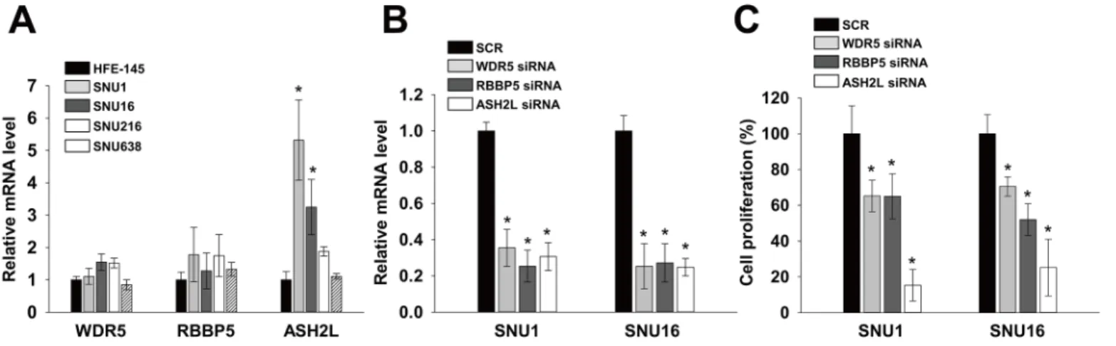
Because DPY30 is a component of WARDs, we examined expressions and roles of other components of WARDs. Real-time PCR showed that mRNA levels of WDR5 and RBBP5 in SNU1, SNU16, SNU216 and SNU638 were similar to those of HFE145. However, the mRNA
level of ASH2L were significantly higher (fold change>3) in SNU1 and SNU16 than in
HFE145 cells (Fig 3A) as DPY30 inFig 1E. In order to investigate whether WRAD components
are associated with proliferation of gastric cancer cells, we knockdowned each WRAD
compo-nent using their specific siRNA and monitored knockdown efficiencies by real-time PCR (Fig
with SCR by 65%-75%. Knockdown of WDR5 and RBBP5 inhibited the proliferation of SNU1
and SNU16 cells as compared with SCR by 30–48% (Fig 3C). However, ASH2L inhibited the
proliferation of SNU1 and SNU16, in which the expression level of ASH2L was high, as com-pared with SCR by 85% and 75%, respectively.
Fig 2. DPY30 regulated the proliferation of gastric cancer cells.(A) Real-time PCR was used to determine the efficiency of knockdown or overexpression of DPY30 in HFE145, SNU1, SNU16, SNU216 and SNU638 cells. Knockdown efficiency was determined after transfecting cells with 100 nM DPY30 siRNA targeting the ORF or scrambled siRNA (SCR). Overexpression efficiencies were determined in DPY30-overexpressing and mock cells. (B) Effect of DPY30 knockdown or overexpression on cell proliferation. A cell viability assay was used to measure cell proliferation in the presence of 1% FBS. For the DPY30 knockdown experiments, cell viability assays were performed five days after transfecting 100 nM DPY30 siRNA or SCR. After three days of culture, cell viability assays were performed on DPY30-over and mock cells. (C) Expression analysis of DPY30 after known-down or overexpression by siRNAs or DPY30 ORF respectively. The ORF-targeting or the 3’-UTR-targeting siRNA was used for the knock-down. (D) Exogenous DPY30-ORF rescued the inhibition of proliferation by the 3’-UTR-targeting DPY30 siRNA. Mock or DPY30-overexpressing cells were transduced with two kinds of DPY30 siRNA (3’ -UTR-targeting or ORF-targeting), then cell viability assay was examined. Values are the means±SDs of three independent experiments performed in triplicate.*,p<0.01 (Student’sttest, versus SCR or Mock).
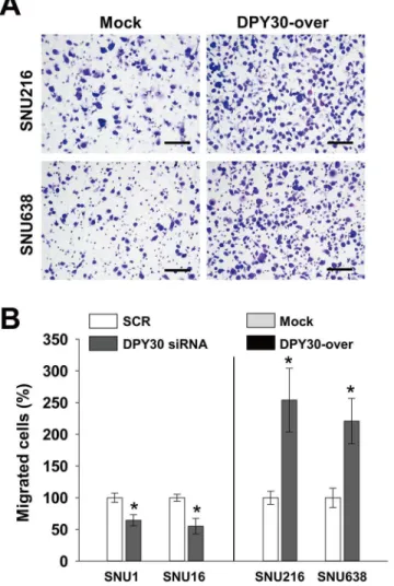
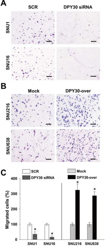
Roles of DPY30 in the migration and invasion of gastric cancer cells
We next examined whether DPY30 regulates the migration of gastric cancer cells. Knockdown of DPY30 decreased the FBS-induced migrations of SNU1 and SNU16 cells versus SCR by 35%
and 45%, respectively (Fig 4). In contrast, DPY30 overexpression increased the FBS-induced
migrations of SNU216 and SNU638 cells versus mock cells by 2.5 and 2.2 fold, respectively (Fig 4). These results led us to examine the role of DPY30 in the invasion of gastric cancer cells. In a Matrigel invasion assay, DPY30 siRNA inhibited the FBS-induced invasions of SNU1 and
SNU16 cells versus SCR siRNA by 65% and 84%, respectively (Fig 5A and 5C). Furthermore,
DPY30 overexpression increased the FBS-induced invasions of SNU216 and SNU638 cells
ver-sus mock cells by 3.2 and 2.9 fold, respectively (Fig 5B and 5C). These results show that DPY30
promotes the migration and invasion of gastric cancer cells.
Discussion
Roles of DPY30 in cancer biology have been poorly characterized although its roles in
differen-tiation of ESCs and hematopoietic progenitor cells had been reported [10,12]. In the present
study, we showed novel roles of DPY30 in gastric cancer. DPY30 is amplified (data not shown)
and highly expressed (Fig 1) in some gastric cancer tissues. Moreover, its knockdown or
over-expression regulated the proliferation, migration and invasion of gastric cancer cells. Alter-ations in the expression, chromosomal translocAlter-ations and somatic mutAlter-ations of genes
encoding subunits of SET1/MLL complexes have been reported in many cancers. These results support the critical roles of members of SET1/MLL complex in cancer progression.
Up-regulation of DPY30 expression was observed only in SNU1 and SNU16 gastric cancer cells among six gastric cancer cells we have examined. However its up-regulation was observed
in more than half of gastric cancer tissues we have examined (Fig 1). It is hard to explain this
discrepancy. However, it is obvious that the number of gastric cancer tissues we have examined is not enough. So, an extensive expression study using much more number of tissues is required to evaluate the clinical value in the future.
Roles of SET1/MLL H3K4 methyltransferase complex in cancer biology is complex. Regula-tory subunits of SET1/MLL complexes, including WRAD, contain both potent oncoproteins
Fig 3. WRAD components regulated proliferation of gastric cancer cells.(A) The mRNA levels of WDR5, RBBP5 and ASH2L in HFE145, SNU1, SNU16, SNU216 and SNU638 cells were determined by real-time PCR, using specific primers for WDR5, RBBP5 and ASH2L. GAPDH was used to normalize data. (B) Knockdown efficiency was determined by real-time PCR. Knockdown efficiency was determined after transfecting cells with 100 nM WDR5, RBBP5 and ASH2L siRNA or scrambled (SCR). (C) Effects of WDR5, RBBP5 and ASH2L knockdown on cell proliferation. A cell viability assay was used to measure cell proliferation in the presence of 1% FBS. 24 hrs after transduction, cell viability assays were performed. Values are the means±SDs of three independent experiments performed in triplicate.*,p<0.01 (Student’sttest, versus HFE145 (A) or SCR (B-C)).
and tumor suppressors. For example, the MEN1 gene encodes menin, an integral subunit of MLL1 and MLL2, and is mutated in patients with multiple endocrine neoplasia type 1 (MEN1)
[18,19]. Furthermore, menin regulates p18 (CDKN2C) and p27 (CDKN1B) by recruiting
MLL1 complex [20]. In contrast, WDR5, a component of WRAD complex, has been reported
to act as an oncoprotein in prostate cancer, in which it is overexpressed and crucial for the
androgen-induced proliferation of tumor cells [21]. ASH2L, which is another component of
WRAD complex and can interact with the oncoprotein MYC [22], is crucial for the
MYC/Ha-RAS-dependent transformation of rat embryo fibroblasts. Moreover, it is overexpressed in
many kinds of tumors and is critical for the proliferation of tumor cells [23]. In the present
study, we showed roles of DPY30 as an oncoprotein in the gastric cancer.
What are molecular mechanisms underlying roles of DPY30 in gastric cancer? Although we did not reveal, several hypotheses can be proposed based on previous studies. One of possible
Fig 4. DPY30 regulated the migration of gastric cancer cells.(A) A Boyden chamber assay was used to measure the migration of gastric cancer cells. 10% FBS was used to induce migration, and mitomycin C (0.01μg/ml) was added to remove the effects of proliferation. Two days after transfection with 100 nM ORF-targeting DPY30 siRNA or scrambled (SCR) siRNA, migration assays (Boyden chamber assay) were performed. Migration assays were carried out on DPY30-over and mock cells after culture for one day. Bar = 100μm. (B) Migrated cells were counted and results are presented as a bar graph. Values are the means±SDs of three independent experiments performed in triplicate.*,p<0.01 (Student’sttest, versus SCR or Mock).
Fig 5. DPY30 regulated the invasion of gastric cancer cells.(A-B) A Matrigel invasion assay was used to measure gastric cancer cell invasion. Presented results are representative of the results obtained. 10% FBS was used to induce invasion, and mitomycin C (0.01μg/ml) was added to remove effects of proliferation. Invasion assays were performed two days after transfection with 100 nM ORF-targeting DPY30 or SCR siRNA. After culture for one day, invasion assay was carried out for the DPY30-over and mock cells. Bar = 100μm. (C) Invasive cells were counted and results are displayed as a bar graph. Values are the means±SDs of three independent experiments performed in triplicate.*,p<0.01 (Student’sttest, versus SCR or Mock).
hypotheses is that DPY30 overexpression leads to the overexpressions of oncogenes via increased H3K4MT activity. Depletion of DPY30 or ASH2L leads to a decrease in H3K4me3, while WDR5 and RBBP5 are crucial for all three kinds of H3K4 (H3K4me1, H3K4me2, and
H3K4me3) [6,8,10,11]. In addition, the overexpression of DPY30 alone can increase
H3K4MT activity [10]. Since H3K4me2/3 is an active mark for transcription [3,24], increased
H3K4MT may directly upregulate many genes, such as, oncogenes, or alternatively indirectly down-regulate tumor suppressors. One previous report supporting this hypothesis is that ASH2L, a critical component of SET1/MLL complex, has been shown to play as an oncoprotein
[25,26]. Another previous report is that DPY30 directly activates the expressions of ID
pro-teins via H3K4 methylation [13,27]. ID proteins inhibited activities of ETS1 and ETS2 which
can induce one of tumor suppressor gene, p16. In the present study, we showed that compo-nents of WRAD (WDR5, RBBP5 and ASH2L) can inhibit proliferation of gastric cancer cells
(Fig 3), suggesting that DPY30 act through the increased H3K4MT activity. More studies are
required to reveal the mechanisms underlying the roles of DPY30 in gastric cancer cells.
Author Contributions
Conceived and designed the experiments: SOO. Performed the experiments: YJL MEH. Ana-lyzed the data: SJB SYK. Contributed reagents/materials/analysis tools: SYK SOO. Wrote the paper: YJL SOO.
References
1. Kouzarides T. Chromatin modifications and their function. Cell. 2007; 128(4):693–705. doi:10.1016/j. cell.2007.02.005PMID:17320507.
2. Bhaumik SR, Smith E, Shilatifard A. Covalent modifications of histones during development and dis-ease pathogenesis. Nature structural & molecular biology. 2007; 14(11):1008–16. doi:10.1038/ nsmb1337PMID:17984963.
3. Berger SL. The complex language of chromatin regulation during transcription. Nature. 2007; 447 (7143):407–12. doi:10.1038/nature05915PMID:17522673.
4. Ruthenburg AJ, Allis CD, Wysocka J. Methylation of lysine 4 on histone H3: intricacy of writing and reading a single epigenetic mark. Molecular cell. 2007; 25(1):15–30. doi:10.1016/j.molcel.2006.12.014 PMID:17218268.
5. Shilatifard A. Chromatin modifications by methylation and ubiquitination: implications in the regulation of gene expression. Annual review of biochemistry. 2006; 75:243–69. doi:10.1146/annurev.biochem. 75.103004.142422PMID:16756492.
6. Dou Y, Milne TA, Ruthenburg AJ, Lee S, Lee JW, Verdine GL, et al. Regulation of MLL1 H3K4 methyl-transferase activity by its core components. Nature structural & molecular biology. 2006; 13(8):713–9. doi:10.1038/nsmb1128PMID:16878130.
7. Ernst P, Vakoc CR. WRAD: enabler of the SET1-family of H3K4 methyltransferases. Briefings in func-tional genomics. 2012; 11(3):217–26. doi:10.1093/bfgp/els017PMID:22652693; PubMed Central PMCID: PMC3388306.
8. Steward MM, Lee JS, O'Donovan A, Wyatt M, Bernstein BE, Shilatifard A. Molecular regulation of H3K4 trimethylation by ASH2L, a shared subunit of MLL complexes. Nature structural & molecular biol-ogy. 2006; 13(9):852–4. doi:10.1038/nsmb1131PMID:16892064.
9. Zhang Y, Reinberg D. Transcription regulation by histone methylation: interplay between different cova-lent modifications of the core histone tails. Genes & development. 2001; 15(18):2343–60. doi:10.1101/ gad.927301PMID:11562345.
10. Jiang H, Shukla A, Wang X, Chen WY, Bernstein BE, Roeder RG. Role for Dpy-30 in ES cell-fate speci-fication by regulation of H3K4 methylation within bivalent domains. Cell. 2011; 144(4):513–25. doi:10. 1016/j.cell.2011.01.020PMID:21335234; PubMed Central PMCID: PMC3572774.
12. Yang Z, Augustin J, Chang C, Hu J, Shah K, Chang CW, et al. The DPY30 subunit in SET1/MLL com-plexes regulates the proliferation and differentiation of hematopoietic progenitor cells. Blood. 2014; 124 (13):2025–33. doi:10.1182/blood-2014-01-549220PMID:25139354.
13. Simboeck E, Gutierrez A, Cozzuto L, Beringer M, Caizzi L, Keyes WM, et al. DPY30 regulates path-ways in cellular senescence through ID protein expression. The EMBO journal. 2013; 32(16):2217–30. doi:10.1038/emboj.2013.159PMID:23872946; PubMed Central PMCID: PMC3746196.
14. Jemal A, Bray F, Center MM, Ferlay J, Ward E, Forman D. Global cancer statistics. CA: a cancer journal for clinicians. 2011; 61(2):69–90. Epub 2011/02/08. doi:10.3322/caac.20107PMID:21296855.
15. Shi Y, Zhou Y. The role of surgery in the treatment of gastric cancer. Journal of surgical oncology. 2010; 101(8):687–92. doi:10.1002/jso.21455PMID:20512944.
16. Varier RA, Timmers HT. Histone lysine methylation and demethylation pathways in cancer. Biochimica et biophysica acta. 2011; 1815(1):75–89. doi:10.1016/j.bbcan.2010.10.002PMID:20951770.
17. Chervona Y, Costa M. Histone modifications and cancer: biomarkers of prognosis? American journal of cancer research. 2012; 2(5):589–97. PMID:22957310; PubMed Central PMCID: PMC3433108.
18. Chandrasekharappa SC, Guru SC, Manickam P, Olufemi SE, Collins FS, Emmert-Buck MR, et al. Posi-tional cloning of the gene for multiple endocrine neoplasia-type 1. Science. 1997; 276(5311):404–7. PMID:9103196.
19. Guru SC, Goldsmith PK, Burns AL, Marx SJ, Spiegel AM, Collins FS, et al. Menin, the product of the MEN1 gene, is a nuclear protein. Proceedings of the National Academy of Sciences of the United States of America. 1998; 95(4):1630–4. PMID:9465067; PubMed Central PMCID: PMC19125.
20. Karnik SK, Hughes CM, Gu X, Rozenblatt-Rosen O, McLean GW, Xiong Y, et al. Menin regulates pan-creatic islet growth by promoting histone methylation and expression of genes encoding p27Kip1 and p18INK4c. Proceedings of the National Academy of Sciences of the United States of America. 2005; 102(41):14659–64. doi:10.1073/pnas.0503484102PMID:16195383; PubMed Central PMCID: PMC1253549.
21. Kim JY, Banerjee T, Vinckevicius A, Luo Q, Parker JB, Baker MR, et al. A role for WDR5 in integrating threonine 11 phosphorylation to lysine 4 methylation on histone H3 during androgen signaling and in prostate cancer. Molecular cell. 2014; 54(4):613–25. doi:10.1016/j.molcel.2014.03.043PMID: 24793694; PubMed Central PMCID: PMC4075454.
22. Yu M, Schreek S, Cerni C, Schamberger C, Lesniewicz K, Poreba E, et al. PARP-10, a novel Myc-inter-acting protein with poly(ADP-ribose) polymerase activity, inhibits transformation. Oncogene. 2005; 24 (12):1982–93. doi:10.1038/sj.onc.1208410PMID:15674325.
23. Luscher-Firzlaff J, Gawlista I, Vervoorts J, Kapelle K, Braunschweig T, Walsemann G, et al. The human trithorax protein hASH2 functions as an oncoprotein. Cancer research. 2008; 68(3):749–58. doi:10. 1158/0008-5472.CAN-07-3158PMID:18245475.
24. Shi Y, Whetstine JR. Dynamic regulation of histone lysine methylation by demethylases. Molecular cell. 2007; 25(1):1–14. doi:10.1016/j.molcel.2006.12.010PMID:17218267.
25. South PF, Fingerman IM, Mersman DP, Du HN, Briggs SD. A conserved interaction between the SDI domain of Bre2 and the Dpy-30 domain of Sdc1 is required for histone methylation and gene expres-sion. The Journal of biological chemistry. 2010; 285(1):595–607. doi:10.1074/jbc.M109.042697PMID: 19897479; PubMed Central PMCID: PMC2804208.
26. Takahashi YH, Westfield GH, Oleskie AN, Trievel RC, Shilatifard A, Skiniotis G. Structural analysis of the core COMPASS family of histone H3K4 methylases from yeast to human. Proceedings of the National Academy of Sciences of the United States of America. 2011; 108(51):20526–31. doi:10.1073/ pnas.1109360108PMID:22158900; PubMed Central PMCID: PMC3251153.