http://dx.doi.org/10.1590/1516-635x1801063-068
of Chickens
Author(s)
Fonseca BBI
Ferreira Júnior AIV
Santos JP dosV
Coelho LRI
Rossi DAI
Melo RTI
Mendonça EPI
Araújo TGIII
Alves RNII
Beletti MEII
I Faculdade de Medicina Veterinária - UFU II Centro de Microscopia Eletrônica - ICBIM -
UFU
III Laboratório de Genética - Instituto de
Ge-nética e Bioquímica - UFU
IV Programa de Pós-graduação em Sanidade e
Produção Animal nos Trópicos - Universida-de Universida-de Uberaba (UNIUBE)
V Curso de Medicina Veterinária - UNIUBE
Mail Address
Corresponding author e-mail address Álvaro Ferreira Júnior
Av. Nenê Sabino, 1801 – Bairro Universitário – Uberaba/MG – Brasil – 38.055-500
Phone: (34) 3319-8913
E-mail: [email protected]
Keywords
Campylobacter jejuni, chicken, explants, TGF-β3, IL-1β.
Submitted: June/2015 Approved: December/2015
ABSTRACT
Campylobacteriosis is a worldwide foodborne zoonosis disease
caused by Campylobacter jejuni. This microorganism is considered a
commensal bacterium in chicken hosts. C. jejuni produces epithelial
cell modifications and induces a cytokine gene transcription innate immunity repertoire. In the present study, we describe the invasiveness, morphological cellular modifications, and transcript level expressions
of innate immune cytokines from C. jejuni-inoculated chicken ileum
explants. C. jejuni was internalized by epithelial ileum cells at 15
minutes postinoculation (p.i.) and was detected intracellularly for 4hs (p.i.). Inoculated explants displayed significant increases in cell height.
C. jejuni induced a significant elevation of Transforming Growth Factor
Beta 3 (TGF-β3) and Interleukin-1β (IL-1β) transcripts. In conclusion,
C. jejuni is internalized in explanted epithelial ileum cells, produces
morphological cell modifications, and induces gene transcription of both anti-inflammatory and pro-inflammatory cytokines.
INTRODUCTION
Campylobacteriosis is a worldwide foodborne bacterial zoonosis
caused by Campylobacter jejuni (De Perio et al., 2013; Guyard-Nicodème
et al., 2015). Contaminated chicken meat is the most common source
of human infection by C. jejuni bacteria (De Carvalho et al., 2013).
The human disease is characterized by a robust intestinal inflammatory
response with bloody, watery diarrhoea (Sjöling et al., 2015). Despite
its commensal behaviour in chicken hosts, C. jejuni is able to invade
the cytoplasm of poultry enterocytes and survive in the mucus layer at
the brush border of epithelia (Van Deun et al., 2008). C. jejuni-induced
modifications of the enterocyte microvillus or mucosal crypts were
described after experimental infection (Awad et al., 2015).
It has been demonstrated that both the addition of C. jejuni antigens
to primary cell cultures of chicken embryo intestines (Li et al., 2008) and
inoculation of these antigens in live chickens (Barjesteh et al., 2013;
Humphrey et al., 2014) cause inflammatory response and early elevation
of the innate immune response of cytokine mRNA expression. Among
chicken cytokines, Interleukin-1β (IL-1β) has pro-inflammatory effects,
while the Transforming Growth Factor-β (TGF-β) has anti-inflammatory
properties (Schat, Kaspers, & Kaiser, 2014).
Animal intestinal explants have been used to investigate the relationship between luminal bacteria and the gastrointestinal immune
system (Low et al., 2006; Skoczek et al., 2014). Human explant models
were used for the determination of cell damage and the expression of pro-inflammatory cytokines in colitis studies and for the investigation of
the interaction between enteropathogenic bacteria, such as Salmonella
2004; Harvey et al., 2014). C. jejuni behaviour and the
chicken innate immune response to this agent have been described by the examination of the intestinal sections of experimentally-infected live chickens
(Lamb-Rosteski et al., 2008). Attempts to investigate
the most significant age of the chicken host for C.
jejuni infection (2 to 3 weeks old) could be relevant
for focusing on 3-week-old chickens (Chaloner et al.,
2014).
In this study, we investigated whether C. jejuni could
be internalized by the host cell, cause morphological alterations in epithelial cells, and induce the transcription of cytokine genes of the innate immunity
repertoire in the experimentally C. jejuni-infected
chicken ileum explants.
MATERIAL AND METHODS
Chicken and ileum explantsFour 25-day-old specific pathogen free (SPF) broilers,
tested negative for C. jejuni, were euthanized for the
collection of ileum explants. Explants were inoculated
with the C. jejuni strain IAL2383 (Fonseca et al., 2014)
at a dose level of 2x10⁷ CFU, diluted in saline solution
at a final volume of 100mL. The control was inoculated
with 100mL of the saline solution. All the experiments
were performed in triplicate. This study was approved by the Ethics Committee on the Use of Animals (CEUA) of the Universidade Federal de Uberlândia (Minas Gerais, Brazil), number 057/09 and number 323/09.
Invasiveness
The invasiveness assay was carried out as
previously described (Van Deun et al., 2008). C.
jejuni-inoculated ileum explants were evaluated 15, 30, and 60 minutes (min.) and 2 and 4hs post-inoculation (p.i.). Non-internalized bacteria were removed by successive washing procedures or by treatment with
the antibiotic gentamicin (100mg/mL). Internalized
bacteria were recovered by the permeabilization of the cytoplasmic membrane with 1% Tritom-X-100.
C. jejuni quantification was performed using real-time
PCR (BaxÒSystem, DuPont, USA).
Morphological epithelial cell alterations Morphological epithelial cell alterations were
detected in C. jejuni -inoculated ileum explants at 15 and
60 minutes p.i. Explants were processed by histology and stained with eosin-haematoxylin. Enterocyte morphology and morphometry were evaluated by optical microscopy at 1000x magnification (Olympus
BX 40 microscope, USA). Morphometric analyses were performed using Image HL software (Western Vision Software, USA).
Cytokine transcripts
Transcripts of cytokines and chemokines (IL-1β,
IL-8, IL-6, Transforming Growth Factor-2 [TGF-2] and CXCL2) were quantified using quantitative real-time PCR (qPCR) and reverse transcription at 15, 30, and 60 minutes, and 2 and 4hs (p.i.). For detection of complementary DNA (cDNA) from the RNA transcript, PCR was performed in real time for each gene using as a reference to the endogenous gene beta actin.
Statistical analysis
The data were statistically analysed using one-way analysis of variance (ANOVA) and Tukey’s test. A p-value ≤ 0.05 was considered significant.
RESULTS AND DISCUSSION
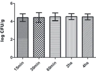
C. jejuni bacteria were internalized by the epithelial
cells of the C. jejuni-inoculated chicken ileum explants
during the first 15 min (p.i.) and the concentration of intracellular bacteria (log CFU/g) was unaltered throughout the invasiveness experiment (Figure 1). The
dynamics of C. jejuni invasion in cultured human cells (De
Melo, Gabbiani, & Pechère, 1989) includes bacterium adherence to the host cell surface and internalization of bacteria around 1h (p.i.), as well as intracytoplasmic bacteria during the interval of time from 3h to 9h (p.i.).
Additionally, it was demonstrated that C. jejuni has the
ability to invade different cultured cell lines and that its
invasiveness is dependent on the host species (Aguilar
et al., 2014). Our results corroborate the occurrence of
bacterium internalization in cultured primary chicken cells, as previously described (Byrne, Clyne, & Bourke, 2007), and may suggest that chicken ileum explants
could be an alternative method to investigate C. jejuni
invasiveness. Curiously, although mucin proteins in
chicken intestines reduces C. jejuni internalization by
the host cell (Alemka et al., 2010), that phenomenon
was apparently not impaired in the C. jejuni-inoculated
chicken ileum explants.
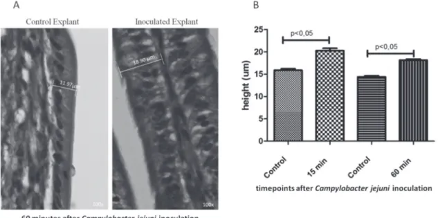
There was a significant increase in the height
of epithelial cells in the C. jejuni inoculated ileum
explants (Figure 2). We did not detect disruptions in
the structure of the microvilli or crypts (Figure 2). In
vitro, the inoculation of C. jejuni in the cell monolayer
has been shown to increase paracellular permeability and the transepithelial flux of the cultured cells
(Lamb-Rosteski et al., 2008). Additionally, C. jejuni disrupts
intercellular junctions, increasing the width of the infected cells compared with the controls (Wine, Chan, & Sherman, 2008). Cytolethal Distending Toxin (CDT) induces a cytoplasmic distension and leads to cell death (Jeon, Itoh, & Ryu, 2005). It seems that the epithelial cell modification observed in the inoculated chicken
ileum explants was an effect of bacteriumon cells.
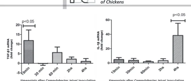
In the present study, we showed a significant
elevation of TGF-β3 and IL1β mRNA transcript
Figure 2 – Morphometry from inoculated explants: (A) micrography of Campylobacter jejuni-inoculated chicken ileum explants at 60 minutes postinfection; (B) average of epithelial cell height from C. jejuni-inoculated chicken ileum explants at two different times.
levels, which reached a peak at 15 min and 4h (p.i.), respectively (Figure 3). No statistical difference was found in the transcript levels of IL-6, IL-8, or of CXCL1 and CXCL2 mRNA transcripts. Bacterial pathogen-associated molecular patterns (PAMPs) recognized by pathogen recognition receptors (PRRs), which are expressed by intestinal epithelial cells, result in cytokine
gene transcription (IL-1β, IL-6, IL-8 and chemokynes)
from the innate immune repertoire (Keeler et al., 2007;
Wigley, 2013). Interestingly, disrupted or live C. jejuni
bacteria have been shown to have different effects on the activation of the PRRs signalizing pathways
(Al-Sayeqh et al., 2010; De Zoete et al., 2010). Smith et
al. (2005) and Li et al. (2008) described an elevated
expression of the proinflammatory cytokines
IL-1β, IL-6, IL-8 transcript levels in cultured avian cells
infected with C. jejuni. In this context, the live C. jejuni
could apparently activate innate immunity signalling pathways in chicken ileum explants.
Commensal bacteria have the ability to suppress the expression of anti-inflammatory cytokine mRNA
(i.e. TGF-β) and also reduce pro-inflammatory (i.e.,
IL-1β) cytokines, whereas pathogenic bacteria increase
both pro-inflammatory and anti-inflammatory cytokine mRNA (Bahrami, Macfarlane, & Macfarlane, 2011). In
this sense, C. jejuni cannot be regarded as merely a gut
commensal bacterium because the infection of broiler chickens has been clearly associated with intestinal
inflammation (Humphrey et al., 2014). Additionally,
the anti-inflammatory or pro-inflammatory cytokine mRNA levels display different concentrations according
(Al-Amri et al., 2008). In the present study, live C. jejuni
may have induced cytokine gene transcription similarly to pathogenic bacteria.
In conclusion, C. jejuni can be internalized by
chicken ileum cells, causes morphological cell modifications, and induces both anti-inflammatory and pro-inflammatory cytokine transcripts.
ACKNOWLEDGEMENTS
This study was financed by the Research Support Foundation in Minas Gerais, Brazil (FAPEMIG), and the Brazilian Research Support Foundation (CNPq).
REFERENCES
Aguilar C, Jiménez-Marín Á, Martins RP, Garrido JJ. Interaction between Campylobacter and intestinal epithelial cells leads to a different proinflammatory response in human and porcine host. Veterinary Immunology and Immunopathology 2014;162(1/2):14-23. Available from: http://www.ncbi.nlm.nih.gov/pubmed/25307769.
Al-Amri AI, Botta GA, Tabbara KS, Ismaeel AY, Al-Mahmeed AE, Qareeballa AY, Bin Dayna KM, Bakhiet MO. Campylobacter jejuni induces diverse kinetics and profiles of cytokine genes in INT-407 cells. Saudi Medical Journal 2008;29(4):514-519. Available from: http://www.ncbi.nlm.nih. gov/pubmed/18382790.
Alemka A, Whelan S, Gough R, Clyne M, Gallagher ME, Carrington SD, et al. Purified chicken intestinal mucin attenuates Campylobacter jejuni pathogenicity in vitro. Journal of Medical Microbiology 2010;59(8):898-903. Available from: http://www.ncbi.nlm.nih.gov/pubmed/20466838.
Al-Sayeqh AF, Loughlin MF, Dillon E, Mellits KH, Connerton IF. Campylobacter jejuni activates NF-kappaB independently of TLR2, TLR4, Nod1 and Nod2 receptors. Microbial Pathogenesis 2010;49(5):294-304. Available from: http://www.ncbi.nlm.nih.gov/pubmed/20599492.
Awad WA, Molnár A, Aschenbach JR, Ghareeb K, Khayal B, Hess C, et al. Campylobacter infection in chickens modulates the intestinal epithelial barrier function. Innate Immunology 2015;21(2):151-160. Available from: http://www.ncbi.nlm.nih.gov/pubmed/24553586.
Bahrami B, Macfarlane S, Macfarlane GT. Induction of cytokine formation by human intestinal bacteria in gut epithelial cell lines. Journal of
Applied Microbiology 2011;110(1):353-363. Available from: http:// www.ncbi.nlm.nih.gov/pubmed/21070518.
Barjesteh N, Hodgins DC, St Paul M, Quinteiro-Filho WM, DePass C, Monteiro MA, et al. Induction of chicken cytokine responses in vivo and in vitro by lipooligosaccharide of Campylobacter jejuni HS:10. Veterinary Microbiology 2013;164(1-2):122-130. Available from: http://www.ncbi.nlm.nih.gov/pubmed/23473646.
Byrne CM, Clyne M, Bourke B. Campylobacter jejuni adhere to and invade chicken intestinal epithelial cells in vitro. Microbiology 2007;153(2):561-569. Available from: http://www.ncbi.nlm.nih.gov/pubmed/17259628.
Carvalho AF de, Silva DM da, Azevedo SS, Piatti RM, Genovez ME, Scarcelli E. Detection of CDT toxin genes in Campylobacter spp. strains isolated from broiler carcasses and vegetables in São Paulo, Brazil. Brazilian Journal Microbiology 2013;44(3):693-699. Available from: http:// www.ncbi.nlm.nih.gov/pubmed/24516435.
Chaloner G, Wigley P, Humphrey S, Kemmett K, Lacharme-Lora L, Humphrey T, et al. Dynamics of dual infection with Campylobacter jejuni strains in chickens reveals distinct strain-to-strain variation in infection ecology. Applied Environmental Microbiology 2014;80(20):6366-6372. Available from: http://www.ncbi.nlm.nih.gov/pubmed/25107966 .
Fonseca BB, Rossi DA, Maia CA, Nalevaiko PC, Melo RT, Cuccato LP, et a. Characterization of the virulence, growth temperature and antibiotic resistance of the Campylobacter jejuni IAL 2383 strain isolated from humans. Brazilian Journal of Microbiology 2014;45(1):271-274. Available from: http://www.ncbi.nlm.nih.gov/pubmed/24948944.
Guyard-Nicodème M, Rivoal K, Houard E, Rose V, Quesne S, Mourand G, et al. Prevalence and characterization of Campylobacter jejuni from chicken meat sold in French retail outlets. International Journal of Food Microbiology 2015;203:8-14. Available from: http://www.ncbi.nlm. nih.gov/pubmed/25770428.
Haque A, Bowe F, Fitzhenry RJ, Frankel G, Thomson M, Heuschkel R, et al. Early interactions of Salmonella enterica serovar typhimurium with human small intestinal epithelial explants. Gut 2004;53(10):1424-1430. Available from: http://www.ncbi.nlm.nih.gov/pubmed/15361488.
Harvey BS, Sia TC, Wattchow DA, Smid SD. Interleukin 17A evoked mucosal damage is attenuated by cannabidiol and anandamide in a human colonic explant model. Cytokine 2014;65(2):236-244. Available from: http://www.ncbi.nlm.nih.gov/pubmed/24238999.
Humphrey S, Chaloner G, Kemmett K, Davidson N, Williams N, Kipar A, et al Campylobacter jejuni is not merely a commensal in commercial broiler chickens and affects bird welfare. mBio 2014;5(4):e01364-0114. Available from: http://www.ncbi.nlm.nih.gov/pubmed/24987092.
Jeon B, Itoh K, Ryu S. Promoter analysis of cytolethal distending toxin genes (cdtA , B, and C ) and effect of a luxS mutation on CDT production in Campylobacter jejuni. Microbiology Immunology 2005;49(7):599-603. Available from: http://www.ncbi.nlm.nih.gov/pubmed/16034202.
Keeler CL, Bliss TW, Lavric M, Maughan MN. A functional genomics approach to the study of avian innate immunity. Cytogenetics Genome Research 2007;117(1-4):139-145. Available from: http://www.ncbi. nlm.nih.gov/pubmed/17675854.
Lamb-Rosteski JM, Kalischuk LD, Inglis GD, Buret AG. Epidermal growth factor inhibits Campylobacter jejuni-induced claudin-4 disruption, loss of epithelial barrier function, and Escherichia coli translocation. Infection Immunology 2008;76(8):3390-3398. Available from: http:// www.ncbi.nlm.nih.gov/pubmed/18490463.
LI YP, Ingmer H, Madsen M, Bang DD. Cytokine responses in primary chicken embryo intestinal cells infected with Campylobacter jejuni strains of human and chicken origin and the expression of bacterial virulence-associated genes. BMC Microbiology 2008;(8):107. Available from: http://www.ncbi.nlm.nih.gov/pubmed/18588667.
Low AS, Dziva F, Torres AG, Martinez JL, Rosser T, Naylor S, et al. Cloning, expression, and characterization of fimbrial operon F9 from enterohemorrhagic Escherichia coli O157:H7. Infection Immunology 2006;74(4):2233-2244. Available from: http://www.ncbi.nlm.nih.gov/ pubmed/16552054.
Melo MA de, Gabbiani G, Pechère JC. Cellular events and intracellular survival of Campylobacter jejuni during infection of HEp-2 cells. Infection Immunology 1989; 57(7):2214-2222. Available from: http:// www.ncbi.nlm.nih.gov/pubmed/2731988.
Perio MA de, Niemeier RT, Levine SJ, Gruszynski K, Gibbins JD. Campylobacter infection in poultry-processing workers, Virginia, USA, 2008-2011. Emerging Infectious Diseases 2013;19(2):286-288. Available from: http://www.ncbi.nlm.nih.gov/pubmed/23347390.
Schat KA, Kaspers B, Kaiser P. Avian immunology. 2nd ed. London: Academic
Press; 2014.
Sjöling Å, Sadeghipoorjahromi L, Novak D, Tobias J. Detection of major diarrheagenic bacterial pathogens by multiplex PCR panels. Microbiology Research 2015;172:34-40. Available from: http://www. ncbi.nlm.nih.gov/pubmed/25542594.
Skoczek DA, Walczysko P, Horn N, Parris A, Clare S, Williams MR, et al. Luminal microbes promote monocyte-stem cell interactions across a healthy colonic epithelium. Journal of Immunology 2014;193(1):439-451. Available from: http://www.ncbi.nlm.nih.gov/pubmed/24907348.
Smith CK, Kaiser P, Rothwell L, Humphrey T, Barrow PA, Jones MA. Campylobacter jejuni-induced cytokine responses in avian cells. Infection Immunology 2005;73(4):2094-2100. Available from: http:// www.ncbi.nlm.nih.gov/pubmed/15784550.
Van Deun K, Pasmans F, Ducatelle R, Flahou B, Vissenberg K, Martel A, et al. Colonization strategy of Campylobacter jejuni results in persistent infection of the chicken gut. Veterinary Microbiology 2008;130(3-4):285-297. Available from: http://www.ncbi.nlm.nih.gov/ pubmed/18187272.
Wigley P. Immunity to bacterial infection in the chicken. Development Comparative Immunology 2013;41(3):413-417. Available from: http:// www.ncbi.nlm.nih.gov/pubmed/23648643.
Wine E, Chan VL, Sherman PM. Campylobacter jejuni mediated disruption of polarized epithelial monolayers is cell-type specific, time dependent, and correlates with bacterial invasion. Pediatric Research 2008;64(6):599-604. Available from: http://www.ncbi.nlm.nih.gov/ pubmed/18679160.