Elevated Atherosclerosis-Related Gene
Expression, Monocyte Activation and
Microparticle-Release Are Related to
Increased Lipoprotein-Associated Oxidative
Stress in Familial Hypercholesterolemia
Morten Hjuler Nielsen1,6*, Helle Irvine2, Simon Vedel3, Bent Raungaard4, Henning Beck-Nielsen5, Aase Handberg6
1Danish PhD School of Molecular Metabolism, University of Southern Denmark, Odense, Denmark,
2Department of Medicine and Cardiology A, Aarhus University Hospital, Aarhus, Denmark,3Department of Radiology, Aarhus University Hospital, Aarhus, Denmark,4Department of Cardiology, Aalborg University Hospital, Aalborg, Denmark,5Department of Endocrinology M, Odense University Hospital, Odense, Denmark,6Department of Clinical Biochemistry, Aalborg University Hospital, Aalborg, Denmark
Abstract
Objective
Animal and in vitro studies have suggested that hypercholesterolemia and increased oxida-tive stress predisposes to monocyte activation and enhanced accumulation of oxidized LDL cholesterol (oxLDL-C) through a CD36-dependent mechanism. The aim of this study was to investigate the hypothesis that elevated oxLDL-C induce proinflammatory monocytes and increased release of monocyte-derived microparticles (MMPs), as well as up-regulation of CD36, chemokine receptors and proinflammatory factors through CD36-dependent path-ways and that this is associated with accelerated atherosclerosis in subjects with heterozy-gous familial hypercholesterolemia (FH), in particular in the presence of Achilles tendon xanthomas (ATX).
Approach and Results
We studied thirty FH subjects with and without ATX and twenty-three healthy control sub-jects. Intima-media thickness (IMT) and Achilles tendon (AT) thickness were measured by ultrasonography. Monocyte classification and MMP analysis were performed by flow cytom-etry. Monocyte expression of genes involved in atherosclerosis was determined by quanti-tative PCR. IMT and oxLDL-C were increased in FH subjects, especially in the presence of ATX. In addition, FH subjects had elevated proportions of intermediate CD14++CD16+ monocytes and higher circulating MMP levels. Stepwise linear regression identified oxLDL-C, gender and intermediate monocytes as predictors of MMPs. Monocyte expression of pro-atherogenic and pro-inflammatory genes regulated by oxLDL-C-CD36 interaction was a11111
OPEN ACCESS
Citation:Hjuler Nielsen M, Irvine H, Vedel S, Raungaard B, Beck-Nielsen H, Handberg A (2015) Elevated Atherosclerosis-Related Gene Expression, Monocyte Activation and Microparticle-Release Are Related to Increased Lipoprotein-Associated Oxidative Stress in Familial Hypercholesterolemia. PLoS ONE 10(4): e0121516. doi:10.1371/journal. pone.0121516
Academic Editor:Alexandre Boissonnas, Centre d'Immunologie et des Maladies Infectieuses, INSERM, FRANCE
Received:December 3, 2014
Accepted:February 2, 2015
Published:April 13, 2015
Copyright:© 2015 Hjuler Nielsen et al. This is an open access article distributed under the terms of the
Creative Commons Attribution License, which permits unrestricted use, distribution, and reproduction in any medium, provided the original author and source are credited.
Data Availability Statement:All relevant data are within the paper.
increased in FH, especially in ATX+ subjects. Monocyte chemokine receptor CX3CR1 was identified as an independent contributor to IMT.
Conclusions
Our data support that lipoprotein-associated oxidative stress is involved in accelerated ath-erosclerosis in FH, particularly in the presence of ATX, by inducing pro-inflammatory mono-cytes and increased release of MMPs along with elevated monocyte expression of oxLDL-C-induced atherosclerosis-related genes.
Introduction
The attachment and subsequent transmigration of circulating monocytes into the subendothe-lial space is facilitated by hypercholesterolemia-induced expression of adhesion molecules on endothelial cells and their secretion of chemoattractant factors. The monocytes differentiate into macrophages, which internalize lipoproteins and become proinflammatory resulting in further recruitment of monocytes and promoting inflammation and progression of atheroscle-rosis as reviewed in [1]. Previous studies have indicated that circulating monocytes are a het-erogenic population composed of at least two distinct subpopulations based on surface expressions of CD14 and CD16 [2]. The major subpopulation expresses high levels of CD14 and low levels of CD16, whereas the minor and more proinflammatory subpopulation ex-presses low levels of CD14 and high levels of CD16 on the cell surface [3]. A subset of CD16-positive monocytes produces high levels of inflammatory mediators and up-regulates a number of chemokine receptors, including CCR2, CX3CR1 and CCR5 which are attributed
non-redundant and independent roles in the development of atherosclerosis [4,5].
Uptake of oxidized LDL cholesterol (oxLDL-C) by the scavenger receptor CD36 in mono-cytes and macrophages leads to an up-regulation of CD36 expression through activation of the transcription factor PPAR-γ, thereby creating a‘vicious’feed-forward cycle of increasing oxLDL-C uptake, ultimately converting the monocyte/macrophage into an atherogenic foam cell as reviewed in [6]. Other effects of oxLDL-C binding to CD36 include activation of the transcription factor NFκB which induces production of proinflammatory cytokines and a proinflammatory phenotype [7].
Familial hypercholesterolemia (FH) is an autosomal codominant genetic disorder of lipo-protein metabolism [8], characterized by elevated plasma levels of LDL cholesterol, a high inci-dence of premature coronary heart disease and extravascular deposits of cholesterol in tendons (tendon xanthomas) [9]. The presence of Achilles tendon xanthomas (ATX) is a marker for high risk of cardiovascular disease among FH patients [10,11] and xanthomas and atheroscle-rosis may result from the same pathophysiological mechanisms. Microparticles (MPs) are vesi-cles (<1μm) shed from the plasma membranes of activated circulating and vascular cells and
are considered to constitute a new inter-cellular signaling system which may be involved in various diseases such as cardiovascular disorders [12].
The overall purpose of the present study was to investigate the involvement of monocytes and lipoprotein-associated oxidative stress in the atherosclerotic process. Our hypothesis was that elevated oxLDL-C in FH induce proinflammatory monocytes and increased release of monocyte-derived microparticles (MMPs), as well as up-regulation of CD36, chemokine recep-tors and proinflammatory facrecep-tors through CD36-dependent pathways, and that this
accelerates atherosclerosis.
To study this, the proportion of CD16-positive monocyte subpopulations in peripheral blood of FH subjects with and without ATX was compared and CD36 surface expression levels determined. Monocyte expression levels of selected genes involved in the atherosclerotic pro-cess and known to be induced by oxLDL-C were determined and related to IMT. Furthermore, as atherosclerosis and Achilles tendon thickening may share common mechanisms we evaluat-ed this relationship in FH. Finally, MMPs were quantifievaluat-ed, and the association between proin-flammatory monocyte subpopulations, oxLDL-C and circulating MMPs was studied.
Materials and Methods
Study population
The study group comprised thirty patients (18 females and 12 males) genetically diagnosed with heterozygous FH and selected on the basis of the presence or absence of ATX according to medical records. Twenty-three healthy controls (15 females and 8 males), as indicated by a medical questionnaire, served as the control group. Exclusion criteria were: Hypertension, dia-betes mellitus, previous history of atherosclerotic disease and the use of lipid-lowering medica-tion (except for FH subjects). FH patients were treated with lipid-lowering therapy at inclusion and underwent an 8-week washout period before blood sampling [13]. The study was con-ducted in agreement with the Helsinki-II declaration and approved by The Central Denmark Region Committees on Health Research (2010–0147) and by the Danish Data Protection Agency (j.no.: 2010-41-4879). Written informed consent was obtained from all participants. Blood pressure, height and body weight were recorded and blood samples were obtained in the fasting state. The following parameters were determined in the routine laboratory: Platelet, leu-kocyte and monocyte counts, as well as hemoglobin levels were determined on a Sysmex XE-5000 Automated Hematology System (EDTA plasma). A Cobas 6000 analyzer (Roche) was used to determine concentrations of glucose, alanine-aminotransferase (ALT), triglycerides, total cholesterol (Total-C), low-density lipoprotein cholesterol (LDL-C), high-density lipopro-tein cholesterol (HCL-C) and apolipoprolipopro-tein B (ApoB) (Li-Heparin plasma). LDL-C concen-trations were estimated from the Friedewald formula [14].
Markers of lipoprotein-associated oxidative stress and inflammation
Oxidized LDL-C (Mercodia, Uppsala, Sweden), C-Reactive Protein (CRP) (MBL International, Woburn, MA, USA), Interleukin-6 (IL-6) (Diaclone, Besancon Cedex, France), Matrix Metal-loproteinase 9 (MMP-9) (Nordic BioSite, Denmark) and Intercellular Adhesion Molecule 1 (ICAM-1) (R&D Systems, Europe) were measured on EDTA plasma. All ELISA measurements were performed according to the manufacturer’s instructions. Soluble CD36 (sCD36) was de-termined by an in-house ELISA method as previously described [15].
Measurement of carotid intima-media thickness
evidence of carotid plaque, care was taken not to include the plaque in the IMT measurement. Plaque was defined according to The Mannheim CIMT Consensus Report [17]. IMT values were calculated as means of the three measured values.
Determination of Achilles tendon xanthomas
Achilles tendons (AT) were examined using the linear-array transducer described above and with the subjects lying in prone position with ankles extended beyond the examination bed and feet at 90° flexion. Special attention was paid to holding the probe perpendicular to the ten-don. Measurements of AT thickness (average of both tendons) were made from longitudinal scans at the point of maximum thickness. In uniformly sized tendons, measurements were taken 20 mm proximally to insertion on Calcaneus. The size of the tendon was considered nor-mal when the maximum thickness was less than 5.8 mm [18].
Determination of monocyte subpopulations and their CD36 expression
To avoid influence of platelet count in regard to CD36 measurements [19], we performed flow cytometry analysis on monocytes isolated from EDTA blood using the Dynabeads FlowComp Human CD14 kit and a magnetic cell sorter (Invitrogen, Carlsbad, CA, USA) following the manufacturer’s instructions. Monocytes used for gene expression analysis were sedimented by centrifugation (350 x g, 10 min.) and stored at minus 80°C in RNAlater solution (Life Technol-ogies, Carlsbad, CA, USA). Flow cytometry on isolated monocytes was performed using a FACSCalibur flow cytometer running Cell Quest software (BD Biosciences). Subsets of classical (CD14++CD16-), intermediate (CD14++CD16+) and non-classical (CD14+CD16++) mono-cytes were defined according to the surface expression pattern of the lipopolysaccharide recep-tor CD14 and the Fcγ-III receprecep-tor CD16 according to the recommendations of the
Nomenclature Committee of the International Union of Immunological Societies [20]. Abso-lute CD36 surface protein expression on each monocyte subset was quantified by the mean fluorescence intensity (MFI) using a mixture of PE-Quantibrite beads (BD Biosciences).
In brief, monocytes (~150.000 cells/100μL) were incubated in the dark (4°C) for 30 min
with the following monoclonal antibodies obtained from Biolegend (San Diego, CA, USA): 3μL peridinin chlorophyll protein complex with cyanin- 5.5 (PerCP-Cy5.5)-conjugated
anti-human CD14 (400μg mL-1IgG1,κ(clone HCD14), 5μL Alexa Fluor 488-conjugated
anti-human CD16 (200μg mL-1IgG1,κ(clone 3G8) and 5μL Phycoerythrin (PE)-conjugated
anti-human CD36 (6.25μg mL-1IgG2a,κ(clone 5–271). Following incubation, 250μL of staining
buffer (PBS + 2% FBS + 0.1% Sodium Azide) was added and samples were kept on ice until analysis. The optimal antibody concentrations were determined experimentally by titration ex-periments and concentration-matched isotype mAbs were used as controls.
A standard curve relating PE fluorescence intensity to PE surface density was then generated using PE-Quantibrite Beads tagged with 4 defined amounts of PE ranging from 200 to 70 000 molecules per bead. Using Flow Jo software (v. 8.8.7, Tree Star, Inc., Oregon, USA) the number of bound CD36 antibody molecules on each monocyte was calculated from the standard curve using the monocyte CD36 MFI, knowing that the fluorochrome-to-protein ratio of the CD36-PE conjugate is constant for each vial in a given lot of reagent and under the assumption that the stoichiometry of anti-CD36 binding to CD36 is 1:1.
Flow cytometric measurements of monocyte-derived microparticles
concentration and processed within 1 hour at room temperature. Platelet-free plasma (PFP) was prepared by a three-step centrifugation procedure and stored at -80°C until analysis. For each analysis, 50-μL of freshly thawed PFP was transferred to a TruCount tube (BD
Biosci-ences, New Jersey, USA) containing a lyophilized pellet which releases a known number of fluorescent beads used for microparticle quantification according to the manufacturer’s in-structions. Subsequently, microparticles were labeled by adding 10μL fluorescein
isothiocya-nate (FITC)-conjugated Lactadherin (83μg mL-1, Haematologic Technologies Inc, Vermont,
USA) [22,23]. To identify MMPs, 5μL peridinin chlorophyll protein complex with cyanin- 5.5
(PerCP-Cy5.5)-conjugated anti-human CD14 (400μg mL-1IgG1,κ(clone HCD14, Biolegend,
San Diego, CA, USA)) was added immediately after Lactadherin-FITC labeling. After 30 min. of incubation (4°C, in the dark), 250μL 0.22μm filtered PBS was added to the labeled sample.
The optimal antibody concentration was determined experimentally by titration experiments and concentration-matched isotype mAbs were used as controls.
A microparticle gate was established by preliminary standardization experiments using a blend of size-calibrated fluorescent beads with sizes ranging from 0.2 to 0.9μm as described in
[21]. The upper and the outer limits of the microparticle gate were established just above the size distribution of 0.9-μm-sized beads in a forward (FSC-A) and side scatter (SSC-A) setting
(log scale) using the“auto-gate”function inside the Flow Jo software. The noise threshold of the instrument was set as the lower limit of the MP gate.
Quantitative real-time RT-PCR
RNA was extracted from isolated monocytes using the RNeasy Micro Kit (Qiagen, Hilden, Ger-many). Subsequently, 4μL of undiluted RNA solution was mixed with 10μL MgCl225 mM,
(Roche A/S, Hvidovre, Denmark), 4μL 10 × PCR buffer II (Roche A/S), 2μL Oligo d(T)16
50μM (DNA Technology A/S, Aarhus, Denmark), 16μL dNTPmix (dATP/dTTP/dCTP/dGTP)
(Pharmacia Biotech, Hilleroed, Denmark), 2μL MuLV (Moloney murine Leukaemia Virus)
re-verse transcriptase, (50 U/ml) (PerkinElmer Denmark A/S, Hvidovre, Denmark) and 2μL
RNase inhibitor, (20 U/ml) (PerkinElmer Denmark A/S) for a final volume of 40μL. cDNA was
synthesized by incubation at 42°C for 30 min and the process was stopped by increasing incuba-tion temperature to 99°C for 5 min. cDNA was either used immediately or stored at -20°C. Quantitative Real-time RT-PCR was performed using the Lightcycler 480 instrument (Roche Di-agnostics, Penzberg, Germany). The mRNA expression of 9 individual genes involved in choles-terol uptake (CD36), reverse cholescholes-terol transport (ABCA1), monocyte migration (CCR2, CCR5, CX3CR1), inflammation (TNF-α), regulatory pathways (PPAR-γ, NFκB) as well as CD16
as an inflammatory marker were measured using the primers listed inTable 1. mRNA expres-sion levels were normalized to the expresexpres-sion of [beta]2M ([beta]2-microglobulin). Primers were
selected to span two exons in order to prevent amplification of genomic DNA.
A PCR mix was made on the basis of the prescription from the supplier. In brief, 3μL sterile
water, 5μL Master Mix, (Roche Diagnostics, Penzberg, Germany), 0.5μL sense and 0.5μL
anti-sense primers and 1μL SYBRgreen (Roche Diagnostics, Penzberg, Germany) were mixed
be-fore adding 1μL target cDNA for a total volume of 10μL. RNA extracted from a bladder
cancer cell line, HCV29 (ATCC, Manassas, Virginia, USA), was used as a calibrator control for PPAR-γand NFκB whereas monocyte-derived RNA extracted buffy coats from healthy do-nors, using the EasySep Human CD14 Positive Selection Kit (STEMCELL Technologies SARL, Grenoble, France), was used as calibrator for CD36, CD16, TNF-α, CCR2, CCR5 and CX3CR1.
annealing (temperatures are given inTable 1) for 5 s and 72°C extension for 10 s. A standard melting curve was used to check the quality of amplification. A calibration curve and positive and negative controls were included in each run. Each calibration curve was composed of serial dilutions of a RNA pool from the appropriate calibrator cell lines. As a negative control for both calibration curves, water was added instead of RNA. All PCR reactions were performed in duplicates on the same plate. Gel electrophoresis and DNA sequencing verified the specificity of each amplified RT-PCR product.
NormFinder software, an algorithm for identifying the optimal normalization gene among a set of candidates, was used to identify the most stable reference gene [24]. Comparing stabili-ty of the household candidate genesβ-actin (Beta-actin), GAPDH (glyceraldehyde-3- phos-phate dehydrogenase), HMBS (Hydroxymethylbilane synthase) and [beta]2M ([beta]2
-microglobulin), respectively, in all 53 participating subjects resulted in the selection of [beta]2M as the most stable household gene.
Statistics
Statistical analyses were carried out using the STATA 11.2 statistical program (StataCorp LP, Texas, USA). The Shapiro-Wilk’s W test was used to test the assumption of normality. Normal and non-normal distributed parameters were compared using two-tailed Student´s t-test and Mann Whitney U-test, respectively. Statistical correlation was analyzed using Spearman's rank correlation for non-normal distributed data. Predictors of IMT and MMP numbers,
Table 1. Characteristics of primers used for RT-PCR.
Gene Target Primer Sequence Annealing temperature (°C) Fragment length (bp)
CD36 Forward 5´-GCT GTG GCA GCT GCA TCC CAT-3´ 65 318
Reverse 5´-AAT CAT GTC GCA GTG ACT TTC CCA A-3´
CD16 Forward 5´-GGC TGT GGT GTT CCT GGA GCC-3´ 65 282
Reverse 5´-TTG AAC ACC CAC CGA GGG GC-3´
CCR2 Forward 5´-AGC CTG ACA TAC CAG GAC TGC CT-3´ 65 228
Reverse 5´-ACA CCA GCG AGT AGA GCG GAG G-3´
CCR5 Forward 5´-GGC CAG AAG AGC TGA GAC ATC CG-3´ 64 153
Reverse 5´-AGG CGG GCT GCG ATT TGC TT-3´
CX3CR1 Forward 5´-TGG GCT TCT TGC TGT TGG TGA GG-3´ 64 220
Reverse 5´-GGT TCC CTT GGC AGT CCA CGC-3´
ABCA1 Forward 5´-GCC CGG CGG TTC TTG TGG AA-3´ 64 220
Reverse 5´-GGT CCG GGT TGG ACC CTG CT-3´
NFkB Forward 5´-ACT CGC CAC CCG GCT TCA GA-3´ 64 133
Reverse 5´-GGC AGT GCC ATC TGT GGT TGA A-3´
PPAR-γ Forward 5´-GCC TTG CAG TGG GGA TGT CTC A-3´ 64 198
Reverse 5´-TCG CCC TCG CCT TTG CTT TGG-3´
TNF-α Forward 5´-CGC TCT TCT GCC TGC TGC ACT-3´ 65 196
Reverse 5´-GCA TTG GCC CGG CGG TTC AG-3´
[beta]2M Forward 5´-AGT GGG ATC GAG ACA TGT AAG CAG-3´ 64 84
Reverse 5´-GCT ACC TGT GGA GCA ACC TGC-3´
Primer sequence, annealing temperature and fragment length of selected target genes. Final concentration of primers was 5 pmol/μL. Abbreviations: CD36, Cluster of Differentiation 36; CD16, Cluster of differentiation 16; CCR2, C-C chemokine receptor type 2; CCR5, C-C chemokine receptor type 5; CX3CR1, CX3C chemokine receptor 1; ABCA1, ATP-binding cassette transporter 1; NFkB, Nuclear Factor-KappaB; PPAR-γ, Peroxisome
proliferator-activated receptor gamma; TNF-α; Tumor necrosis factor-alpha; [beta]2M, [beta]2M, [beta]2-microglobulin.
respectively, were identified by stepwise linear regression (backward selection) using a signifi-cance level of 10%. Variables were log transformed prior to regression. P values are two-sided and considered significant when<0.05.
Results
Characterization of study participants
Table 2shows the clinical characteristics of the study groups. Routine laboratory measure-ments were all within age- and gender-specific reference intervals and no significant differences were observed between groups except for increased levels of total cholesterol, LDL-C, ApoB and oxLDL-C in FH compared to control subjects as well as in FH ATX+ compared to FH ATX- subjects. HDL-C levels were lower in FH subjects compared to controls, but comparable in the two FH study groups. Plasma levels of the inflammatory markers IL-6, hs-CRP, MMP-9, ICAM-1 and sCD36 as a potential marker of plaque instability were not affected by FH, except for IL-6 which was significantly increased (p = 0.022). The presence of ATX did not affect plas-ma levels of any of these bioplas-markers (Table 2).
Table 2. Characteristics of study participants.
Characteristics Controls (n = 23) FH (n = 30) p-valuea FH ATX-(n = 14) FH ATX+ (n = 16) p-valueb
Men/women 8/15 12/18 NS 7/7 5/11 NS
Age (years) 47.0±10.1 45.5±9.1 NS 43.1±10.2 47.5±7.8 NS
BMI (kg/m2) 23.7±3.3 25.5±5.2 NS 25.1±4.2 25.9±5.9 NS
Systolic bp (mm Hg) 120.2±10.8 124.9±14.3 NS 122.4±13.7 127.1±14.8 NS
Diastolic bp (mm Hg) 74.6±6.7 77.4±6.5 NS 76.0±5.5 78.7±7.1 NS
Platelets (109/L) 239.8±50.7 248.6±60.1 NS 247±55.7 250±65.4 NS
Leukocytes (109/L) 5.1±1.2 5.4±1.9 NS 5.0±1.6 5.7±2.2 NS
Monocytes (109/L) 0.4±0.1 0.5±0.1 NS 0.4±0.1 0.5±0.2 NS
Hemoglobin (mmol/L) 8.7±0.6 9.0±0.8 NS 9.1±1.0 8.9±0.7 NS
Total-C (mmol/L) 5.1±0.7 9.1±1.8 <0.0001 8.2±1.1 9.9±2.0 <0.010
LDL-C (mmol/L) 2.9±0.5 7.0±1.8 <0.0001 6.0±1.1 7.9±1.8 0.003
HDL-C (mmol/L) 1.7±0.4 1.5±0.4 0.014 1.5±0.5 1.4±0.4 NS
oxLDL-C (Units/L) 50.0±11.8 100.3±24.2 <0.0001 83.7±13.6 114.8±22.1 0.0001
ApoB (g/L) 0.8±0.1 1.7±0.4 <0.0001 1.4±0.3 1.8±0.3 0.002
Glucose (mmol/L) 5.4±0.5 5.5±0.4 NS 5.5±0.4 5.5±0.4 NS
ALT (U/L) 19.0 (14.0–21.0) 19.0 (17.0–34.0) NS 20.5 (18–32) 18.5 (15.0–36.5) NS
TG (mmol/L) 1.0 (0.8–1.2) 1.1 (0.8–1.8) NS 1.3 (0.7–1.8) 1.1 (1.0–1.6) NS
IL-6 (pg/ml) 0.6 (0.0–0.7) 0.8 (0.3–1.6) 0.022 0.7 (0.3–1.0) 1.0 (0.3–2.0) NS
hs-CRP (μg/ml) 1.0 (0.8–1.9) 1.5 (0.6–4.2) NS 1.3 (0.4–3.9) 1.9 (0.7–5.2) NS
sCD36 (AU) 0.4 (0.3–0.5) 0.4 (0.3–0.5) NS 0.4 (0.2–0.5) 0.4 (0.3–0.6) NS
MMP-9 (pg/ml) 0.9 (0.6–1.2) 1.0 (0.7–2.1) NS 1.0 (0.7–1.8) 1.1 (0.7–2.6) NS
ICAM-1 (pg/ml) 1.1 (1.0–1.5) 1.3 (1.1–1.5) NS 1.3±0.2 1.4±0.5 NS
Normal and non-normally distributions are shown as the mean±standard deviation and as the median (interquartile range), respectively.
aControls vs. FH subjects;
bFH subjects without ATX vs. FH subjects with ATX.
Abbreviations: BMI, Body mass index; ALT, Alanin-aminotransferase, TG, Triglycerides; ApoB, Apolipoprotein B; Total-C, Total cholesterol; LDL-C, LDL Cholesterol; HDL-C, HDL Cholesterol; oxLDL-C, oxidized LDL-C; bp, blood pressure; hs-CRP, High Sensitivity C-reactive Protein; IL-6, Interleukin 6; sCD36; soluble CD36, MMP-9, Matrix metalloproteinase 9; ICAM-1, Intercellular Adhesion Molecule 1; AU, arbitrary units; NS, non-significant.
AT thickness and oxLDL-C
Mean AT thickness was increased in FH compared to control subjects (6.19 ± 1.92 mm vs. 4.78 ± 0.62 mm, p = 0.001). Absence or presence of ATX was confirmed and accordingly AT thickness was higher in the defined FH ATX+ group compared to the FH ATX- group
(7.28 ± 2.08 mm vs. 4.95 ±0.35 mm, p = 0.0003). Notably, no gender- or age-related differences in AT thickness were observed within the groups. AT thickness correlated with oxLDL-C in FH subjects (Rho = 0.58, p = 0.0008) but not in controls. Likewise, AT thickness and IMT cor-related in the FH group only (Rho = 0.57, p = 0.001).
Association between intima media thickness and oxLDL-C
Mean carotid IMT was increased in FH subjects compared with the control group (0.64 ±0.12 mm vs. 0.58 ±0.07 mm, p = 0.033) and in FH ATX+ compared to FH ATX- subjects (0.69 ±0.10 mm vs. 0.57 ±0.10 mm, p = 0.002). IMT was not affected by age or gender in either group. As-suming that the impact of oxLDL-C on IMT is independent of FH, we observed a significant correlation between oxLDL-C and IMT (Rho = 0.27, p = 0.045) within the study population.
Monocyte count and monocyte subpopulations
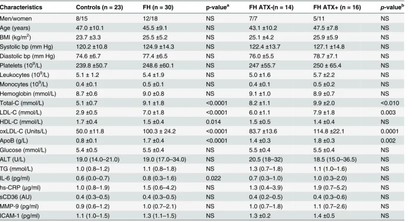
Monocyte counts were significantly associated to IMT in all study subjects (Rho = 0.32, p = 0.019), as well in FH (Rho = 0.40, p = 0.028) and FH ATX+ (Rho = 0.52, p = 0.037) subjects when considered separately. Monocyte subpopulations were heterogeneous in regard to CD16 expression (Fig 1) and unrelated to IMT.
Fig 1. Flow cytometric analysis of monocyte subpopulations.Representative example of monocyte subsets depicted as classical CD14++CD16- monocytes (blue color), intermediate CD14++CD16 + monocytes (red color), and nonclassical CD14+CD16++ monocytes (green color).
The percentage of intermediate monocytes was higher in FH subjects compared to controls ((2.28% (1.34–3.12) vs. (1.39% (1.02–2.38), p = 0.034) whereas no significant difference be-tween FH ATX- and FH ATX+ subjects was found ((2.33% (1.32–2.82) vs. (2.28% (1.61–3.26), p = 0.739). Likewise, the percentage of non-classical monocytes tended to be higher in FH sub-jects compared to controls ((0.58% (0.40–0.98)) vs. (0.42% (0.29–0.74), p = 0.061) whereas no difference between FH ATX+ and FH ATX- subjects was found ((0.52% (0.39–1.10) vs. 0.72% (0.4–0.98), p = 0.884). Assuming that the impact of oxLDL-C on monocyte differentiation is independent of FH, it was found that the percentage of intermediate but not of non-classical monocytes correlated with plasma oxLDL-C (Rho = 0.34, p = 0.014, N = 53).
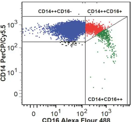
CD36 surface expression within monocyte subpopulations
In general, CD36 surface expression was demonstrated on all monocyte subpopulations. A rep-resentative flow cytometric plot of CD36 expression on monocyte subpopulations is shown in Fig 2A. Non-classical monocyte CD36 expression was elevated in FH compared to control sub-jects (48026 ±8007 vs. 43149 ±10091, p = 0.029,Fig 2B) whereas CD36 expression was unaf-fected by FH in the remaining subpopulations. The presence of ATX did not affect CD36 expression in any of the monocyte subpopulations and CD36 expression showed no correlation to oxLDL-C levels. However, CD36 expression differed among monocyte subpopulations in both FH and healthy controls, revealing a significantly reduced CD36 expression on both of the two CD16-positive monocyte subpopulations compared to the classical CD16-negative monocytes (Fig 2B).
Expression of pro-atherogenic and pro-inflammatory genes in FH
monocytes
The putative proinflammatory and proatherogenic phenotype of circulating monocytes was in-vestigated at the transcriptional level by quantitative real-time RT-PCR. As illustrated inFig 3A, FH subjects had significantly increased mRNA expression of CD36 (p = 0.008), CX3CR1
(p = 0.031) and PPAR-γ(p = 0.016). Moreover, FH ATX+ subjects had increased expression of CD36 (p = 0.046), CCR2 (p = 0.042), CCR5 (p = 0.018), CX3CR1 (p = 0.028), ABCA1
(p = 0.009) and NFκB (p = 0.018) when compared to FH ATX- subjects (Fig 3B).
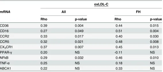
CD36 mRNA and protein levels were unrelated. Correlation analysis was applied in order to test the putative association of oxLDL-C with monocyte expression of genes involved in the atherosclerotic process. Within the entire study population, as well as in FH subjects, oxLDL-C was positively correlated with the expression of CD36, CD16, CCR2, CCR5, CX3CR1 and
NFκB but not PPAR-γ, TNF-αor ABCA1 (Table 3).
Possible contributors to IMT
The relationship between IMT and the expression levels of the pro-atherogenic and pro-inflam-matory genes in monocytes was investigated. Within the entire study population IMT correlat-ed positively with the expression of CD36, CCR2, CCR5, CX3CR1, NFκB and TNF-αbut not
with PPAR-γ, CD16 or ABCA1. In FH subjects, IMT correlated significantly with the expres-sion of CCR2, CCR5, CX3CR1 and NFκB and approached significance with CD36 (p = 0.053)
contributor to IMT within the entire study population (R2= 0.30; p<0.0001) as well as in FH
subjects only (R2= 0.23; p = 0.0008).
Indication of MMP release from proinflammatory monocytes
Total MMP numbers were higher in FH subjects (10 (7–14) MMPs /μL plasma) compared
with controls (5 (3–11) MMPs /μL plasma), p = 0.004 and unaffected by the presence of ATX.
The hypothesis that cholesterol fractions, particularly oxLDL-C, promote the release of MMPs from proinflammatory monocytes was tested. Considering that the impact of cholesterol frac-tions on MMP release is independent of FH, correlation analysis revealed a positive correlation between MMP numbers and LDL-C (Rho = 0.41; p = 0.003) and oxLDL-C (Rho = 0.35;
Fig 2. CD36 expression on monocyte subpopulations.(A) Representative flow cytometric plot of CD36 expression on monocyte subpopulations. (B) Box plot analysis of CD36 surface expression levels on classical (blue color), intermediate (red color) and nonclassical (green color) monocytes in healthy controls and FH subjects. Median (horizontal dotted line), 25% and 75% quartiles (box), 10% and 90% quartiles (whiskers). Filled circles represent outliers.*p<0.010;**p<0.005;***p<0.0001.
Fig 3. Real-time quantitative RT-PCR analysis on isolated monocytes.Expression of pro-atherogenic and pro-inflammatory genes in FH and control subjects (A) and in FH subjects with and without ATX (B). Real-time quantitative RT-PCR was performed on total RNA obtained from freshly isolated monocytes. The amount of mRNA for the gene of interest was normalized relative to [beta]2M mRNA, and the graphs were
generated with the relative median values obtained after normalization. Mann-Whitney U test was used to compare the transcriptional expression of genes and statistically significant difference (p<0.05) is indicated by*.
p = 0.010) whereas HDL-C correlated negatively (Rho = -0.34; p = 0.014). MMP numbers and monocyte counts were not correlated. Within the entire study population, MMP numbers cor-related with the percentage of both intermediate (Rho = 0.37, p = 0.007) and non-classical monocytes (Rho = 0.34, p = 0.012) whereas in FH subjects, only the correlation with the inter-mediate monocytes remained significant (Rho = 0.45, p = 0.013).
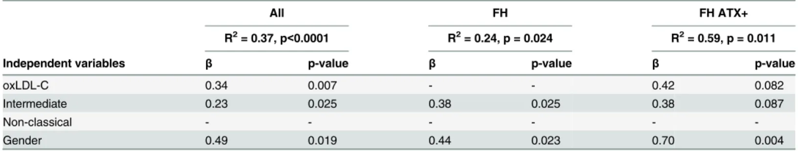
The possible significance of proinflammatory monocytes and lipoprotein-associated oxida-tive stress for circulating MMP numbers was investigated using stepwise linear regression anal-ysis. Gender was included as a variable as it is known to affect MP levels [21]. Regression analysis identified oxLDL-C, intermediate monocyte percentage and gender as independent
Table 3. Correlation analysis of oxLDL-C and expression levels of genes involved in the atheroscle-rotic process.
oxLDL-C
mRNA All FH
Rho p-value Rho p-value
CD36 0.39 0.004 0.44 0.015
CD16 0.27 0.049 0.51 0.004
CCR2 0.33 0.017 0.40 0.030
CCR5 0.32 0.021 0.48 0.008
CX3CR1 0.37 0.007 0.45 0.013
PPAR-γ 0.20 NS -0.11 NS
NFkB 0.29 0.032 0.46 0.010
TNF-α 0.25 NS 0.18 NS
ABCA1 0.22 NS 0.33 NS
mRNA levels are normalized to the expression levels of the housekeeping gene [beta]2M and given in
arbitrary units. NS; non significant. A p-value0.05 was considered statistically significant.
Table 4. Correlation analysis of IMT and expression levels of genes involved in the atherosclerotic process.
IMT
mRNA All FH
Rho p-value Rho p-value
CD36 0.40 0.003 0.36 0.053
CD16 0.26 NS 0.14 NS
CCR2 0.41 0.003 0.39 0.031
CCR5 0.44 0.001 0.40 0.027
CX3CR1 0.49 0.0002 0.42 0.022
PPAR-γ 0.26 NS 0.08 NS
NFkB 0.47 0.0004 0.36 0.047
TNF-α 0.29 0.039 0.15 NS
ABCA1 0.14 NS 0.23 NS
mRNA levels are normalized to the expression levels of the housekeeping gene [beta]2M and given in
predictors of MMP levels, both within the entire study population (R2= 0.37; p<0.0001) and
in FH ATX+ subjects (R2= 0.59; p = 0.011) (Table 5).
Discussion
The results presented herein suggest an association between the clinical manifestations of FH and oxLDL-C, a well-recognized marker of lipoprotein-associated oxidative stress and oxida-tive stress in the arterial wall [25]. We report that IMT is related to AT thickness in FH and to the transcriptional level of genes related to an oxLDL-C-induced response and chemokine re-ceptor expression in monocytes, and that circulating MMPs are associated with the percentage of intermediate monocytes and plasma oxLDL-C.
Relationship between lipoprotein-associated oxidative stress, AT
thickness and subclinical atherosclerosis
IMT is a well-established measure of subclinical atherosclerosis [16]. Like previous studies, we demonstrate increased IMT in FH, particularly in the presence of ATX [11,26]. The formation of ATX has been proposed to involve uptake of oxLDL-C by macrophages within the tendons [9]. Apart from a recent study showing that macrophages from FH ATX+ subjects are more sensitive to the actions of oxLDL-C compared with cells from FH ATX- subjects [27], the rea-son why only some FH subjects develop ATX is largely unknown. The present finding of an as-sociation between oxLDL-C and AT thickness in FH supports a key role of oxLDL-C for the development of ATX. The apparently discordant finding by Tsouli et al. [28] that autoantibod-ies against oxLDL-C, but not elevated plasma oxLDL-C, correlated with AT thickness in FH subjects may be related to differences in the clinical definition of ATX since our study use a well-defined AT thickness threshold value like in [18] whereas Tsouli and coworkers divided FH subjects into three groups according to the tendon echostructure grade.
The observed association between IMT values and plasma oxLDL-C is in agreement with pre-vious publications [29,30]. Interaction between CD36 and oxLDL-C promote a inflammato-ry and atherogenic response [31] and the increased expression of atherogenic and pro-inflammatory genes in FH monocytes, particularly observed in monocytes derived from FH sub-jects with ATX, as well as their correlation with plasma oxLDL-C and IMT demonstrated herein, supports the important contribution of oxLDL-C in the progression of atherosclerosis.
Proinflammatory monocytes are increased in hypercholesterolemia
An association between monocyte counts, subclinical carotid atherosclerosis and plaque for-mation was previously reported from a study of a healthy population [32] and the association
Table 5. Stepwise linear regression analysis (backward selection) to study the independent variables predicting MMPs in FH subjects.
All FH FH ATX+
R2= 0.37, p
<0.0001 R2= 0.24, p = 0.024 R2= 0.59, p = 0.011
Independent variables β p-value β p-value β p-value
oxLDL-C 0.34 0.007 - - 0.42 0.082
Intermediate 0.23 0.025 0.38 0.025 0.38 0.087
Non-classical - - -
-Gender 0.49 0.019 0.44 0.023 0.70 0.004
The following independent variables were considered for the model: oxLDL-C (U/L), intermediate monocytes (percentage), non-classical monocytes (percentage) and gender.β= standardized regression coefficient. A p-value0.1 was considered statistically significant.
between IMT and monocyte counts; especially in FH ATX+ subjects as presented here, sup-ports this potential influence of monocyte number on the development of macrovascular com-plications. A possible link between CD16-positive monocytes and hypercholesterolemia was previously suggested in early 1996 by Rothe and colleagues [33]. To the best of our knowledge, the present study is the first report of increased proportions of intermediate monocytes in FH subjects. However, a relation between subclinical atherosclerosis and any of the investigated CD16-positive subpopulations was not confirmed in our study, thus somewhat in contrast to a large cohort study by Rogacev et al. showing that intermediate monocytes were independently predicting cardiovascular events [34]. A plausible explanation for this disparity is that the de-sign of this study used vascular insults as primary endpoint as opposed to the present study group of FH patients without any clinical symptoms of cardiovascular disease. Moreover, the flow cytometric approaches used to describe the monocyte subpopulations are not identical as Rogacev and coworkers analyzed monocytes in whole blood whereas the flow cytometric data presented herein are based on isolated monocytes. Although the precise function of intermedi-ate monocytes in vivo remains somewhat uncertain, a possible function under hyperlipidemic conditions could be the detoxifying of oxLDL-C along the endothelial layer as proposed by Mosig and colleagues [35] or they could display a protective role by enhancing the clearance of oxLDL-C from the circulation. Finally, a higher proportion of intermediate monocytes may simply reflect a response to an inflammatory environment in FH.
Increased expression of chemokine receptors involved in monocyte
migration
Pathological mechanisms leading to plaque development have been widely studied in murine models of atherosclerosis. Results from mice studies indicate that monocyte recruitment into plaques is dependent on the chemokine receptors CCR2, CCR5 and CX3CR1 [5,36,37]. In vitro
studies indicate that this recruitment and long-range crawling behavior on the resting endothe-lium is mediated by the interaction of monocyte CX3CR1 with CX3CL2 (fractalkine) locally
ex-pressed on activated endothelium [4,38,39]. The present study demonstrates an increased CX3CR1 expression in FH subjects, particularly in FH ATX+ subjects, supporting a possible
in-volvement of monocyte CX3CR1 in subclinical atherosclerosis during pathological conditions
such as hypercholesterolemia. The increased expression levels of all the investigated chemokine receptors in FH subjects with ATX suggest simultaneous roles in atherosclerosis progression as suggested earlier [5,37,39].
CD36 surface expression on monocyte subpopulations
CD36 plays an important role in monocyte uptake of oxLDL-C leading to foam cell formation and thickness of the vessel wall [40]. A previous study reported increased CD36 surface expres-sion on both CD16-positive and CD16-negative monocytes derived from homozygous FH sub-jects when compared to healthy controls [35]. In the present study of heterozygous FH
In line with earlier studies [43,44] we observed an overall and significantly lower CD36 sur-face expression on intermediate compared to classical monocyte and on non-classical com-pared to the intermediate monocytes. Although intermediate and non-classical monocytes express fewer surface CD36 compared to classical monocytes, these subpopulations may take up significantly more oxLDL-C as reported in [35,44,45]. Increased pressure on this mecha-nism like increased oxidative stress and oxLDL-C may thus induce accelerated cholesterol ac-cumulation and local inflammation in the vessel wall.
Overall, these findings suggest that part of the background for accelerated atherosclerosis in FH, particular in the presence of ATX, may be increased lipoprotein-associated oxidative stress leading to elevated CD36 expression and proinflammatory monocytes and thus potentially ac-celerated cholesterol accumulation [41].
A putative link between subclinical atherosclerosis and expression of
pro-atherogenic and pro-inflammatory genes in circulating monocytes
Most of the current knowledge on the pathological mechanisms at the cellular level behind ath-erosclerosis development is based on in vitro studies and animal models. In the present study we have investigated some of these mechanisms in humans before the development of overt atherosclerosis. Our results from FH patients demonstrate significant relationships between IMT and monocyte expression of genes involved in cholesterol uptake (CD36), monocyte mi-gration (CCR2, CCR5 and CX3CR1) and regulatory pathways (PPAR-γ, NFκB). These results
support the widely acknowledged model of macrophage activation and atherosclerosis progres-sion as reviewed in [40,46] operating in individuals without symptoms of cardiovascular dis-ease. Moreover, we identified CX3CR1 as an independent predictor of IMT. Thus, the
transcriptional signature found in circulating and activated monocytes may mimic that of acti-vated macrophages within the arterial wall. Our findings suggest that some pathophysiological mechanisms in monocytes related to atherosclerosis development, as proposed by animal mod-els and in vitro studies, may be similar in human monocytes.
A possible link between MMPs and proinflammatory monocytes
Previous reports of increased plasma levels of MMPs in subjects with acute coronary syndrome and with high autoantibodies against oxLDL-C [47], and of an increased release of MMPs upon cholesterol enrichment of human monocyte/macrophages [48], suggest a link between li-poprotein-associated oxidative stress, monocyte/macrophage activation and MMP release and may also reflect some of the underlying mechanism involved in the atherosclerotic process. We herein report on an association between plasma MMPs, oxLDL-C and the subset of intermedi-ate monocytes known to posses a high capacity for oxLDL-C uptake. To our knowledge, this is the first report to suggest an association between MMP number and intermediate monocytes. Our data support a model in which MMPs are released from this monocyte subpopulation upon stimulation by oxLDL-C. Release of MMPs may enhance inflammation and cause endo-thelial dysfunction [49,50]. Activation of monocytes is associated with vascular endoendo-thelial damage, thus high concentrations of MMPs may indicate vascular complications in FH sub-jects, especially those with ATX.
this treatment may have resulted in less advanced atherosclerosis than would have been the case without treatment. As another limitation, we explored atherosclerosis-related gene expres-sion at mRNA level only, except for CD16 and CD36, which were analyzed by flow cytometry. Hence, post-transcriptional and/or posttranslational regulation of the investigated genes can-not be excluded. RNA analysis was applied on CD14 purified monocytes and extraction of RNA from sorted monocyte subsets would have been ideal, yet technically challenging due to overlapping expression of monocyte markers. However, in future studies flow cytometric anal-ysis of each of the investigated chemokine receptors on monocyte subsets could be investigated. Finally, a limited sample size may be the reason why markers such as inflammatory markers are elevated in FH patients with ATX compared with those without ATX, and in FH patients compared with controls without reaching statistical significance.
A major strength of our study is that it provides an extensive dataset on human monocyte heterogeneity and transcriptional signature in hypercholesterolemia before the development of atherosclerosis. Moreover, the finding of a possible link between CD16-positive intermediate monocytes, lipoprotein-associated oxidative stress (oxLDL-C) and circulating MMPs encour-age further clinical studies on this topic.
Conclusions
In this study a number of mechanisms based on animal models and in vitro studies and pro-posed to constitute the pathophysiological mechanisms behind the role of monocytes in ath-erosclerosis have been demonstrated in patients with FH. Our data suggest that lipoprotein-associated oxidative stress is involved in accelerated atherosclerosis in FH, particularly in the presence of ATX, possibly by inducing pro-inflammatory monocytes and increased release of MMPs along with elevated monocyte expression of oxLDL-C-induced and atherosclerosis-related genes.
Acknowledgments
All studies were performed at the Department of Clinical Biochemistry, Aarhus University Hospital, and at the FACS Core Facility, The Faculty of Health, Aarhus University, Denmark. We thank Anne Lone Larsen for her superb ELISA skills, and Charlotte Christie Petersen for her excellent technical assistance with regard to MP determinations. A special thank to Boe Sandahl Sørensen for helping with the quantitative PCR setup.
Author Contributions
Conceived and designed the experiments: MHN AH. Performed the experiments: MHN SV. Analyzed the data: MHN AH. Contributed reagents/materials/analysis tools: HBN HI BR. Wrote the paper: MHN AH. Recruited the study participants: MHN HI BR. Followed FH pa-tients during lipid-lowering wash-out period: HI. Performed ultrasound measurements: SV.
References
1. Libby P (2007) Inflammatory mechanisms: the molecular basis of inflammation and disease. Nutr Rev 65: S140–146. PMID:18240538
2. Ziegler-Heitbrock HW (1996) Heterogeneity of human blood monocytes: the CD14+ CD16+ subpopula-tion. Immunol Today 17: 424–428. PMID:8854561
3. Frankenberger M, Sternsdorf T, Pechumer H, Pforte A, Ziegler-Heitbrock HW (1996) Differential cyto-kine expression in human blood monocyte subpopulations: a polymerase chain reaction analysis. Blood 87: 373–377. PMID:8547664
5. Tacke F, Alvarez D, Kaplan TJ, Jakubzick C, Spanbroek R, et al. (2007) Monocyte subsets differentially employ CCR2, CCR5, and CX3CR1 to accumulate within atherosclerotic plaques. J Clin Invest 117: 185–194. PMID:17200718
6. Gurnell M (2003) PPARgamma and metabolism: insights from the study of human genetic variants. Clin Endocrinol (Oxf) 59: 267–277. PMID:12919147
7. Tak PP, Firestein GS (2001) NF-kappaB: a key role in inflammatory diseases. J Clin Invest 107: 7–11.
PMID:11134171
8. Rader DJ, Cohen J, Hobbs HH (2003) Monogenic hypercholesterolemia: new insights in pathogenesis and treatment. J Clin Invest 111: 1795–1803. PMID:12813012
9. Tsouli SG, Kiortsis DN, Argyropoulou MI, Mikhailidis DP, Elisaf MS (2005) Pathogenesis, detection and treatment of Achilles tendon xanthomas. Eur J Clin Invest 35: 236–244. PMID:15816992
10. Civeira F, Castillo S, Alonso R, Merino-Ibarra E, Cenarro A, et al. (2005) Tendon xanthomas in familial hypercholesterolemia are associated with cardiovascular risk independently of the low-density lipopro-tein receptor gene mutation. Arterioscler Thromb Vasc Biol 25: 1960–1965. PMID:16020744 11. Kiortsis DN, Argyropoulou MI, Xydis V, Tsouli SG, Elisaf MS (2006) Correlation of Achilles tendon
thick-ness evaluated by ultrasonography with carotid intima-media thickthick-ness in patients with familial hyper-cholesterolemia. Atherosclerosis 186: 228–229. PMID:16516218
12. Leroyer AS, Tedgui A, Boulanger CM (2008) Role of microparticles in atherothrombosis. J Intern Med 263: 528–537. doi:10.1111/j.1365-2796.2008.01957.xPMID:18410596
13. Stein EA, Black DM (2002) Lipoprotein changes with statins. Curr Atheroscler Rep 4: 14–18. PMID:
11772417
14. Friedewald WT, Levy RI, Fredrickson DS (1972) Estimation of the concentration of low-density lipopro-tein cholesterol in plasma, without use of the preparative ultracentrifuge. Clin Chem 18: 499–502.
PMID:4337382
15. Handberg A, Levin K, Hojlund K, Beck-Nielsen H (2006) Identification of the oxidized low-density lipo-protein scavenger receptor CD36 in plasma: a novel marker of insulin resistance. Circulation 114: 1169–1176. PMID:16952981
16. de Groot E, van Leuven SI, Duivenvoorden R, Meuwese MC, Akdim F, et al. (2008) Measurement of ca-rotid intima-media thickness to assess progression and regression of atherosclerosis. Nat Clin Pract Cardiovasc Med 5: 280–288. doi:10.1038/ncpcardio1163PMID:18332891
17. Touboul PJ, Hennerici MG, Meairs S, Adams H, Amarenco P, et al. (2004) Mannheim intima-media thickness consensus. Cerebrovasc Dis 18: 346–349. PMID:15523176
18. Descamps OS, Leysen X, Van Leuven F, Heller FR (2001) The use of Achilles tendon ultrasonography for the diagnosis of familial hypercholesterolemia. Atherosclerosis 157: 514–518. PMID:11472754 19. Dzik WH, Cserti-Gazdewich CM, Ssewanyana I, DeLelys M, Preffer FI (2010) When monocytes and
platelets compete: The effect of platelet count on the flow cytometric measurement of monocyte CD36. Cytometry Part B: Clinical Cytometry 78B: 81–87.
20. Ziegler-Heitbrock L, Ancuta P, Crowe S, Dalod M, Grau V, et al. (2010) Nomenclature of monocytes and dendritic cells in blood. Blood 116: e74–80. doi:10.1182/blood-2010-02-258558PMID:20628149 21. Nielsen MH, Beck-Nielsen H, Andersen MN, Handberg A (2014) A flow cytometric method for
charac-terization of circulating cell-derived microparticles in plasma. J Extracell Vesicles 3.
22. Shi J, Heegaard CW, Rasmussen JT, Gilbert GE (2004) Lactadherin binds selectively to membranes containing phosphatidyl-L-serine and increased curvature. Biochim Biophys Acta 1667: 82–90. PMID:
15533308
23. Shi J, Shi Y, Waehrens LN, Rasmussen JT, Heegaard CW, et al. (2006) Lactadherin detects early phosphatidylserine exposure on immortalized leukemia cells undergoing programmed cell death. Cy-tometry A 69: 1193–1201. PMID:17123296
24. Andersen CL, Jensen JL, Orntoft TF (2004) Normalization of real-time quantitative reverse transcrip-tion-PCR data: a model-based variance estimation approach to identify genes suited for normalization, applied to bladder and colon cancer data sets. Cancer Res 64: 5245–5250. PMID:15289330 25. Le N (2014) Lipoprotein-Associated Oxidative Stress: A New Twist to the Postprandial Hypothesis. Int
J Mol Sci 16: 401–419. doi:10.3390/ijms16010401PMID:25548897
26. Jarauta E, Junyent M, Gilabert R, Plana N, Mateo-Gallego R, et al. (2008) Sonographic evaluation of Achilles tendons and carotid atherosclerosis in familial hypercholesterolemia. Atherosclerosis.
28. Tsouli SG, Kiortsis DN, Lourida ES, Xydis V, Tsironis LD, et al. (2006) Autoantibody titers against OxLDL are correlated with Achilles tendon thickness in patients with familial hypercholesterolemia. J Lipid Res 47: 2208–2214. PMID:16888320
29. Wegner M, Piorunska-Stolzmann M, Araszkiewicz A, Zozulinska-Ziolkiewicz D, Naskret D, et al. (2012) Does oxidized LDL contribute to atherosclerotic plaque formation and microvascular complications in patients with type 1 diabetes? Clin Biochem 45: 1620–1623. doi:10.1016/j.clinbiochem.2012.08.019
PMID:22960236
30. Caparevic Z, Kostic N, Ilic S, Radojkovic J, Marina D, et al. (2009) [Oxidized LDL and C-reactive protein level in relation to carotid intima-media thickness in population with risk factors for atherosclerosis]. Srp Arh Celok Lek 137: 140–145. PMID:19459559
31. Silverstein RL, Febbraio M (2009) CD36, a Scavenger Receptor Involved in Immunity, Metabolism, An-giogenesis, and Behavior. Sci Signal 2: re3-. doi:10.1126/scisignal.272re3PMID:19471024
32. Chapman CM, Beilby JP, McQuillan BM, Thompson PL, Hung J (2004) Monocyte count, but not C-re-active protein or interleukin-6, is an independent risk marker for subclinical carotid atherosclerosis. Stroke 35: 1619–1624. PMID:15155967
33. Rothe G, Gabriel H, Kovacs E, Klucken J, Stohr J, et al. (1996) Peripheral blood mononuclear phago-cyte subpopulations as cellular markers in hypercholesterolemia. Arterioscler Thromb Vasc Biol 16: 1437–1447. PMID:8977447
34. Rogacev KS, Cremers B, Zawada AM, Seiler S, Binder N, et al. (2012) CD14++CD16+ monocytes in-dependently predict cardiovascular events: a cohort study of 951 patients referred for elective coronary angiography. J Am Coll Cardiol 60: 1512–1520. doi:10.1016/j.jacc.2012.07.019PMID:22999728 35. Mosig S, Rennert K, Krause S, Kzhyshkowska J, Neunubel K, et al. (2009) Different functions of
mono-cyte subsets in familial hypercholesterolemia: potential function of CD14+ CD16+ monomono-cytes in detoxi-fication of oxidized LDL. Faseb J 23: 866–874. doi:10.1096/fj.08-118240PMID:19001052
36. Combadiere C, Potteaux S, Gao JL, Esposito B, Casanova S, et al. (2003) Decreased Atherosclerotic Lesion Formation in CX3CR1/Apolipoprotein E Double Knockout Mice. Circulation 107: 1009–1016.
PMID:12600915
37. Combadiere C, Potteaux S, Rodero M, Simon T, Pezard A, et al. (2008) Combined Inhibition of CCL2, CX3CR1, and CCR5 Abrogates Ly6Chi and Ly6Clo Monocytosis and Almost Abolishes Atherosclero-sis in Hypercholesterolemic Mice. Circulation 117: 1649–1657. doi:10.1161/CIRCULATIONAHA.107.
745091PMID:18347211
38. Fong AM, Robinson LA, Steeber DA, Tedder TF, Yoshie O, et al. (1998) Fractalkine and CX3CR1 me-diate a novel mechanism of leukocyte capture, firm adhesion, and activation under physiologic flow. J Exp Med 188: 1413–1419. PMID:9782118
39. Barlic J, Zhang Y, Foley JF, Murphy PM (2006) Oxidized lipid-driven chemokine receptor switch, CCR2 to CX3CR1, mediates adhesion of human macrophages to coronary artery smooth muscle cells through a peroxisome proliferator-activated receptor gamma-dependent pathway. Circulation 114: 807–819. PMID:16908772
40. Collot-Teixeira S, Martin J, McDermott-Roe C, Poston R, McGregor JL (2007) CD36 and macrophages in atherosclerosis. Cardiovasc Res 75: 468–477. PMID:17442283
41. Nagy L, Tontonoz P, Alvarez JG, Chen H, Evans RM (1998) Oxidized LDL regulates macrophage gene expression through ligand activation of PPARgamma. Cell 93: 229–240. PMID:9568715
42. Huh HY, Pearce SF, Yesner LM, Schindler JL, Silverstein RL (1996) Regulated expression of CD36 during monocyte-to-macrophage differentiation: potential role of CD36 in foam cell formation. Blood 87: 2020–2028. PMID:8634453
43. Wong KL, Tai JJ, Wong WC, Han H, Sem X, et al. (2011) Gene expression profiling reveals the defining features of the classical, intermediate, and nonclassical human monocyte subsets. Blood 118: e16–
31. doi:10.1182/blood-2010-12-326355PMID:21653326
44. Randolph GJ, Sanchez-Schmitz G, Liebman RM, Schakel K (2002) The CD16(+) (FcgammaRIII(+)) subset of human monocytes preferentially becomes migratory dendritic cells in a model tissue setting. J Exp Med 196: 517–527. PMID:12186843
45. Mosig S, Rennert K, Buttner P, Krause S, Lutjohann D, et al. (2008) Monocytes of patients with familial hypercholesterolemia show alterations in cholesterol metabolism. BMC Med Genomics 1: 60. doi:10. 1186/1755-8794-1-60PMID:19040724
46. Moore KJ, Tabas I (2011) Macrophages in the pathogenesis of atherosclerosis. Cell 145: 341–355.
doi:10.1016/j.cell.2011.04.005PMID:21529710
47. Matsumoto N, Nomura S, Kamihata H, Kimura Y, Iwasaka T (2004) Increased level of oxidized LDL-de-pendent monocyte-derived microparticles in acute coronary syndrome. Thromb Haemost 91: 146–
48. Liu ML, Reilly MP, Casasanto P, McKenzie SE, Williams KJ (2007) Cholesterol Enrichment of Human Monocyte/Macrophages Induces Surface Exposure of Phosphatidylserine and the Release of Biologi-cally-Active Tissue Factor-Positive Microvesicles. Arterioscler Thromb Vasc Biol 27: 430–435. PMID:
17158353
49. Aharon A, Tamari T, Brenner B (2008) Monocyte-derived microparticles and exosomes induce procoa-gulant and apoptotic effects on endothelial cells. Thromb Haemost 100: 878–885. PMID:18989533 50. Wang JG, Williams JC, Davis BK, Jacobson K, Doerschuk CM, et al. (2011) Monocytic microparticles
activate endothelial cells in an IL-1beta-dependent manner. Blood 118: 2366–2374. doi:10.1182/