Identification of Glucose Transporters in
Aspergillus
nidulans
Thaila Fernanda dos Reis
2, João Filipe Menino
4, Vinícius Leite Pedro Bom
2, Neil Andrew Brown
2, Ana
Cristina Colabardini
2, Marcela Savoldi
2, Maria Helena S. Goldman
3, Fernando Rodrigues
4, Gustavo
Henrique Goldman
1,2*1 Laboratório Nacional de Ciência e Tecnologia do Bioetanol – CTBE, Campinas, São Paulo, Brazil, 2 Faculdade de Ciências Farmacêuticas de Ribeirão Preto, Universidade de São Paulo, São Paulo, Brazil, 3 Faculdade de Filosofia, Ciências e Letras de Ribeirão Preto, Universidade de São Paulo, São Paulo, Brazil,
4 Life and Health Sciences Research Institute (ICVS), School of Health Sciences, University of Minho, Braga, Portugal
Abstract
To characterize the mechanisms involved in glucose transport, in the filamentous fungus Aspergillus nidulans, we have identified four glucose transporter encoding genes hxtB-E. We evaluated the ability of hxtB-E to functionally complement the Saccharomyces cerevisiae EBY.VW4000 strain that is unable to grow on glucose, fructose, mannose or galactose as single carbon source. In S. cerevisiae HxtB-E were targeted to the plasma membrane. The expression of HxtB, HxtC and HxtE was able to restore growth on glucose, fructose, mannose or galactose, indicating that these transporters accept multiple sugars as a substrate through an energy dependent process. A tenfold excess of unlabeled maltose, galactose, fructose, and mannose were able to inhibit glucose uptake to different levels (50 to 80 %) in these s. cerevisiae complemented strains. Moreover, experiments with cyanide-m -chlorophenylhydrazone (CCCP), strongly suggest that hxtB, -C, and –E mediate glucose transport via active proton symport. The A. nidulans ΔhxtB, ΔhxtC or ΔhxtE null mutants showed ~2.5-fold reduction in the affinity for glucose, while ΔhxtB and -C also showed a 2-fold reduction in the capacity for glucose uptake. The ΔhxtD mutant had a 7.8-fold reduction in affinity, but a 3-7.8-fold increase in the capacity for glucose uptake. However, only the ΔhxtB mutant strain showed a detectable decreased rate of glucose consumption at low concentrations and an increased resistance to 2-deoxyglucose.
Citation: dos Reis TF, Menino JF, Bom VLP, Brown NA, Colabardini AC, et al. (2013) Identification of Glucose Transporters in Aspergillus nidulans. PLoS ONE 8(11): e81412. doi:10.1371/journal.pone.0081412
Editor: Mary Bryk, Texas A&M University, United States of America
Received June 5, 2013; Accepted October 12, 2013; Published November 25, 2013
Copyright: © 2013 dos Reis et al. This is an open-access article distributed under the terms of the Creative Commons Attribution License, which permits unrestricted use, distribution, and reproduction in any medium, provided the original author and source are credited.
Funding: The authors would like to thank the Fundação de Amparo a Pesquisa do Estado de São Paulo and Conselho Nacional de Desenvolvimento Científico e Tecnológico, Brazil for financial support. The funders had no role in study design, data collection and analysis, decision to publish, or preparation of the manuscript.
Competing interests: The authors declare that no competing interests exist and FR also declares that they are a PLOS ONE Editorial Board member. * E-mail: [email protected]
Introduction
Glucose represents the main source of carbon and energy for most heterotrophic organisms, in turn influencing the regulation of cell growth, metabolism and development [1]. When glucose is available, the synthesis of enzymes specific for the use of alternative, less preferred, carbon sources are repressed by a mechanism termed carbon catabolite repression (CCR) [2]. The action of the orthologous transcriptional repressors Mig1 and CreA/1, in Saccharomyces cerevisiae and filamentous fungi respectively, is central to CCR [3–6]. Subsequently, the sensing of extracellular and intracellular glucose, in addition to glucose transport, which occurs via facilitated diffusion [7] represent key events in the regulation of carbohydrate metabolism.
degradation [19-21]. This process results in the protein kinase A (PKA) mediated hyperphosphorylation of Rgt1, releasing it from the promoter regions of HXT genes, allowing their transcription [22]. Interestingly, the Snf3 and Rgt2 sensors induce the transcription of specific HXT genes.
Hxt proteins form part of the sugarporter family within the Major Facilitator Superfamily (MSF) group [23]. In S. cerevisiae, twenty proteins have been classified as hexose transport proteins, with different Hxt proteins being transcriptionally induced depending upon the concentration of glucose available. Individual transporters have specific functions, since they all possess different substrate affinities or specificities such as (i) low-affinity Hxt1p and Hxt3p [Km(glucose) 100 mM]; (ii) moderate to low affinity Hxt2p and Hxt4p [Km(glucose), 10 mM]; and (iii) high affinity Hxt6p and Hxt7p [Km(glucose) 1–2 mM] [24]. Differences in individual
HXT gene expression are not only dependent upon the concentration of available glucose but also upon osmotic pressure, starvation, and the physiological state of the cell [1,15,16,25-32].
Although considerable progress has been made in the understanding of how S. cerevisiae senses glucose, the equivalent knowledge of how filamentous fungi sense the presence of, and uptake, sugar is lacking. Only a single putative glucose sensor, rco-3, has been described in
Neurospora crassa [33]. In addition, only a few glucose transporters have been characterized, such as the high affinity glucose transporters in Amanita muscaria AmMst1, in
Uromyces fabae HXT1, in Tuber borchii TBHX and N. crassa hgt-1 [34-39]. In the hemibiotrophic plant pathogen
Colletotrichum graminicola several low and high affinity glucose transporters have been characterized and demonstrated infection phase specific regulation [40]. In Aspergilli, the A. niger mstA gene was shown to encode a high affinity glucose transporter [41] while the A. nidulans hxtA and mstE genes were characterized as a high affinity hexose transporter and a low affinity glucose transporter, respectively [42,43]. Recently, a high affinity glucose transporter, Hxt, was identified in
Fusarium oxysporium that is able to transport glucose and xylose [44].
In order to characterize the mechanisms involved with glucose transport in the filamentous fungus A. nidulans, we have identified and characterized four putative glucose transporter homologues. To characterize their kinetic properties, we have expressed each homologue in a S.
cerevisiae strain that cannot grow on D-glucose as a single carbon source. A. nidulans null mutants for these genes were analyzed for their ability to transport glucose. Using the aforementioned approaches, we were able to classify these genes as glucose transporters.
Results
Identification of glucose transporter homologues in A. nidulans
A BLASTp search of the A. nidulans genome (http:// www.aspgd.org) using several genes from different fungal species that have been functionally identified as encoding glucose transporters [33-44] revealed four open reading frames as the best hits, with significant similarity to most of them (Table 1). The proteins of the four potential homologues, AN1797, AN10891, AN8737, and AN6669 (here named hxtB-E) were predicted to be from 527 to 535-amino acids in length and all belonged to the sugar porter subfamily of the Major Facilitator Superfamily (MFS). The HxtB and HxtD proteins contained 12 transmembrane segments (Figures 1A and C), while HxtC and HxtE contained only 10 helices (Figures 1B and D). All four Hxt transporters possessed a short C-terminal tail (Figure 1 A-D). Subsequently, the transcription of the four hxt
genes when A. nidulans is grown in the presence of either 1 or 0.1 % glucose was confirmed via RT-qPCR (Figure 2). A putative high-affinity glucose transporter, hxtA, which has increased mRNA accumulation when A. nidulans is grown in the presence of low glucose concentrations or during carbon starvation was used as a control [42]. As previously described,
hxtA showed higher mRNA accumulation at 0.1 % glucose (Figure 2A), while hxtB, hxtC, hxtD, and hxtE also showed higher levels of mRNA accumulation in 0.1 % than in 1.0 % glucose (Figures 2B- E).
Table 1.A. nidulans putative glucose transporters identified as possible homologues of fungal glucose transporters.
Genes Species AN1797 (hxtB) AN10891 (hxtC) AN8737 (hxtD) AN6669 (hxtE)
Identity (%) e-value Identity (%) e-value Identity (%) e-value Identity (%) e-value
Rco3 N. crassa 49 0.0 94 1e-173 41 4e-125 42 6e-134
Hgt1 N. crassa 30 1e-64 0 0 29 4e-63 30 6e-69
Mst1 A. muscaria 51 2e-159 50 2e-175 47 3e-152 48 7e-156
Hxt1 T. borchii 59 0.0 60 0.0 47 1e-157 47 1e-152
Hxt1 C. graminearum 60 0.0 67 0.0 48 8e-158 47 1e-156
MstA A. niger 80 0.0 0.0 0 81 0.0 47 3e-139
Hxt1 U. fabae 47 2e-149 50 4e-145 43 3e-133 43 3e-133
HxtA A. nidulans 29 2e-62 28 2e-58 29 3e-63 28 2e-60
MstE A. nidulans 33 2e-93 32 3e-90 32 2e-88 33 2e-88
Hxt1 F. oxysporum 29 6e-47 32 2e-47 28 3e-51 28 4e-52
doi: 10.1371/journal.pone.0081412.t001
To gain more insight into the function of the hxtB-E genes during A. nidulans sexual development, we examined their expression during the sexual cycle (Figure 3). First, asexual spore development was synchronized by transferring a thin mycelial mat filtered from liquid culture to an agar plate [45]. The exposure of cells to an air interphase induces development and conidiophores formation by 24–48 h. To induce sexual development, we incubated the mycelia for 11 days. By the third day, young cleistothecia could be observed. By the sixth and eleventh days, immature and mature ascospores could be detected respectively (data not shown). Total RNA was isolated at the different stages of sexual development and analyzed by real-time qPCR to determine transcript levels of nsdD and
hxtB-D genes (Figure 3). The nsdD gene encodes a predicted GATA-type zinc-finger transcription factor required for sexual development [46]. As expected, the nsdD gene showed increased mRNA accumulation during sexual development (Figure 3A). The hxtB-E showed increased mRNA accumulation during vegetative growth (control), but they showed decreased mRNA accumulation in both asexual development (the first 48 h) and sexual development (Figure 3B-E).
Characterization of the hxtB-E Genes in S. cerevisiae To show the functionality of the putative A. nidulans glucose transporter-encoding genes, we evaluated functional complementation of hxtB-E in the S. cerevisiae strain EBY.VW4000, which is unable to grow on glucose, fructose, mannose or galactose as the sole carbon source [47].
Subsequently, hxtB-E were cloned into the centromeric modified vector pRH195 under the control of the HXT7
promoter and terminator. Transformants were selected in maltose liquid medium, and serial dilutions of logarithmically growing cells were spotted in onto YNB agar plates containing either one of the following carbon sources: glucose, fructose, mannose or galactose, at a range of different concentrations. Maltose was used as a positive control for growth and a transformant carrying the empty plasmid was used as negative control, where no growth was observed on medium containing sugars that do not sustain the EBY.VW4000 strain (Figure 4). The drop-out assay showed that the expression of HxtB, HxtC or HxtE was able to restore the growth of EBY.VW4000 on glucose, indicating that the corresponding genes encode glucose transporters (Figure 4). Moreover the strains expressing HxtB, HxtC or HxtE were also able to grow on fructose, mannose or galactose indicating that the encoded transporters accept multiple sugars as a substrate. However, their growth was inhibited at higher sugar concentrations, such as 2.0 % (Figure 4). The S. cerevisiae strain expressing hxtD
was unable to grow on glucose or fructose and displayed very little growth on mannose or galactose (Figure 4). In S. cerevisiae, HxtB-E were confirmed to be targeted to the plasma membrane (Figure 5). Thus, the inability of hxtD to restore EBY.VW4000 growth on glucose cannot be explained by the incorrect targeting of the protein.
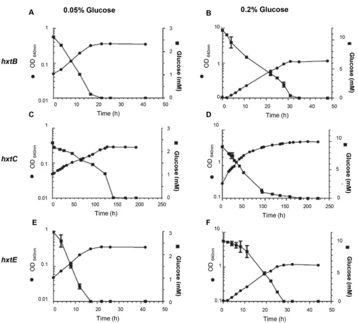
Subsequently, we concentrated our attention on the growth rate and glucose consumption of the HxtB, HxtC, and HxtE strains in YNB medium with 0.05 or 0.2% (w/v) glucose, during
Figure 1. Transmembrane helices prediction for the A. nidulans HxtB-E transporters (predicted via TMHMM; http:// www.cbs.dtu.dk/services/TMHMM/) and long C-terminal tails. The HxtB (A) and HxtD (C) contain 12 helices, while HxtC (B) and HxtE (D) contain 10 helices.
doi: 10.1371/journal.pone.0081412.g001
shake-flask aerobic batch cultivations. The S. cerevisiae strain expressing hxtD was excluded due to the absence of growth on glucose. S. cerevisiae expressing the hxtB or hxtE genes demonstrated highest growth rates at the glucose concentrations evaluated (Figure 6A, Table 2). The strain expressing the hxtC gene grew very slowly at 0.05 and 0.2 % (w/v) glucose (Figure 6B, Table 2) and no growth improvement was observed at higher glucose concentration (2%, w/v; data
not shown). These findings were confirmed by the glucose consumption profile (Figures 6A-B). In addition, no ethanol production was detected in any of the glucose concentrations tested, for any of the strains expressing hxtB, hxtC or hxtE
(data not shown).
The HxtB, -C, and –E transporters were also able to accept other sugars as substrates (Figure 4). Thus, to confirm this physiological data, we studied the uptake of [14C]glucose in the
Figure 2. The A. nidulans hxtA-E mRNA accumulation levels during growth in 0.1 or 1 % glucose. The wild-type strain was grown for 24 or 48 hours in MM liquid medium supplemented with either 0.1 or 1 % glucose. Real-time qPCR for hxtA-E (A-E) genes.
doi: 10.1371/journal.pone.0081412.g002
absence or presence of either fructose, mannose or galactose as potential transport competitors (Figure 7A-C). As expected, a 10-fold excess of unlabeled glucose drastically inhibited the transport of radiolabeled glucose in the S. cerevisiae cells expressing hxtB,-C, and –E (Figures 7A-C). A tenfold excess of unlabeled maltose, galactose, fructose and mannose were also able to inhibit to different levels (50 to 80 %) of radiolabeled glucose transport in the S. cerevisiae cells expressing hxtB,-C, and –E (Figures 7A-C). These results suggest that HxtB, -C, and –E have different substrate affinities.
To determine whether the mechanism by which HxtB, HxtC and HxtE transport glucose was by passive facilitated diffusion or active proton symport, we evaluated the sensitivity of each transporter to cyanide-m-chlorophenylhydrazone (CCCP), an uncoupler of transmembrane proton gradients. Upon the addition of CCCP, [14C]glucose uptake was affected in S.
cerevisiae cells expressing hxtB, -C, and –E, demonstrating a 80, 70 and 55 % reduction in the respective strains (Figure 7D). Taken together, these data suggested that HxtB, HxtC, and HxtE mediated glucose transport via active proton symport.
14C-glucose transport in the null mutants of hxtB-E
A. nidulans hxtB-E null alleles were generated using an in vivo S. cerevisiae fusion-based approach (see Materials and Methods). Several primary transformants that had homologous integration of either pyrG (hxtD) or pyroA (hxtB,-C,-E) at the
hxtB-E loci were isolated and one of each gene was selected for further characterization.
Since previous studies have described that glucose uptake in germinating conidia (incubated with 1.0 % glucose) is an energy dependent process [6], we evaluated the impact of each deletion on conidia germination at a this glucose
Figure 3. The A. nidulans hxtA-E mRNA accumulation levels during asexual and sexual development. Asexual spore development was synchronized by transferring a thin mycelial mat filtered from liquid culture (grown stationary at 37 °C for 24 hours; C=control) to an agar plate. To induce sexual development, we incubated the mycelia for 11 days (0–2 days: conidiophore development and asexual development; 2–11 days: cleistothecia development and sexual development; and 6–11 days: the presence of ascospores). Real-time qPCR for hxtA-E (A-E) genes.
doi: 10.1371/journal.pone.0081412.g003
concentration. In fact under these conditions, we found that glucose uptake in A. nidulans obeyed a single saturation kinetic with a Km = 10.7 ± 0.9 mM and a Vmax = 2.1 ± 0.1 µmol of
glucose h–1 per 2.5 × 10⁷ conidia (Figure 8). The ΔhxtB mutant
strain showed both a decreased affinity for glucose (Km = 25.3
± 3.4 mm) and a reduction in transport capacity (Vmax = 1.16 ± 0.06 µmol of glucose per hour per 2.5 × 10⁷ conidia; Figure
8A). The same behaviour was also observed for ΔhxtC mutant strain that showed both a decreased affinity for glucose and speed of transport compared to the wild-type strain (a Km =
26.0 ± 2.7 mm and a Vmax = 1.20 ± 0.05 µmol of glucose per hour per 2.5 × 10⁷ conidia (Figure 8B). Interestingly, in the case
of the ΔhxtD mutant, we also found alterations in the glucose uptake system, but this time an increase in Km and Vmax values
to 77.6 ± 12.1 µm and 6.1 ± 0.5 µmol of glucose per hour per 2.5 × 10⁷ conidia (Figure 8C). Despite the fact that the
introduction of HxtD to the EBY.VW4000S strain did not restore growth on glucose, its deletion in A. nidulans resulted in the loss of both glucose affinity and transport speed. The deletion of HxtE in A. nidulans resulted in a decrease in glucose affinity, but had little impact on the speed of transport (Figure 8D; Km =
23.1 ± 2.4 mm and a Vmax = 2.2 ± 0.1 µmol of glucose per hour per 2.5 × 10⁷ conidia)
Taking into consideration the impact of each HxtB-E deletions on glucose uptake, we evaluated the growth of the null hxtB-E mutants compared to the wild-type strain on solid MM supplemented with a single carbon sources, such as glucose, xylose, maltose, glycerol, mannose, fructose, acetate,
Figure 4. Comparative growth analyses of the S. cerevisiae cells expressing one of the four hxtB-E transporters. Tenfold dilutions (left to right) of S. cerevisiae cells (strain EBY.VW4000) expressing the indicated hxt cDNA or harbouring the empty expression vector were spotted on agar medium and incubated for 144 hour at 30 °C on plates containing the indicated carbon source.
doi: 10.1371/journal.pone.0081412.g004
rhamnose, casein, carboxymethylcellulose, inulin, guar, peptone, and pectin at 30, 37, and 44 °C. The four strains showed the same growth and conidiation as the wild-type strain under all the tested conditions (data not shown).
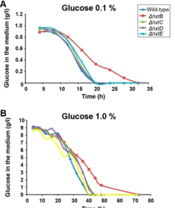
Finally, we investigated the glucose consumption by growing the wild-type and the ΔhxtB-E in liquid MM medium with either 0.1 % or 1% of glucose (Figure 9). When grown in MM+0.1 % glucose, all the strains showed a comparable rate in glucose consumption, except for the ΔhxtB mutant strain which showed a delayed consumption of glucose (Figure 9A). The same behaviour was observed for the ΔhxtB mutant strain in MM+1.0 % glucose (Figure 9B). The lower affinity for glucose in the
ΔhxtB mutant strain was emphasized by its increased resistance to carbon catabolite repression, when the wild-type and the mutant strains were grown in increasing concentrations of xylose+2 mM 2-deoxyglucose (2DG), which is a toxic glucose analogue (Figure S2). Taken together, these results suggest that the lack of hxtB results in the less efficient transport of glucose under low concentration.
Discussion
Understanding how filamentous fungi can transport and sense glucose is of a topic of substantial interest to industrial mycology. As a preliminary step to identify genes involved in these processes within A. nidulans, we characterized four genes that showed homology to other functionally characterized fungal glucose transporters. These four genes named hxtB-E are from the sugar porter subfamily of the MFS transporters. Despite hxtB-E demonstrating high identity with the S. cerevisiae glucose sensors, Snf3p and Rgt2p (data not shown), the absence of an extended cytosolic tail, essential for the intracellular signaling role in S. cerevisiae [15,16,20],
suggests that the A. nidulans proteins were transporters. However, HxtB-E possessed an extended cytosolic region within the center of the respective proteins, which could play a signaling role. Rgt2 and Snf3 have an approximately 50 amino acids long central region, while HxtB-E have 91, 129, 88, and 99 amino acids long central regions, respectively. The lack of a glutamine-rich region that acts as a mediator of protein-protein interaction, indicative of a signaling molecule, such as within the central cytosolic region of in RCO3 glucose sensor from N. crassa, implies otherwise [33,48]. Subsequently, biochemical and molecular assays enabled the classification of these Hxt proteins as glucose transporters. No transporters with extended cytosolic regions at either the N- or C-terminus were found in any Aspergilli whose genomes are available (data not shown). Thus, it is possible as suggested [49,50] that the global expression of transporters during the A. nidulans
isotropic growth phase, i.e., during spore germination [43,49–51] might operate as a general system for sensing solute availability.
The previously characterized high affinity glucose transporter HxtA was shown to be transcriptionally induced under glucose starvation and sexual development [42]. In contrast, hxtB-E
showed decreased mRNA accumulation during sexual development. In A. nidulans, hxtB, -C, -D, and -E also demonstrated increased mRNA accumulation when exposed to low glucose concentrations. The hexose transport-deficient S. cerevisiae strain (EBY.VW4000) has been an important tool for characterizing new hexose transporters of other fungi, such as four transporters from the hemibiotrophic plant pathogen
Colletotrichum graminicola (CgHXT2, CgHXT3, CgHXT4 and CgHXT5) [40] and TBHXT1 transporter from the ascomycete
Tuber borchii [36]. Subsequently, the ability of HxtB, -C, and –E to complement the growth defect of this strain on glucose,
Figure 5. Subcellular localization of hxtB-E in S. cerevisiae. Subcellular localization of hxtB-E in S. cerevisiae grown in: 0.2% glucose, 2% glucose or 2% maltose, was determined by fluorescence microscopy. Scale bar, 5 μm.
doi: 10.1371/journal.pone.0081412.g005
galactose, fructose, mannose, and sucrose, confirmed these
proteins to be hexose transporters, while competition experiments showed them to possess a higher affinity forglucose. In contrast, HxtD was unable to restore growth,
Figure 6. Evaluation of the growth rate, glucose consumption and kinetic parameters for S. cerevisiae cells (strain EBY.VW4000) expressing the hxtB (A and B) –C (C and D), and –E genes (E and F) grown on either 0.05 or 0.2 % glucose. doi: 10.1371/journal.pone.0081412.g006
Table 2. Specific growth rates (μ; h-1) of S. cerevisiae EBY.VW4000 strain expressing either hxtB, hxtC or hxtE genes grown
in YNB medium with glucose at 0.05% or 0.2% (w/v) as the only carbon source.
μ (h-1)
0.05% 0.2%
hxtB 0.118 ± 0.001 0.084 ± 0.002
hxtC 0.019 ± 0.002 0.016 ± 0.001
hxtE 0.110 ± 0.0002 0.098 ± 0.0011
doi: 10.1371/journal.pone.0081412.t002
despite being localized to the S. cerevisiae cell membrane, on any of the tested sugar sources. It is possible glucose transport by HxtD may involve transporter cooperation with other transporter proteins interacting with each other to produce specific phenotypes aiming to achieve a high rate of glucose influx [52]. Evidence of such transport cooperation has already been demonstrated when coexpressing Candida intermedia GXS1 glucose/xylose symporter and GXF1 glucose/xylose facilitator [53].
Few Aspergilli glucose transporters that have been functionally characterized [6,41-43]. As previously shown, D-Glucose uptake in germinating wild-type A. nidulans conidia is an energy-requiring process mediated by transport systems with differing affinities for glucose [6]: a low-affinity system (km ~ 1.4 mM), and intermediate-affinity system (Km~400mM), and a high-affinity system (Km ~ 16 mM). To investigate the involvement of hxtB-E in the glucose transport system and metabolism in A. nidulans we generated and characterized the corresponding null mutants. We were not able to see any
relevant phenotypic differences in these mutants when compared to the wild-type strain, except for the ΔhxtB mutant strain that showed a decreased rate of glucose consumption at low concentrations and an increased resistance to 2-DG. This indicates that although A. nidulans possesses other transporters capable of compensating for the absence of these transporters, the absence of HxtB has a measurable effect on glucose metabolism at low concentration. We detected a reduction on the glucose uptake for ΔhxtB-E mutants, with the loss of at least twice (ΔhxtB, ΔhxtC, and ΔhxtE) and seven-fold (ΔhxtD) affinity for glucose. Glucose uptake experiments using the S. cerevisiae strains expressing hxtB, -C, and –E, performed in the presence of CCCP, which blocks transmembrane proton gradients, strongly indicated that these
A. nidulans transporters also act as energy-dependent glucose/H+ symporters. Many other glucose transporters, identified in filamentous fungi, such as U. fabae HXT1 [35], glomeromycotan GpMST1 [37], Glomus MST2 [54], and four
Figure 7. Substrate specificities of the indicated Hxt transporters. Substrate specificities of HxtB (A), HxtC (B), and HxtE (C) were determined in S. cerevisiae cells (strain EBY.VW4000) expressing the respective cDNA. Relative transport levels were determined in the absence of a competitor or in the presence of a tenfold excess of unlabeled glucose or a tenfold excess of unlabeled maltose, galactose, fructose, or mannose (n=3, ±, standard deviation). The results are expressed as the percentage of inhibition of the transport of radiolabelled glucose. (D) Sensitivities of the HxtB-E transporters to the uncoupler CCCP in the absence or presence of 250 μM CCCP (n=3, ±, standard deviation).
doi: 10.1371/journal.pone.0081412.g007
transporters CgHXT1-4 from C. graminicola [40] have all been shown to be energy-dependent.
The presented study set out to improve the understanding of glucose metabolism in A. nidulans via studying the role of four possible glucose transporters. The data described provides a clear molecular and biochemical characterization of four genes involved with glucose uptake in A. nidulans.
Materials and Methods
Strains, media and culture methods
The genetic backgrounds of the A. nidulans strains used in this study are described in Table 3. Two basic types of media were used, i.e. complete and minimal. Three variants of complete media were used: YAG (2% w/v glucose, 0.5% w/v yeast extract, 2% w/v agar, trace elements), YUU (YAG supplemented with 1.2 g/liter [each] of uracil and uridine), and liquid YG or YG+UU medium with the same composition but without agar. A modified minimal media (original high-nitrate salts, trace elements, 2% w/v agar, pH 6.5) containing either 2%, 1%, 0.1% glucose (w/v) or no carbon source were used. Trace elements, vitamins, and nitrate salts were included as described by 55. A. nidulans strains were grown at 37°C unless indicated otherwise.
The S. cerevisiae sugar transporter knockout strain EBY.VW4000 (CEN.PK2-1C Δhxt1-17 Δstl1 Δagt1 Δydl247w Δyjr160c Δgal2) [45] was used for the in vivo complementation phenotype assays. The S. cerevisiae SC9721 strain (MATα his3-Δ200 URA3-52 leu2Δ1 lys2Δ202 trp1Δ63) acquired from the Fungal Genetic Stock Center (FGSC) was used for in vivo
recombination. Yeast strains were cultivated at 30°C in synthetic medium (SC, 0.67% Difco yeast nitrogen base without amino acids, 0.083 % amino acid drop out mix) supplemented with glucose or another specific carbon source.
Construction of A. nidulans hxtB-E null mutants
Standard genetic techniques for A. nidulans were used for all strain constructions and genetic transformation [55,56]. DNA manipulations were performed according to [57]. All PCR reactions were performed using Phusion High-Fidelity DNA polymerase (New England Biolabs), except for the amplification of whole cassettes where TaKaRa Ex Taq DNA Polymerase
(Clontech USA) was used. All the primers used in this work are listed in Table S1.
Deletion cassettes for ΔhxtB, C and E (AN6669, AN10891 and AN1797, respectively) were constructed by in vivo
recombination in S. cerevisiae as previously described [58]. Briefly, a construct consisting of a 1.0-kb region of the 5´-UTR
Figure 8. Km values for glucose in the A. nidulans wild-type and ΔhxtB-E mutant strains. Uptake rates for [14C] glucose
germinating conidia of the wild-type and ΔhxtB-E (A-D) mutant strains were determined at the indicated substrate concentrations at pH 7.0. Michaelis-Menten plots of the same data are shown (n=3, ±, standard deviation).
doi: 10.1371/journal.pone.0081412.g008
and 3´-UTRs (primers P1-6 and P7-12 respectively) flanking each of the target genes and the A. fumigatus pyroA gene (P13 and P14; used as a selective marker for pyridoxine prototrophy) was constructed by in vivo recombination in S. cerevisiae. The 5´-UTR, 3´-UTR and pyroA fragments plus the
linearized pRS426 vector cut with EcoRI and BamHI, were purified from agarose gel and transformed into S. cerevisiae
SC9721 strain using the lithium acetate method [59]. The external 5´-UTR Forward and 3´-UTR Reverse primers possessed cohesive ends with the vector pRS426 and the
Figure 9. The speed of glucose consumption during growth of the wild-type and ΔhxtB-E A. nidulans mutant strains in different glucose concentrations. The wild-type and mutant strains were grown in MM+0.1 % glucose (A) or MM+1.0 % glucose and the residual glucose concentration (g/l) was determined.
doi: 10.1371/journal.pone.0081412.g009
Table 3. Plasmids and A. nidulans and S. cerevisiae strains used in this work.
Plasmids/Strains Genotype Reference
pRS426 ampR lacZ URA3 [63,]
pCDA21 Zeo::pyr ampR [64]
pRH195 * pBluescript II SK+, TRP1, CEN6, ARSH4+ PHXT7-XKS1-THXT7 [65]
TNO2A3 pyroA4 pyrG89; chaA1; ΔnKuA::argB [60]
ΔhxtB pyroA4 pyrG89; chaA1; ΔnKuA::argB; ΔhxtB::pyroA4 This work
ΔhxtC pyroA4 pyrG89; chaA1; ΔnKuA::argB; ΔhxtC::pyroA4 This work
ΔhxtD pyroA4 pyrG89; chaA1; ΔnKuA::argB; ΔhxtD::pyrG This work
ΔhxtE pyroA4 pyrG89; chaA1; ΔnKuA::argB; ΔhxtE::pyroA4 This work
SC9721 MATa his 3-D200 URA 3-52 leu2D1 lys 2D202 trp 1D63 FGSC
EBY.VW4000
MATK leu2-3,112 ura3-52 trp1-289 his3-v1 MAL2-8c SUC2 hxt17v hxt13v : :loxP hxt15v: :loxP hxt16v: :loxP hxt14v : :loxP hxt12v: :loxP hxt9v: :loxP hxt11v: :loxP hxt10v: :loxP hxt8v : :loxP hxt514v: :loxP hxt2v: :loxP hxt367v : :loxP gal2v stl1v : :loxP agt1v : :loxP ydl247wv: :loxP yjr160cv: :loxP
[47]
*. The original vector pRH195 carries the XKS1 gene which was released after digestion with SpeI and SalI. The resultant vector without the XKS1 gene was used in this work for compl ementation assays.
doi: 10.1371/journal.pone.0081412.t003
internal primers 5´-UTR R and 3´-UTR F contained cohesive ends with 5´ and 3´sequence of pyro gene. All cassettes were PCR-amplified from genomic DNA extracted from the respective S. cerevisiae transformant, purified and used to transform A. nidulans strain TNO2a3 (ΔnkuA) strain [60], according to [56]. Transformants were scored for their ability to grow on minimal medium without pyridoxine and homologous integration confirmed by PCR (Figure S1). The deletion cassette for ΔhxtD was acquired from the FGSC. This cassette carried the pyrG gene as a selective marker for uridine and uracil prototrophy. The deletion cassette was PCR amplified using specific primers (P15 and P16)
S. cerevisiae genomic DNA was extracted by using the protocol described by 61. All cassettes were PCR-amplified using TaKaRa Ex Taq DNA Polymerase (Clontech) and used for transformation of wild-type A. nidulans strain TNO2a3 (ΔnkuA) strain [60] according to [56]. Transformants were scored for their ability to grow on minimal medium without uridine and uracil and checked by PCR to confirm their homologue integration.
RNA extraction and Real-time PCR reactions
Asexual spore development was synchronized by transferring a thin mycelial mat, filtered from liquid culture, to an agar plate. To induce sexual development, we incubated the mycelia for 11 days (0–2 days: conidiophore development and asexual development; 2–11 days: cleistothecia development and sexual development; 6–11 days: presence of ascospores). Mycelia were harvested, washed twice with dH2O and
immediately frozen in liquid nitrogen. The mycelia were then lyophilized, disrupted by grinding in liquid nitrogen and total RNA was extracted using the RNeasy Plant Mini Kit (Qiagen). To check RNA integrity, 10 µg of RNA was fractionated in 2.2 M formaldehyde, 1.2% agarose gel, stained with ethidium bromide, and visualized under UV-light. A total of 20 µg of RNA were treated with RNAse-free DNAse (Promega), purified with RNeasy Mini Kit (Qiagen) and then quantified on a NanoDrop 2000 Thermo Scientific). The SuperScript III First Strand Synthesis system (Invitrogen) and oligo(dT) primers were used for cDNA synthesis, according to the manufacturer’s protocol. All RT-qPCR reactions were performed using an ABI 7500 Fast Real-Time PCR System (Applied Biosystems) and Taq-Man™ Universal PCR Master Mix kit (Applied Biosystems). The RT-qPCR reactions and calculations were performed according to [62]. The primers and Lux™ fluorescent probes (Invitrogen) used in this work are described in Table S1.
Constructions for S. cerevisiae complementation assays
The sugar transporter deletion strain EBY.VW4000 was used for the S. cerevisiae complementation assays [45]. More than 20 sugar transporters and sensors including HXT1-17 and
GAL2 have been deleted from this strain [45]. For this reason, the strain is unable to grow on D-glucose, but it can grow on maltose, as a single carbon source. The hxtB-E ORFs were PCR amplified from A. nidulans cDNA using specific primers P29-30, P31-32, P33-34 and P35-36, respectively (Table S1). Note that the reverse primers included the stop codon. The
forward and reverse primers (P29-36) possessed cohesive ends for the modified vector pRH195 (under the control of the
HXT7 promoter and terminator) which was double digested with SpeI and SalI to liberate the XKS1 gene and linearize the vector. The purified linearized plasmid and PCR-amplified sugar transporter ORFs were transformed into S. cerevisiae
EBY.VW4000 strain by lithium acetate method [59], where they underwent in vivo recombination. Transformants were selected for tryptophan prototrophy on a SC medium supplemented with tryptophan and 2% maltose (SC-Trp). Genomic DNA of single colonies was isolated as described by 62 and the specific ORFs were PCR amplified using specific primers. Single transformed colonies were analyzed for their ability to grow on SC-Trp medium supplemented with either 2% glucose or 0.2% glucose.
The subcellular localization of hxtB-D in S. cerevisiae was checked by constructing hxtB-E::GFP cassettes. Thus, these ORFs were tagged with GFP at their C-terminal. The GFP gene was separated from the target ORF by the Spacer-GFP [63,]. Briefly, each ORF were PCR amplified from cDNA of the
A. nidulans A4 strain using primers P29 and 37 (hxtB), P31 and 38 (hxtC), P33 and 39 (hxtD) and finally P35 and 40 (hxtE) (Table S1). The forward primers included the Spacer-GFP sequence and omitted the stop codon. The forward and reverse primers possessed cohesive ends with the vector modified pRH195, which was double digested with SpeI and
SalI for linearization. The GFP gene containing the stop codon was amplified from pMCB17apx (kindly provided by Vladimir P. Efimov; primers P41 and P42) (Table S1) and the forward primer possessed cohesive ends with the modified vector pRH195 which was double digested with SpeI and SalI for linearization. In order to get in vivo recombination in S. cerevisiae, the linearized modified pRH195 plasmid was purified from agarose gel and transformed into S. cerevisiae
EBY.VW4000 strain with PCR-amplified sugar transporter ORFs and GFP gene by lithium acetate method [59]. The transformants were selected for tryptophan prototrophy on a SC medium supplemented with tryptophan and 2% maltose (SC-Trp). Genomic DNA of single colonies was isolated as described by 59 and the specific ORFs were PCR amplified using specific primers. Single transformed colonies were analyzed for their ability to grow on SC-Trp medium supplemented with either 2% glucose or 0.2% glucose.
Liquid growth conditions for S. cerevisiae
S. cerevisiae EBY.VW4000 expressing hxt-B, -C, -D and -E were grown in YNB medium supplemented with different carbon sources. The cultures were performed in flasks containing a 2:1 ratio of gas to liquid phase in an orbital shaker (160 rpm) at 26°C. Growth was monitored via OD measurements at 640 nm, while aliquots were taken at each time point to evaluate the concentration of glucose and ethanol in the medium.
Estimation of glucose and ethanol concentrations using S. cerevisiae strains
Glucose and ethanol concentrations in the media were assayed by high-performance liquid chromatography, using a
Refractive Index detector and a HyperREZ XP Organic Acids (8µm 100mm x 7.70mm) column at 57°C. The column was eluted with 2.5 mM of sulphuric acid at a flow rate of 0.7 ml/ min. The sample was injected through Gilson 234 auto-injector, with a retention time for glucose of 7.43 min and for ethanol of 15.29 min.
Estimation of cell dry weight for S. cerevisiae
The dry weight of S. cerevisiae cells (DW) from the different transformants was determined using pre-weighed aluminium caps. After removal of the medium by centrifugation, the cellular samples were washed with 4 volumes of ice-cold dH2O
and transferred to the aluminium caps for drying overnight at 80°C before being reweighed. Parallel samples varied by less than 1%.
Evaluation of free glucose in the extracellular culture medium
For the glucose uptake assay, a total of 1x 10⁷ spores were
inoculated in 100 ml of MM containing 1% or 0.1% glucose, maintained at 37°C in an orbital shaker. Aliquots (3 ml) of the supernatant were collected after 4, 8, 12, 16, 20, 24, and 48 hours and stored at -20°C. The enzymatic kit Glucose GOD-PAP Liquid Stable Mono-reagent (LaborLab Laboratories Ltda) was used to measure free glucose in the medium, according to the manufacturer’s specifications.
A. nidulans glucose uptake assay
Glucose uptake rates were measured by assaying the incorporation of D-[U-14C] glucose [289.0 mCi/mmol (10.693
GBq)/mmol] (Perkin Elmer Life Sciences) in germinating conidia at various D-glucose concentration according to [6] with modifications. Briefly, 1.2 x 10⁹ conidia were inoculated into
600 ml MM containing 1% D-glucose (w/v) as a carbon source. Incubation was carried out for 6 h at 37°C in an orbital shaker at 180 rpm. Germinating conidia were harvested by filtration over nitrocellulose filters (Fisherbrand) mounted in a vacuum manifold and washed twice with ice-cold water to eliminate traces of glucose. For glucose transport analysis, aliquots of 250 μl (of 2.5 x 10⁷ germinating conidia) containing D-glucose
[0.1-100mM] were dispensed into 2 ml tubes plus 1 μl of radiolabelled 14C-glucose (0.2 μCi) and incubated at 37°C. After
incubation for 30 to 60 seconds, uptake was immediately quenched by the addition of 1.5 ml ice-cold water and filtration over nitrocellulose filters (Fisherbrand) mounted in a vacuum manifold, followed by two consecutive washes with 1.5 ml of ice-cold water. Filters were subsequently transferred to 8 ml of ScintiSafeTM Econo1 scintillation liquid (Fisher Scientific). The D-[U-14C] glucose taken up by cells was measured using Tri-Carb® 2100TR Liquid Scintillation Counter.
CCCP assays
For CCCP (carbonylcyanide m-chlorophenylhydrazone) assays using S. cerevisiae strains, 500 ml of SC-Trp medium supplemented with 0.2 % glucose was incubated at 30°C with EBY.WV4000 strain harboring one of the hxtB, hxtC or hxtE
genes. Cultures started from an initial OD640 0.1 and were
grown until reached OD640 ~ 0.6. Cells were harvested by
centrifugation (4000 rpm), washed twice with 50 ml ice-cold water and resuspended in 1.250 ml of water. A total of 400 μl of cells was diluted in 800 μl of water and aliquots of 40 μl incubated at 30°C for 5 min to allow temperature equilibration. Subsequently, 10 μl of water containing 250 μM of CCCP were added 5 minutes before or concomitantly with 0.2 μCi of 14
C-glucose. Subsequently, the reaction was immediately stopped by quenching with 1.5 ml ice-cold water and filtration over nitrocellulose filters (Fisherbrand) mounted in a vacuum manifold, followed by two consecutive washes with 1.5 mL of ice-cold water. Filters were subsequently transferred to 8 ml of ScintiSafeTM Econo1 scintillation liquid (Fisher Scientific). The D-[U-14C] glucose taken up by cells was measured using Tri-Carb® 2100TR Liquid Scintillation Counter.
Supporting Information
Figure S1. PCR confirmation of homologue integrations for A. nidulans mutants ΔhxtB, ΔhxtC, ΔhxtD and ΔhxtE.
(TIF)
Figure S2. Growth phenotypes of A. nidulans wild-type and ΔhxtB-E mutants grown on different concentrations of xylose (A) or xylose plus 0.2 mM 2-deoxy-glucose (2-DG).
(TIF)
Table S1. Primers and probes used in this work.
(DOC)
Acknowledgements
We would like to thank Dr. Roberto do Nascimento Silva for the help in the Michaelis-Menten kinetics, Dr. Eckardt Boles for providing the EBY.VW4000 yeast strain, and the editor and the two anonymous reviewers for their comments and suggestions.
Author Contributions
Conceived and designed the experiments: GHG FR. Performed the experiments: TFR JFM MS ACC VLPB. Analyzed the data: GHG FR MHSG. Wrote the manuscript: GHG FR TFR NAB.
References
1. Ozcan S, Johnston M (1999) Function and regulation of yeast hexose transporters. Microbiol Mol Biol Rev 63: 554-569. Review. PubMed: 10477308.
2. Gancedo JM (1998) Yeast carbon catabolite repression. Microbiol Mol Biol Rev 62(2): 334-361. PubMed: 9618445.
3. Turcotte B, Liang XB, Robert F, Soontorngun N (2010) Transcriptional regulation of nonfermentable carbon utilization in budding yeast. FEMS Yeast Res 10(1): 2-13. doi:10.1111/j.1567-1364.2009.00555.x. PubMed: 19686338.
4. Bailey C, Arst HN (1975) Carbon catabolite repression in Aspergillus nidulans. Eur J Biochem 51: 573-577. doi:10.1111/j. 1432-1033.1975.tb03958.x. PubMed: 168071.
5. Dowzer CEA, Kelly JM (1991) Analysis of the creA gene, a regulator of carbon catabolite repression in Aspergillusnidulans. Mol Cell Biol 11: 5701-5709. PubMed: 1922072.
6. MacCabe AP, Miró P, Ventura L, Ramón D (2003) Glucose uptake in germinating Aspergillusnidulans conidia: involvement of the creA and sorA genes. Microbiology 149: 2129-2136. doi:10.1099/mic.0.26349-0. PubMed: 12904552.
7. Kim JH, Polish J, Johnston M (2003) Specificity and regulation of DNA binding by the yeast glucose transporter gene repressor Rgt1. Moll. Cell Biol 23: 5208-5216.
8. Kruckeberg AL (1996) The hexose transporter family of
Saccharomycescerevisiae. Arch Microbiol 166: 283–292. doi:10.1007/ s002030050385. PubMed: 8929273.
9. Boles E, Hollenberg CP (1997) The molecular genetics of hexose transport in yeasts. FEMS Microbiol Rev 21: 85–111. doi:10.1111/j. 1574-6976.1997.tb00346.x. PubMed: 9299703.
10. Reifenberger E, Boles E, Ciriacy M (1997) Kinetic characterization of individual hexose transporters of Saccharomycescerevisiae and their relation to the triggering mechanisms of glucose repression. Eur J Biochem 245: 324-333. doi:10.1111/j.1432-1033.1997.00324.x. PubMed: 9151960.
11. Kruckeberg AL, Walsh MC, Van Dam K (1998) How do yeast cells sense glucose? Bioessays 20(12): 972-976. PubMed: 10048296. 12. Rolland F, Winderickx J, Thevelein JM (2001) Glucose sensing
mechanisms in eukaryotic cells. Trends Biochem Sci 26(5): 310-317. doi:10.1016/S0968-0004(01)01805-9. PubMed: 11343924.
13. Marshall-Carlson L, Celenza JL, Laurent BC, Carlson M (1990) Mutational analysis of the SNF3 glucose transporter of Saccharomyces cerevisiae . Mol Cell Biol 10(3): 1105-1115. PubMed: 2406560. 14. Neigeborn L, Schwartzberg P, Reid R, Carlson M (1986) Null mutations
in the SNF3 gene of Saccharomyces cerevisiae cause a different phenotype than do previously isolated missense mutations. Mol Cell Biol 6: 3569–3574. PubMed: 3540596.
15. Ozcan S, Dover J, Rosenwald AG, Wölfl S, Johnston M (1996) Two glucose transporters in Saccharomycescerevisiae are glucose sensors that generate a signal for induction of gene expression. Proc Natl Acad Sci U S A 93: 12428–12432. doi:10.1073/pnas.93.22.12428. PubMed: 8901598.
16. Ozcan S, Dover J, Johnston M (1998) Glucose sensing and signaling by two glucose receptors in the yeast Saccharomyces cerevisiae. EMBO J 17: 2566–2573. doi:10.1093/emboj/17.9.2566. PubMed: 9564039.
17. Zaman S, Lippman SI, Zhao X, Broach JR (2008) How Saccharomyces responds to nutrients. Annu Rev Genet 42: 27-81. doi:10.1146/ annurev.genet.41.110306.130206. PubMed: 18303986.
18. Polish JA, Kim JH, Johnston M (2005) How the Rgt1 transcription factor of Saccharomycescerevisiae is regulated by glucose. Genetics 169: 583–594. doi:10.1534/genetics.104.034512. PubMed: 15489524. 19. Flick KM, Spielewoy N, Kalashnikova TI, Guaderrama M, Zhu Q et al.
(2003) Grr1 dependent inactivation of Mth1 mediates glucose induced dissociation of Rgt1 from HXT gene promoters. Mol Biol Cell 14: 3230– 3241. doi:10.1091/mbc.E03-03-0135. PubMed: 12925759.
20. Moriya H, Johnston M (2004) Glucose sensing and signaling in
Saccharomyces cerevisiae through the Rgt2 glucose sensor and casein kinase I. Proc Natl Acad Sci of the USA 101: 1572–1577. doi: 10.1073/pnas.0305901101. PubMed: 14755054.
21. Spielewoy N, Flick K, Kalashnikova TI, Walker JR, Wittenberg C (2004) Regulation and recognition of SCFGrr1 targets in the glucose and amino acid signaling pathways. Mol Cell Biol 24: 8994–9005. doi: 10.1128/MCB.24.20.8994-9005.2004. PubMed: 15456873.
22. Palomino A, Herrero P, Moreno F (2006) Tpk3 and Snf1 protein kinases regulate Rgt1 association with Saccharomyces cerevisiae
HXK2 promoter. Nucleic Acids Res 34: 1427–1438. doi:10.1093/nar/ gkl028. PubMed: 16528100.
23. Scarborough A (1970) Sugar transport in Neurosporacrassa. II. A second glucose transport system. J Biol Chem 245: 3985-3987. PubMed: 5492960.
24. Reifenberger E, Freidel K, Ciriacy M (1995) Identification of novel HXT genes in Saccharomycescerevisiae reveals the impact of individual hexose transporters on glycolytic flux. Mol Microbiol 16: 157–167. doi: 10.1111/j.1365-2958.1995.tb02400.x. PubMed: 7651133.
25. Wendell DL, Bisson LF (1993) Physiological characterization of putative highaffinity glucose transport protein Hxt2 of Saccharomycescerevisiae
by use of antisynthetic peptide antibodies. J Bacteriol 175: 7689–7696. PubMed: 8244939.
26. Wendell DL, Bisson LF (1994) Expression of high-affinity glucose transport protein Hxt2p of Saccharomycescerevisiae is both repressed and induced by glucose and appears to be regulated posttranslationally. J Bacteriol 176: 3730–3737. PubMed: 8206851. 27. Hirayarna T, Maeda T, Saito H, Shinozaki K (1995) Cloning and
characterization of seven cDNAs for hyperosmolarity-responsive (HOR) genes of Saccharomycescerevisiae . Mol Gen Genet 249: 127–138. doi:10.1007/BF00290358. PubMed: 7500933.
28. Rep M, Krantz M, Thevelein JM, Hohmann S (2000) The transcriptional response of Saccharomycescerevisiae to osmotic shock. J Biol Chem 275: 8290–8300. doi:10.1074/jbc.275.12.8290. PubMed: 10722658. 29. Diderich JA, Schepper M, van Hoek P, Luttik MAH, van Dijken JP et al.
(1999) Glucose Uptake Kinetics and Transcription of HXTGenes in Chemostat Cultures of Saccharomycescerevisiae. J Biol Chem 274: 15350–15359. doi:10.1074/jbc.274.22.15350. PubMed: 10336421. 30. Ko CH, Liang H, Gaber RF (1993) Roles of multiple glucose
transporters in Saccharomycescerevisiae . Mol Cell Biol 13: 638–648. PubMed: 8417358.
31. Theodoris G, Fong NM, Coons DM, Bisson LF (1994) High-copy suppression of glucose transport defects by HXT4 and regulatory elements in the promoters of the HXT genes in Saccharomyces cerevisiae . Genetics 137: 957–966. PubMed: 7982576.
32. Luyten K, Riou C, Blondin B (2002) The hexose transporters of
Saccharomyces cerevisiae play different roles during enological fermentation. Yeast 19: 713–726. doi:10.1002/yea.869. PubMed: 12185841.
33. Madi L, McBride SA, Bailey LA, Ebbole DJ (1997) rco3, a gene involved in glucose transport and conidiation in Neurospora crassa. Genetics 146: 499508.
34. Nehls U, Wiese J, Guttenberger M, Hampp R (1998) Carbon allocation in ectomycorrhizas: identification and expression analysis of an Amanita muscaria monosaccharide transporter. Mol Plant Microbe Interact 11(3): 167-176. doi:10.1094/MPMI.1998.11.3.167. PubMed: 9487692.
35. Voegele RT, Struck C, Hahn M, Mendgen K (2001) The role of haustoria in sugar supply during infection of broad bean by the rust fungus Uromycesfabae. Proc Natl Acad Sci U S A 98(14): 8133-8138. doi:10.1073/pnas.131186798. PubMed: 11390980.
36. Polidori E, Ceccaroli P, Saltarelli R, Guescini M, Menotta M et al. (2007) Hexose uptake in the plant symbiotic ascomycete Tuberborchii Vittadini: biochemical features and expression pattern of the transporter TBHXT1. Fungal Genet Biol 44(3): 187-198. doi:10.1016/j.fgb. 2006.08.001. PubMed: 17005424.
37. Schüssler A, Martin H, Cohen D, Fitz M, Wipf D (2006) Characterization of a carbohydrate transporter from symbiotic glomeromycotan fungi. Nature. 444(7121): 933-936. doi:10.1038/ nature05364. PubMed: 17167486.
38. Delgado-Jarana J, Moreno-Mateos MA, Benítez T (2003) Glucose uptake in Trichoderma harzianum: role of gtt1. Eukaryot Cell 2(4): 708-717. doi:10.1128/EC.2.4.708-717.2003. PubMed: 12912890. 39. Xie X, Wilkinson HH, Correa A, Lewis ZA, Bell-Pedersen D et al. (2004)
Transcriptional response to glucose starvation and functional analysis of a glucose transporter of Neurosporacrassa. Fungal Genet Biol 41: 1104-1119. doi:10.1016/j.fgb.2004.08.009. PubMed: 15531214. 40. Lingner U, Münch S, Deising HB, Sauer N (2011) Hexose transporters
of a hemibiotrophic plant pathogen: functional variations and regulatory differences at different stages of infection. J Biol Chem 286(23): 20913-20922. doi:10.1074/jbc.M110.213678. PubMed: 21502323. 41. Vankuyk PA, Diderich JA, MacCabe AP, Hererro O, Ruijter GJ et al.
(2004) Aspergillus niger mstA encodes a high-affinity sugar/H+ symporter which is regulated in response to extracellular pH. Biochem J 379(2): 375-383. doi:10.1042/BJ20030624.
42. Wei H, Vienken K, Weber R, Bunting S, Requena N, Fischer R (2004) A putative high-affinity hexose transporter, hxtA, of Aspergillusnidulans
is induced in vegetative hyphae upon starvation and in ascogenous hyphae during cleistothecium formation. Fungal Genet Biol 41: 148-156. doi:10.1016/j.fgb.2003.10.006. PubMed: 14732261. 43. Forment JV, Flipphi M, Ramón D, Ventura L, MacCabe AP (2006)
Identification of the mstE gene encoding a glucose inducible, low affinity glucose transporter in Aspergillusnidulans. J Biol Chem 281: 8339-8346. doi:10.1074/jbc.M508198200. PubMed: 16418173.
44. Ali SS, Nugent B, Mullins E, Doohan FM (2013) Insights from the fungus Fusariumoxysporium point to high affinity glucose transporters as targets for enhancing ethanol production from lignocelluloses. PLOS ONE 8: e54701. doi:10.1371/journal.pone.0054701. PubMed: 23382943.
45. Boylan TM, Mirabito PM, Willett CE, Zimmerman CR, Timberlake WE (1987) Isolation and physical characterization of three essential conidiation genes from Aspergillus nidulans. Mol Cell Biol 7: 3113– 3118. PubMed: 2823119.
46. Han KH, Han KY, Yu JH, Chae KS, Jahng KY, Han DM (2001) The nsdD gene encodes a putative GATA-type transcription factor necessary for sexual development of Aspergillus nidulans. Mol Microbiol 41: 299-309. doi:10.1046/j.1365-2958.2001.02472.x. PubMed: 11489119.
47. Wieczorke R, Krampe S, Weierstall T, Freidel K, Hollenberg CP et al. (1999) Concurrent knock-out of at least 20 transporter genes is required to block uptake of hexoses in Saccharomyces cerevisiae. FEBS Lett 464: 123–128. doi:10.1016/S0014-5793(99)01698-1. PubMed: 10618490.
48. Okazawa H (2007) Glutamine/Asparagine-Rich Regions in Proteins and Polyglutamine Diseases. Protein Reviews 6: 451-463. doi: 10.1007/978-0-387-36534-3_22.
49. Tazebay UH, Sophianopoulou V, Scazzocchio C, Diallinas G (1997) The gene encoding the major proline transporter of Aspergillusnidulans
is upregulated during conidiospore germination and in response to proline induction and amino acid starvation. Mol Microbiol 24: 105-117. doi:10.1046/j.1365-2958.1997.3201689.x. PubMed: 9140969. 50. Amillis S, Cecchetto G, Sophianopoulou V, Koukaki M, Scazzocchio C,
Diallinas G (2004) Transcription of purine transporter genes is activated during the isotropic growth phase of Aspergillusnidulans conidia. Mol Microbiol. 2004. APR;52(1): 205-216.
51. Fekete E, Karaffa L, Seiboth B, Fekete E, Kubicek CP, Flipphi M (2012) Identification of a permease gene involved in lactose utilisation in
Aspergillusnidulans. Fungal Genet Biol 49: 415-425. doi:10.1016/j.fgb. 2012.03.001. PubMed: 22445777.
52. Young E, Lee S-M, Alper H (2010) Optimising pentose utilisation in yeast: the need for novel tools and approaches. Biotechnol Biofuels 3: 24. doi:10.1186/1754-6834-3-24. PubMed: 21080929.
53. Leandro MJ, Spencer-Martins I, Gonçalves P (2008) The expression in
Saccharomycescerevisiae of a glucose/xylose symporter from Candida intermedia is affected by the presence of a glucose/xylose facilitator. Microbiology 154: 1646-1655. doi:10.1099/mic.0.2007/015511-0. PubMed: 18524919.
54. Helber N, Wippel K, Sauer N, Schaarschmidt S, Hause B et al. (2011) A versatile monosaccharide transporter that operates in the arbuscular mycorrhizal fungus Glomus sp is crucial for the symbiotic relationship
with plants. Plant Cell. 23(10): 3812-3823. doi:10.1105/tpc.111.089813. PubMed: 21972259.
55. Käfer E (1977) Meiotic and mitotic recombination in Aspergillus and its chromosomal aberrations. Adv Genet 19: 33-131. doi:10.1016/ S0065-2660(08)60245-X. PubMed: 327767.
56. Osmani SA, May GS, Morris NR (1987) Regulation of the mRNA levels of nimA, a gene required for the G2-M transition in Aspergillus nidulans. J Cell Biol 104(6): 1495-1504. doi:10.1083/jcb.104.6.1495. PubMed: 3294854.
57. Sambrook J, Russell DW (2001) Molecular Cloning: A Laboratory Manual, 3rd edn. New York: Cold Spring Harbor Laboratory Press. 58. Colot HV, Park G, Turner GE, Ringelberg C, Crew CM et al. (2006) A
high-throughput gene knockout procedure for Neurospora reveals functions for multiple transcription factors. Proc Natl Acad Sci U S A 103: 10352–10357. doi:10.1073/pnas.0601456103. PubMed: 16801547.
59. Schiestl RH, Gietz RD (1989) High efficiency transformation of intact yeast cells using single stranded nucleic acids as a carrier. Curr Genet 16: 339-346. doi:10.1007/BF00340712. PubMed: 2692852.
60. Nayak T, Szewczyk E, Oakley CE, Osmani A, Ukil L et al. (2006) A versatile and efficient gene-targeting system for Aspergillus nidulans. Genetics 172: 1557-1556. PubMed: 16387870.
61. Goldman GH, dos Reis Marques E, Duarte Ribeiro DC, de Souza Bernardes LA, Quiapin AC et al. (2003) Expressed sequence tag analysis of the human pathogen Paracoccidioides brasiliensis yeast phase: identification of putative homologues of Candida albicans
virulence and pathogenicity 988 genes. Eukaryot Cell 2: 34-48. doi: 10.1128/EC.2.1.34-48.2003. PubMed: 12582121.
62. Semighini CP, Marins M, Goldman MHS, Goldman GH (2002) Quantitative analysis of the relative transcript levels of ABC transporter
Atr genes in Aspergillusnidulans by Real-Time Reverse Transcripition-PCR assay. Appl Environ Microbiol 68: 1351–1357. doi:10.1128/AEM. 68.3.1351-1357.2002. PubMed: 11872487.
63. Teepe AG, Loprete DM, He Z, Hoggard TA, Hill TW (2007) The protein kinase C orthologue PkcA plays a role in cell wall integrity and polarized growth in Aspergillus nidulans. Fungal Genet Biol 44: 554-562. doi:10.1016/j.fgb.2006.10.001. PubMed: 17118679. 64. Christianson TW, Sikorski RS, Dante M, Shero JH, Hieter P (1992) A
two-component system that regulates an osmosensing MAP kinase cascade in yeast. Gene, V. 110: 119-122. doi: 10.1016/0378-1119(92)90454-W. PubMed: 1544568.
65. Chaveroche MK, Ghigo JM, D’Enfert C (2000) A rapid method for efficient gene replacement in the filamentous fungus Aspergillus nidulans. Nucleic Acids Res 28: E97-E104. doi:10.1093/nar/28.1.97. PubMed: 11071951.
66. Hector RE, Dien BS, Cotta MA, Qureshi N (2011) Engineering industrial Saccharomyces cerevisiae strains for xylose fermentation and comparison for switchgrass conversion. J Ind Microbiol Biotechnol 38: 1193–1202. doi:10.1007/s10295-010-0896-1. PubMed: 21107642.