www.biogeosciences.net/10/1155/2013/ doi:10.5194/bg-10-1155-2013
© Author(s) 2013. CC Attribution 3.0 License.
Biogeosciences
Geoscientiic
Geoscientiic
Geoscientiic
Geoscientiic
An unknown oxidative metabolism substantially contributes to soil
CO
2
emissions
V. Maire1,*, G. Alvarez1,2, J. Colombet3, A. Comby1, R. Despinasse4, E. Dubreucq5, M. Joly1, A.-C. Lehours3,
V. Perrier5, T. Shahzad1,**, and S. Fontaine1
1INRA, UR 874 UREP, 63100 Clermont-Ferrand, France
2Clermont Universit´e, VetAgro Sup, 63000, Clermont-Ferrand, France 3University of Clermont Ferrand, UMR 6023 LMGE, 63177 Aubi`ere, France 4INRA, UMR 1095 UBP, 63100 Clermont-Ferrand, France
5Montpellier SupAgro, UMR 1208 IATE, 34060 Montpellier, France
*present address: Department of Biological Sciences, Macquarie University, NSW 2109, Australia **present address: Department of Environmental Sciences, Government College University Faisalabad, 38000-Allama Iqbal Road Faisalabad, Pakistan
Correspondence to:S. Fontaine ([email protected]) Received: 31 May 2012 – Published in Biogeosciences Discuss.: 18 July 2012
Revised: 25 January 2013 – Accepted: 27 January 2013 – Published: 21 February 2013
Abstract.The respiratory release of CO2from soils is a
ma-jor determinant of the global carbon cycle. It is traditionally considered that this respiration is an intracellular metabolism consisting of complex biochemical reactions carried out by numerous enzymes and co-factors. Here we show that the endoenzymes released from dead organisms are stabilised in soils and have access to suitable substrates and co-factors to permit function. These enzymes reconstitute an extracellular oxidative metabolism (EXOMET) that may substantially con-tribute to soil respiration (16 to 48 % of CO2released from soils in the present study). EXOMETand respiration from liv-ing organisms should be considered separately when study-ing effects of environmental factors on the C cycle because EXOMETshows specific properties such as resistance to high temperature and toxic compounds.
1 Introduction
Knowledge of the metabolic pathways through which or-ganic carbon (C) is oxidised into carbon dioxide (CO2) in soils is fundamental to understanding the global C cycle and its interactions with climate (Fontaine et al., 2007; Heimann and Reichstein, 2008). Organic C is the building block of liv-ing organisms and is de facto highly stable in absence of
cata-lysts. For instance, the physical oxidation of one molecule of glucose requires a temperature greater than 500◦C. In soils, microorganisms set up a complex cascade of biochemical re-actions mediated by numerous enzymes making the oxidisa-tion of organic C possible at low temperature.
CO2. Respiration fluxes and ATP production in soils are usually positively correlated with activity of dehydrogenase endoenzymes (e.g., Casida et al., 1964), but only some of them carry a decarboxylase function (e.g., pyruvate dehydro-genase, oxoglutarate dehydrogenase) that cut the carboxyl function of organic C molecules to liberate CO2.
The function of respiration-carrying endoenzymes de-pends on various co-factors that must be regenerated (e.g., NAD+), on being located adjacent to other enzymes and on physiological properties of the cell (e.g., redox poten-tial) (Krebs, 1981; Rich, 2003). Given this complexity and the typical fragility of respiratory enzymes, it is tradition-ally considered that respiration (second step of C minerali-sation process) is strictly an intracellular metabolism process (Burns, 1982; Makoi and Ndakidemi, 2008). Besides, from an evolutionary point of view, soil microorganisms have no interest in provoking extracellular respiration since the en-ergy resulting from this respiration (ATP) may escape from the cell that provided enzymes (Allison et al., 2011). How-ever, some studies have shown that substantial emission of CO2 can persist for several weeks in soils where microbial life has been reduced by exposition to toxics (ClCH3, or-ange acridine) or irradiation to undetectable value (Peterson, 1962; Ramsay and Bawden, 1983; Lensi et al., 1991; Trevors, 1996; Kemmitt et al., 2008). Such emission of CO2 could not be explained by an activity of surviving microbes that would be present in low quantity in soils, unless considering unrealistically high cell respiratory activity. Neither could it be explained by the activity of extracellular enzymes previ-ously secreted by soil microorganisms since these enzymes only solubilize organic C and do not release CO2. To date, the cause of CO2emission in soils where microbial life has been minimised seems to be unknown and questions our ba-sic knowledge of biology.
The objective of this study is to understand the cause of CO2 emission in soils where microbial life has been suppressed. We hypothesise that an extracellular oxidative metabolism (EXOMET) can be reconstituted by respiratory endoenzymes released from dead organisms in soils and that this EXOMETcan substantially contribute to soil CO2 emis-sion. The manuscript is organised according to four key ques-tions:
1. Can an oxidative metabolism occur in an extracellular context?
2. How long can this EXOMET persist within soil? What is the role of soil particles for EXOMET-carrying en-zymes?
3. How much does EXOMETcontribute to CO2emission from living soil?
4. What are the specific properties of EXOMET?
First, to assess the possibility of an EXOMET, we in-cubated a cell-free yeast-extract containing respiratory
en-zymes with glucose in sterilised water and soil. The EXOMET induced by the yeast-extract was quantified by measuring CO2and O2 fluxes in water and soil microcosms. Second, the protective role of soil particles (minerals and humus) for respiratory enzymes was tested by incubating three en-zymes involved in glycolysis and the Krebs cycle in five top soils sampled from different regions of the world (Table 1). The third part of the manuscript is devoted to the quantifica-tion of EXOMETcontribution to the CO2emissions from five studied soils. This contribution was quantified with a method combining modelling and incubations of sterilised and non-sterilised soils. Finally, we studied some EXOMETproperties by incubating soils exposed to high temperature, high pres-sure and toxic compounds.
2 Materials and methods
2.1 Soil sampling and sterilisation
For each of the five studied sites (Table 1), twenty indepen-dent soil samples were collected from the 0–20 cm soil layer. Soil samples from each site were pooled to make a composite sample per site. Soil was sieved at 2 mm and was then used to determine pH, texture and organic matter content and to conduct incubation experiments. The five soils presented tex-tures from sandy-silted to silty-clay soils, pH ranging from 4.3 to 8.6 and three types of land use (grassland, forest and crops, Table 1). The soil from Theix was used for all inves-tigations, whereas the other soils were used to generalise the key findings of this study.
Some incubation experiments involved the use of sterilised soils. We tested different methods of soil sterilisation (γ -irradiation, autoclaving and dry heating) during preliminary investigations. Irradiation was chosen among other sterilis-ing methods for its efficiency to kill soil micro-organisms and for its moderate effect on soil enzymes (see S1 for de-tails of investigations on soil sterilisation). The preservation of soil enzymes was important for quantifying the EXOMET contribution to soil CO2emissions (see the Sect. 2.4). Soils were sterilised byγ-irradiation at 45 kGy (60Co, IONISOS, ISO14001, France).
2.2 Demonstration of EXOMETby incubating a cell-free
yeast-extract in sterilised water and soil
2.2.1 Production of cell-free yeast-extract
Pichia pastorisX33 (Invitrogen, Clare et al., 1991) cells were cultured at 28◦C in a 1.5 L capacity Applikon bioreactor con-taining synthetic medium described by Boze et al. (2001). This medium contained 80 µg L−1 D-biotin, 40 g L−1 glyc-erol and mineral solutions (FM21 and PTM1). The pH of the medium was regulated at 5 using a NH4OH solution (15 %,
Table 1.Site characteristics and soil properties.
Sites
Bugac Laqueuille Ponta Grossa Sorø Theix
Country Hungary France Brazil Denmark France
Land use Grassland Grassland Crops Beech forest Grassland
Clay/Silt/Sand (%) 10/5/85 19/55/26 14/6/80 10/22/68 26/25/49
pH 8.6 5.3 7.0 4.3 6.2
OM content (g C kg−1) 44.0 121.9 14.8 33.7 39.0
CEC (cmol+kg−1) 10.2 25.1 3.9 9.5 21.5
over 30 % saturation by stirring up to 1800 rpm and aeration (an injection of 1 L of air per litre of medium per minute). Data acquisition and bioprocess control were carried out us-ing the Applikon biocontroller software. Biomass concentra-tion of culture samples was determined by weighing washed cells after drying at 105◦C to constant weight.
The yeast culture was harvested at the end of the growth phase by centrifugation at 25 000x g for 10 min at 5◦C to yield a hard pellet. To remove the culture media, the sediment was washed and re-decanted three times in potassium phosphate buffer (0.1 M, pH=6.5 which corresponded to the pH of the Theix soil used for the experiment, Vyeast/Vbuffer=1/10). The sediment was re-suspended in a small volume of potassium phosphate buffer (Vyeast/Vbuffer=2/1) before cells were disrupted with a French pressure cell press (100 MPa, Vanderheiden et al., 1970). Unbroken cells and large cell debris were removed from the yeast extract by fourteen successive centrifugations at 25 000x gand 5◦C. The yeast extract was filtered under sterile conditions at 0.2 µm to obtain a cell-free extract. Di-rect microscopic observation confirmed sterility of the yeast extract (S1). The cell-free yeast extract was immediately in-corporated into water and soil microcosms under sterile con-ditions. The cell-free yeast extract contained 28.5 mg protein (Biuret method, Okutucu et al., 2007) and 0.77 unit L−1 of malate dehygrogenase (MDH).
2.2.2 Yeast extract incubation in sterilised water
and soil
The cell-free yeast extract (YE) was incubated in sterilised water (W) or in the irradiated-soil (S) of Theix (Table 1). Ex-perimental microcosms consisted of 5 mL of cell-free yeast extract and 1 mL of13C labelled glucose with or without 20 g ofγ-irradiated-soil (S+G+YE andW+G+YE treatments, respectively) placed in 250 mL airtight flasks. A second dose of glucose was applied after twenty days of incubation in or-der to determine the persistence of the EXOMET. Water with glucose and irradiated-soil with or without13C labelled glu-cose were incubated as control samples (W+G,SandS+G
treatments, respectively). The13C labelled glucose solution (δ13C=3712 ‰) contained 60 mg C-glucose mL−1and was
sterilised by filtration at 0.2 µm. Three replicates per treat-ment were prepared. Soils were incubated at a water poten-tial of−100 kPa. Water and soil microcosms were incubated at 30◦C for 53 days. The EXOMETinduced by the cell-free yeast extract was quantified by measuring the concentration of CO2,13CO2and O2in water and soil microcosms through-out the 53 days of incubation (see Sect. 2.6 for details on flux measurements). All manipulations were done under sterile conditions and the sterility of microcosms was verified af-ter 13 and 53 days of incubation through direct fluorescence microscopic observations and TSA-FISH method (see Sup-plement S1).
2.3 Soil stabilisation of oxidative metabolism enzymes
2.3.1 Enzyme incubation
Glucose
Glucose-6-phosphate
Phosphoglucono-δ-lactone
Fructose-6-phosphate
6-phospho gluconate
G6P dehydrogenase
NADP+ NADPH + H+
Mg2+ Mg2+
H2O
Lactonase
G6P isomerase
Glucose hexokinase ATP
ADP
GLYCOLYSIS
PENTOSE PHOSPHATE PATHWAY
Glucose
Glucose-6-phosphate
Phosphoglucono-δ-lactone
Fructose-6-phosphate
6-phospho gluconate
G6P dehydrogenase
NADP+ NADPH + H+
Mg2+ Mg2+
H2O
Lactonase
G6P isomerase
Glucose hexokinase
ATP
ADP GLYCOLYSIS
PENTOSE PHOSPHATE PATHWAY
Acetyl-CoA CoA-SH
CO2
NADH + H+ NAD
Pyruvate dehydrogenase Dihydrolipoyl transacetylase Dihydrolipoate dehydrogenase
Oxaloacetate Citrate
CoA-SH
Citrate synthase L-Malate
NADH + H+
NAD+
Malate dehydrogenase
KREBS CYCLE
Mg2+
B
. Glucose-6-phosphate isomerase (G6PI EC 5.3.1.9)A
. Glucose hexokinase (GHK E.C. 2.7.1.1)C
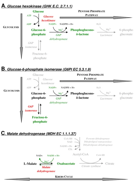
. Malate dehydrogenase (MDH EC 1.1.1.37)Fig. 1.Enzymatic reactions mediated by the two enzymes involved in glycolysis ((A)GHK: glucose hexokinase,(B)G6PI: glucose-6-phosphate isomerase) and the enzyme involved in the Krebs cycle ((C)MDH: malate dehydrogenase) used to quantify the protective role of soil particles on respiratory enzymes. This protective role was studied in the non-irradiated-soil from Theix. The principle of the enzymatic activity measurement was to set up a system of enzymatic reactions where the studied enzyme (red) was the limiting factor. For doing this, we added all substrates, cofactors and intermediary enzymes in excess (green). The activity of the studied enzyme (GHK, G6PI, MDH) was quantified by measuring the formation or the consumption of NADH by spectrometry. Reactions surrounding the studied enzymatic (grey) are given for information.
above experiment and by using the G6PI as a model enzyme (see Supplement S2).
2.3.2 Principle of enzyme activity measurement
The principle of the enzymatic activity measurement was to set up a system of enzymatic reactions where the stud-ied enzyme (red in Fig. 1) was the limiting factor (Nardi et
conditions as opposed to leaves. In particular, concentrations of buffer and Mg++solution were increased in order to better control soil pH and precipitate humic acids that could hamper the quantification of NADPH by spectrometry (both humic acids and NADPH absorb at 340 nm). For each of the three enzymes, the enzymatic reaction system and the method of enzyme activity measurement were as follows:
– GHK (Fig. 1a): Soil with or without GHK was amended with 300 µL of solution containing a buffer (Bicine-NaOH, 100 mM, pH=8.5), glucose (1.5 mM), NADP+ (1.5 mM), Mg++(32 mM) and glucose-6-phosphate de-hydrogenase (EC‘1.1.1.49, Sigma-Aldrich ref G5885). Production of NADPH following the chemical trans-formation of glucose in phosphoglucono-δ-lactone was measured by spectrometry at 340 nm.
– G6PI (Fig. 1b): Soil with or without G6PI was amended with 300 µL of solution containing a buffer (Bicine-NaOH, 100 mM, pH=8.5), fructose-6-phosphate
(1.5 mM), NADP+ (1.5 mM), Mg++ (32 mM) and glucose-6-phosphate dehydrogenase (2.5 U L−1). Production of NADPH following the chemical transfor-mation of fructose-6-phosphate to phosphoglucono-δ -lactone was measured by spectrometry at 340 nm.
– MDH (Fig. 1c): Soil with or without MDH was amended with 300 µL of solution containing a buffer (TRIS 100 mM, pH=6.7), oxaloacetate (1.5 mM), NADH (1.5 mM) and Mg++(32 mM). Consumption of NADH following the chemical transformation of ox-aloacetate to L-malate was measured by spectrometry at 340 nm.
2.3.3 Activity of total, soluble and soil-immobilised
enzymes
For each endoenzyme, activity of total, soluble and soil-immobilised enzymes was estimated at different times be-tween 20 min and 35 days of enzyme incubation in soils. At each harvest date, two independent sets of soils were used to quantify activity of total (TotalEnz) and soluble enzymes (SoluEnz). Activity of soil-immobilised enzymes was esti-mated by difference (ImmEnz=TotalEnz−SoluEnz). Ac-tivity of soluble enzyme (SolEnz) was quantified after their extraction from soil. For extraction, 80 mg soil samples were mixed with the 300 µl of the buffer solution containing sub-strates, co-factors and intermediate enzymes (see Sect. 2.3.2) and shaken during 5 min. Then, samples were centrifuged at 11 000x gduring 3 min. The supernatant containing sol-uble enzymes, co-factors and substrates was transferred into a micro-plate, where activity of soluble enzyme activity was measured during 3 min. The production rate of NADPH (for GHK and G6PI) and the consumption rate of NADH (for MDH) consecutive to the activity of soluble enzymes were quantified by spectrometry at 340 nm. For
measument of total enzyme activity (TotalEnz), the enzymatic re-action was made into the soil in presence of soluble and soil-immobilised enzymes. To this end, soil-enzyme mixture was incubated with substrates and cofactors (see Sect. 2.3.2) dur-ing 45 min. At different times between 5 and 45 min of incu-bation with substrates, independent samples were harvested and centrifuged at 11 000x g during 3 min. The NADPH concentration in the supernatant was determined by spec-trophotometry at 340 nm. The production of NADPH (for GHK and G6PI) or the consumption of NADH (for MDH) during the 45 min incubation of soil with substrates corre-sponded to the activity of total enzymes.
2.3.4 Kinetic analysis of total enzyme activity
Different exponential models were tested to fit the decrease in total enzyme activity (Total Enz) with time. The sole ex-ponential model able to explain kinetics of total enzymatic activity considers three pools of enzymes:
Y (t )=a·exp(−b·t )+c·exp(−d·t )+f·exp(−g·t ) (1) wheret is the time of incubation,a,candf represent the sizes andb,d andgthe decay rates of fast, intermediate and slow pools, respectively. Decay rate of the fast pool was so rapid that it could not be characterised precisely in this exper-iment. Consequently, sizeaand decay ratebof the fast pool were fixed to the amount of enzymatic activity lost during the first 20 min of incubation and to 1/5 min−1. Then, param-etersc,d,f andgof intermediate and slow pools of enzymes could be estimated using a classical nonlinear regression pro-cedure. We verified that uncertainty on decay rate of the fast pool has negligible effect on estimation of parameters of the intermediate and slow pools. This lack of effect on the es-timation of parameters is explained by the strong difference between decay rate of the fast pool and those of intermediate and slow enzyme pools (data not shown). The half-lives of the enzyme pools were calculated as (ln2)/(decay rate).
2.4 Contribution of EXOMETto soil respiration
Time (days)
0 10 20 30 40
0 10 20 30 40 50
Time (days)
0 10 20 30 40
R
a
ti
o
Aj
/
A0
(
%
)
0 10 20 40 50 60 70
Total enzyme Soluble enzyme Immobilized enzyme
A
Time (days)
0 10 20 30 40
R
a
ti
o
Aj
/
A0
(
%
)
0 10 20 30 40 50 60 70
B
C
r2= 0.99***
Y(t) = a eb t+ c ed t+ f eg t
GHK G6PI MDH
r2= 0.99***
Y(t) = a eb t+ c ed t+ f eg t r
2= 0.99***
Y(t) = a eb t+ c ed t+ f eg t
Fig. 2.Activity of three specific enzymes involved in glycolysis and the Krebs cycle following their incorporation in the non-irradiated-soil from Theix. Enzyme activity along time (Aj) is expressed as % of the initial activity of enzymatic solution (A0) applied to the soil. The
activities of total (dark circles), soluble (white circles) and immobilised enzymes (grey circles) are distinguished. Full, dashed and dotted lines represent the fit of the kinetic model on total enzyme activity, its confidence and predictive error intervals at 5 % P-level, respectively. (A)G6PI: glucose-6-phosphate isomerase;(B)GHK: glucose hexokinase;(C)MDH: malate dehydrogenase.
2.4.1 Model of carbon flux
In the model, CO2 emission from non-irradiated soil (Rni) is represented by the sum of living respiration (Rl) and EXOMET (Rx) (Eq. 2). After irradiation, a fractionk of Rl is converted to EXOMET via the soil stabilisation of respi-ratory enzymes released by the killed organisms. As a result, the sum of EXOMET(Rx)andk·Rldetermines CO2emission from irradiated soil (Ri) (Eq. 3). The model reads as follows
Rni=Rl+Rx (2)
Ri=k·Rl+Rx (3)
By fixing the sameRxin the irradiated and non-irradiated soils, the model assumes that irradiation has no effect on the pre-existent EXOMETRx. We discuss here two examples where irradiation could modifyRx. First, the γ-irradiation by denaturing part of soil enzymes could decreaseRx. In this case, it is easy to show that the EXOMETcontribution to soil CO2 emissions is underestimated by the model. Neverthe-less, this underestimation of EXOMETis likely to be moder-ate since the effect ofγ-irradiation on soil enzymes is typ-ically low (see review of McNamara et al., 2003). Second, by suppressing the microbial uptake of organic substrates, the irradiation could increase the availability of these sub-strates for EXOMET increasingRx. In this case, the current model would overestimate the EXOMETcontribution to soil CO2emissions. However, EXOMETand living respiration are not likely to be in competition for organic substrates. Indeed, EXOMET may have preferential access to organic substrate since EXOMET-carrying enzymes are adsorbed on soil parti-cles including organic matter. Moreover, most of the soil mi-crosites where EXOMETcan proceed are likely to deprived of microorganisms. Indeed, enzymes responsible for EXOMET may diffuse in most soil pores whereas living soil
microor-ganisms, due to their size, occupy less than 0.5 % of the soil pore space (Paul and Clark, 1989).
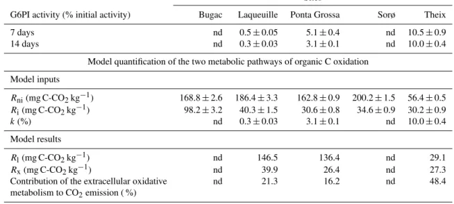
Parameterkvaries between 0–1 and depends on the frac-tion of respiratory enzymes released by dead organisms that is stabilised in irradiated-soils. In order to estimate k, we incubated the glucose-6-phosphate isomerase (G6PI) in the irradiated-soils of Theix, Ponta Grossa, Laqueuille, Sorøand Bugac for 14 days. This enzyme was selected because it showed the smallest stabilised fraction among the three res-piratory enzymes tested in the soil from Theix (Fig. 2). The total G6PI activity was measured throughout the incubation as described in the Sect. 2.3.2. For each soil, we fixed the parameterk equal to the fraction of stabilised G6PI activ-ity assuming that the reaction sustained by the G6PI was the limiting reaction for the EXOMET. Other enzymatic re-actions could be more limiting for extracellular oxidative metabolism. In this case, k would have a lower value and the calculated EXOMET would increase which would sig-nify that the model underestimates the contribution of the EXOMETto soil CO2emissions. Moreover, parameterkhas a limited bearing on the calculation of EXOMET. For example, for the soil from Theix, we calculated that a 50 % variation ofkcaused a 6 % deviation in EXOMET.
2.4.2 Soil incubation experiment
Experimental microcosms consisted of 30 g (dry mass basis) samples of fresh sieved soils placed in 250 mL flask. Sets of irradiated and non-irradiated soils were prepared for the five studied soils. Soils wereγ-irradiated as previously de-scribed. Soils were incubated in a dark chamber at 30◦C and with a water potential of −100 kPa for 21 days.
irradiated-soils under sterile conditions throughout the incu-bation. The sterility was verified by a combination of meth-ods including electron microscopy and molecular tracing of functional RNA-producing microorganisms (S1). The C flux model was constrained with CO2emissions from irradiated (Ri) and non-irradiated-soils (Rni)corresponding to the in-cubation period 13–21 days. The 0–13 day period was ex-cluded because soil stabilisation of respiratory enzymes re-quired several days (Fig. 2 and S3).
2.5 Some EXOMETproperties
Given that we consider EXOMET to be carried out by soil-protected slow-cycling enzymes rather than microbe-dependent living respiration with tight physiological con-straints, we predicted that EXOMET would show specific properties. First, we suggest that EXOMET persist in the long-term (>100 days) without microbial production of new enzymes. To test this hypothesis, the irradiated soil from Theix was incubated at 30◦C and with a water potential of
−100 kPa for 332 days. The CO2emission rate was regularly
measured during the incubation period in order to calculate the EXOMEThalf-life. Second, we suggest that EXOMET re-sist high temperature, pressure and toxic compounds. To test this hypothesis, we exposed the irradiated-soil from Theix to additional treatments: 150◦C for two hours, autoclaving (137◦C and 2.4 105Pa for 45 min) or chloroform vapours for 24 h. All microcosms were then incubated at 30◦C and a wa-ter potential of−100 kPa for 21 days. Four replicates were prepared for each treatment. The CO2 emission from soils exposed to temperature, pressure or toxic compounds were compared to that of control soil (only irradiated) in order to quantify treatments effects on EXOMET.
2.6 Flux measurements
In the incubation experiments where gas fluxes were stud-ied (Sects. 2.2, 2.4 and 2.5), two sets of microcosms were prepared in order to quantify gas exchange and to determine the13C content of released CO
2. In one set of microcosms, the released CO2was trapped in NaOH. The13C abundance of CO2was analysed by IRMS after precipitating carbonates with an excess of BaCl2and filtration. CO2and O2gas con-centrations in the other set of microcosms were measured by gas spectrometry (Agilent 3000 µGC, Agilent Technology, Lyon). Concentration of CO2in atmosphere of flasks was al-ways maintained below 3 % (17 % of O2in flasks where CO2 was trapped), except at two dates during the yeast extract in-cubation experiment. In these latter cases, concentration of CO2reached 4 % (day 10) and 22 % (day 0.75) signifying the diffusion of O2 may have limited the oxidative metabolism (EXOMET) induced by yeast extract.
2.7 Data analysis
All statistical tests were performed with the Statgraphics Plus software (Manugistics, Rockville, MD, USA). General lin-ear model (GLM) procedures, using the LSD method in post ANOVA multiple mean comparison tests, were employed to test effects of soil, treatment (irradiation, substrate and yeast-extract amendment, exposure to high temperature, autoclav-ing and toxic compounds) and time factors on CO2emission and O2consumption.When model residuals did not follow a normal distribution, the variables were log-transformed. Nonlinear regressions were used to analyse kinetics of CO2, O2concentration and enzymatic activity.
3 Results
3.1 Demonstration of EXOMETby incubating a cell-free
yeast-extract in sterilised water and soil
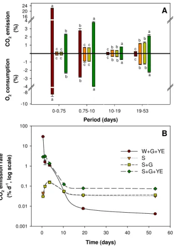
The sterility of microcosms was maintained throughout the experiment (S1). The emission of CO2 was null in wa-ter with glucose (W+G, data not shown). Despite sterili-sation, soils with and without glucose (S andS+G) still emitted CO2throughout the incubation (Fig. 3) confirming previous observations (Peterson, 1962; Ramsay and Baw-den, 1983; Lensi et al., 1991; Trevors, 1996). The supply of glucose had no effect on soil CO2 emissions (S versus
S+G). In contrast, the combined supply of glucose and cell-free yeast-extract containing respiratory enzymes trig-gered sudden and enormous respiration fluxes in water and soil (W+G+YE andS+G+YE, respectively). The CO2 emission fromW+G+YE andS+G+YE treatments re-spectively represented 725 and 72 times that of control soil (after eighteen hours of incubation, Fig. 3). The released CO2originated from added13C labelled glucose and unla-belled substrates present in yeast-extract and soil (S4). More-over, CO2 emissions mirrored the consumption of O2 for all treatments and dates, with the exception ofW+G+YE treatment at eighteen hours of incubation (respiratory quo-tient=2, Fig. 3a). In this particular case, the availability of O2was reduced, metabolic activity was intense and the pres-sure in microcosms rose to 1.25 atm, all of which indicated the presence of fermentative metabolism.
Period (days)
0-0.75 0.75-10 10-19 19-53
C O2 e m is s io n (% ) 0 1 2 3 4 16 20 24 W+G+YE S S+G S+G+YE O2 c o n s u m p ti o n (% ) -10 -8 -4 -3 -2 -1 0 a b a b
c c a
a b b
A
c c cb b c
a c
b b a cb b
a c b c b c c 0 a -4 -8 0
B
Time (days)0 10 20 30 40 50 60
C O2 e m is s io n r a te (% d -1 , lo g s c a le ) 0.001 0.01 0.1 1 10 100 W+G+YE S S+G S+G+YE B
Fig. 3. (A) Total (labelled+unlabelled) CO2 emission and O2 consumption from irradiated-soil (S); irradiated-soil+labelled glucose (S+G); irradiated-soil+labelled glucose+ yeast-extract (S+G+YE); water+labelled glucose+yeast-extract (W+G+YE) for four periods of incubation. Results are ex-pressed as % of microcosm atmosphere. Water with labelled glucose was also incubated as control, but CO2 emission from this control was null and not reported. Letters at each sampling date indicate the ANOVA-based differences at 5 % P-level among treatments. (B) Total (labelled+unlabelled) CO2 emission rate from water and soil microcosms. Symbols are the same as Fig. 3a.
EXOMETover 53 days (Fig. 3). Moreover, our results show that fermentative metabolism is another intracellular process that can be reconstituted outside the cell when the availability of O2for the EXOMETis low.
The EXOMETwas significantly higher in water than in soil during the first eighteen hours of incubation (W+G+YE ver-susS+G+YE, Fig. 3b), which can be explained by enzyme inhibitors present in soil (Burns, 1982). However, EXOMET continuously decreased with time in water whereas it sta-bilised in soil at a rate representing 247 % of CO2emissions from sterile control soil at day 20. The higher EXOMET in soil than in water after 20 days (Fig. 3b) cannot be explained by an exhaustion of C-substrate in water since the second glucose dose had no effect (S5). These results indicate that
Table 2.Size and half-time of fast, intermediate and slow pools of respiratory enzymes in the non-irradiated-soil from Theix. GHK: glucose hexokinase, G6PI: glucose-6-phosphate isomerase, MDH: malate dehydrogenase. Pool size and half-time were ob-tained by fitting the kinetic model presented in Sect. 2.3 to total enzyme activity (Fig. 2).
Enzyme
Fast pool Intermediate pool Slowpool
Size Half-life Size Half-life Size Half-life (%) (min) (%) (h) (%) (days)
GHK 35.8 <14 57.6 8.9 6.6 43.3 G6PI 66.4 <14 28.8 26.4 4.8 31.5 MDH 38.3 <14 47.6 17.1 14.1 495.1
the soil particles permit EXOMET persistence, possibly by preserving the respiratory enzymes from denaturation and proteolysis.
3.2 Soil stabilisation of oxidative metabolism enzymes
Kinetic analysis of total enzyme activities indicated the ex-istence of fast-, intermediate- and slow-cycling pools of en-zymes in the soil from Theix (Fig. 2, Table 2). Between 36 and 66 % of the initial enzymatic activity was lost within minutes following enzyme addition to the soil (the difference between initial activity and that 20 min after enzyme addi-tion, Fig. 2). The half-life of this fast pool was too fast to be determined precisely, but is<14 min (Table 2). Given the ra-pidity of their inactivation, enzymes of the fast-cycling pool were likely denatured by physico-chemical processes (Burns, 1982). A second pool of enzymes representing 29–58 % of the initial activity was inactivated more slowly with a half-life of 9–26 h. Soil proteolytic activity may have contributed to the degradation of this pool (Sarkar et al., 1989). Finally, 5–14 % of the initial enzymatic activity was retained in a highly stable form with a half-life of 32–495 days. Figure 2 shows that this long-term persistence of enzymatic activities exclusively relied on enzymes immobilised on soil particles (humus and minerals). These results confirm the protective role of soil particles for respiratory enzymes against physico-chemical denaturation and proteolysis. Our investigations in the other soils gave consistent results with 0.8–3.3 % of the initial enzymatic activity stabilised (S2).
3.3 Contribution of EXOMETto soil respiration
Table 3.Activity of glucose-6-phosphate isomerase (G6PI) and model quantification of CO2 emissions from living organisms (Rl)and extracellular oxidative metabolism (Rx)for the five studied soils.RniandRirepresent CO2emissions from non-irradiated and irradiated
soil, respectively, for the incubation period 13–21 days.kis the fraction ofRlconverted in extracellular oxidative metabolism (EXOMET) after irradiation. Contribution (%) of EXOMETto soil CO2emission was calculated asRx/Rni·100. nd = not determined
Sites
G6PI activity (% initial activity) Bugac Laqueuille Ponta Grossa Sorø Theix
7 days nd 0.5±0.05 5.1±0.4 nd 10.5±0.9
14 days nd 0.3±0.03 3.1±0.1 nd 10.0±0.4
Model quantification of the two metabolic pathways of organic C oxidation
Model inputs
Rni(mg C-CO2kg−1) 168.8±2.6 186.4±3.3 162.8±0.9 200.2±1.5 56.4±0.5
Ri(mg C-CO2kg−1) 98.2±3.2 40.3±1.5 30.6±0.8 34.6±0.9 30.2±0.9
k(%) nd 0.3±0.03 3.1±0.1 nd 10.0±0.4
Model results
Rl(mg C-CO2kg−1) nd 146.5 136.4 nd 29.1
Rx(mg C-CO2kg−1) nd 39.9 26.4 nd 27.3
Contribution of the extracellular oxidative nd 21.3 16.2 nd 48.4
metabolism to CO2emission ( %)
R
e
la
ti
v
e
C
O2
e
m
is
s
io
n
s
(I
rr
a
d
ia
te
d
s
o
il
/
N
o
n
i
rr
a
d
ia
te
d
s
o
il
)
0.0 0.2 0.4 0.6 0.8 1.0 1.2 1.4
Day 2 Day 6 Day 13 Day 21 a
b
c c ab c
d a
b
c
c a
b c
c a
b
c c
A B C D E
Ponta Grossa (Brazil)
Soro (Danemark)
Bugac (Hungary)
Laqueuille (France)
Theix (France)
Fig. 4. Relative CO2 emission between irradiated and non-irradiated soil for the five studied soils. Day 2, Day 6, Day 13 and Day 21 represent days of incubation. Letters indicate the differ-ences at 5 % level between measurements over time (lower case) and across soils (upper case) based on repeated-measures analysis of variance.
(Table 3 and S3). For the three soils wherek could be de-termined, modelling results indicated that EXOMET substan-tially contributed to CO2emissions from the non-irradiated-soils (Table 3). EXOMETwas responsible for 16, 21 and 48 % of CO2emissions from the soils of Ponta Grossa, Laqueuille and Theix, respectively. These unexpected contributions of EXOMETindicate the presence of a large quantity of respira-tory enzymes in soils outside cells.
3.4 Some EXOMETproperties
Significant CO2emissions were maintained throughout the 332 day incubation of irradiated-soil from Theix (Fig. 5). We calculated that the half-life of EXOMET was 165 days. This result confirms the idea that EXOMETcan persist in the long-term without microbial production of new enzymes. This per-sistence reflects soil stabilisation of respiratory enzymes re-leased by ancient generations of microbial populations.
Although the treatments applied to the irradiated-soil from Theix (high temperature, pressure and chloroform) are known to denature unprotected enzymes and be lethal for most microorganisms (Koffler et al., 1957; Kashefi and Lov-ley, 2003; Rainey et al., 2005; Lopez-Garcia, 2007), signif-icant CO2emissions persisted during the 21 day incubation period. Based on soil CO2emissions (Fig. 6), we estimated that 50, 20 and 10 % of EXOMET were resistant to chloro-form, 150◦C and autoclaving, respectively. Thus, soil par-ticles not only protect enzymes against denaturation (e.g., L¨ahdesm¨aki and Pnspanen, 1992), but also allow the mainte-nance of complex oxidative metabolism in conditions where life is generally impossible.
4 Discussion and perspectives
Time (days)
0 100 200 300
C
O2
e
m
is
s
io
n
r
a
te
(m
g
C
k
g
-1 s
o
il
d
-1 )
0,0 0,5 1,0 1,5 2,0 2,5 3,0 3,5 4,0
Y (t) = 3.68 (±0.09).e-[0.0042 (±0.0003).t ]
P < 0.001 r2 = 0.97 half-life = 165 days
Fig. 5.CO2emission rate from the irradiated-soil from Theix incu-bated during 238 days. Half-life (t1/2) was calculated from a simple
exponential model.
Time (days)
0 5 10 15 20 25
C
u
m
u
la
te
d
C
O2
e
m
is
s
io
n
(m
g
C
k
g
-1 s
o
il
)
0 25 50 75 100 125 150
Irradiated-soil + CHCl3
+ 150°C + Autoclaving
Fig. 6. Cumulated CO2 emission from the soil from Theix af-ter exposure to irradiation (Irradiated-soil), irradiation and 150◦C (+150◦C), irradiation and chloroform fumigation (+CHCl3), and irradiation and autoclaving (+Autoclaving).
to the protective role of soil particles. The concentration of endoenzymes around soil particles may facilitate exchanges of co-substrates and co-factors between enzymes, which are necessary for the cascade of reactions implied in oxidative metabolism. At the end of the respiratory chain, the transfer of electrons to O2may be carried out by soil particles due to their electric charge (Trevors et al., 1982).
Our results suggest that the EXOMET can substantially (16–48 %) contribute to soil CO2emissions. This unexpected contribution of EXOMET suggests the presence of a large quantity of respiratory enzymes in soils outside cells. This enzymatic pool may result from the long-term accumulation of enzymes released from dead organisms and stabilised on soil particles (Fig. 2). Thus, CO2emissions from soils are ap-parently driven by two major oxidative metabolisms: (1) the
well-known respiration of soil biota, (2) an EXOMETcarried out by enzymes released from dead organisms and stabilised by soil particles. These key results deserve further experi-ments that will verify assumptions of our model designed to quantify the EXOMET or ideally will propose an alternative independent method for this quantification.
Living respiration and EXOMETshould be considered sep-arately when studying effects of environmental factors on the C cycle because they do not likely obey to the same laws and respond differently to environmental factors. Soil microor-ganisms have tight physiological constraints comprising spe-cific environmental conditions (temperature, moisture, ab-sence of toxic compounds) and needs in energy and nutri-ents. These needs explain why soil CO2emissions are con-trolled by the availability of fresh energy-rich C and nutrients to soil microorganisms (Fontaine et al., 2003; Blagodatskaya et al., 2007; Pascault et al., 2013). In contrast, the EXOMET -carrying enzymes have few physiological constraints and are highly resistant to toxics, high temperature and pressure due to their protection by soil particles (section Sect. 3.4). This EXOMET can explain why part of soil CO2emissions is in-dependent of microbial biomass size, community structure or specific activity (Kemmitt et al., 2008) and is resistant to toxic compounds and extreme environments (chloroform, high temperature, pressure, and irradiation) (Peterson, 1962; Ramsay and Bawden, 1983; Lensi et al., 1991; Trevors, 1996; Kemmitt et al., 2008). Moreover, the EXOMETcould explain the flush of CO2 emission from soils submitted to freeze-thaw or wet-dry cycles since these treatments promote mi-crobial death and release of respiratory enzymes in soils (Henry, 2007; Borken and Matzner, 2009; Kim et al., 2012). Finally, the long-term persistence of EXOMET-carrying en-zymes (Figs. 2 and 5) signifies that current CO2emissions from soils partly depend on past microbial activities. This memory of soils suggests a delay between the modification of microbial activities and its consequence on soil respira-tion. Such a delay must be taken into account when studying effects of environmental factors on soil respiration to avoid an underestimation of modification of soil functioning.
(Table 3) despite the fact that it contains clay in proportion comparable to the soil from Theix and large amounts of or-ganic matter that can stabilise enzymes (Burns, 1982; Sarkar et al., 1989). However, the low pH of this soil (pH 5.3, Ta-ble 1) likely reduces the activity of G6PI since this enzyme (like all those of glycolysis) requires alkaline conditions to function. Thus, the capacity of soil particles to promote an EXOMET may not only rely on their ability to protect en-zymes, but also on the specific physico-chemical require-ments (e.g., pH) of each enzyme. This idea is supported by results from our incubation of three different endoenzymes in the soil from Theix. In this acid soil (pH=6.2), the pro-portion of stabilised enzyme activity was twice as high in the MDH case (pH optimal=6.7) compared to the G6PI and GHK cases (pH optimal=8.5) (Fig. 2).
Further experiments are necessary to better understand the molecular mechanisms at play and predict the EXOMET across soils. Processes leading to microbial death and re-lease of endoenzymes in soils (virus infection, predation, cell death due to stress like drought) must be identified. In marine and freshwater ecosystems, viral infection of micro-bial cells may be an important way by which endoenzymes are released in the environment since between 10 and 40 % of bacterial cells are lysed by viruses (see the review Wein-bauer, 2004; Colombet et al., 2006). To our knowledge, such quantification does not exist for terrestrial ecosystems. The endoenzymes stabilised by soil particles (humus, minerals) could be identified by soil proteomics (Wallenstein et al., 2008) whereas the chain of biochemical reactions involved in EXOMETcould be precisely characterised by metabolomics (Baudoin et al., 2001; Wallenstein et al., 2008, 2010). Fur-ther experiments should also explore possible interactions between cellular respiration and EXOMET, such as the com-petition for soluble substrate use. Nevertheless, the EXOMET is likely to be common in many soil types as consistent re-sults were found in the five contrasted studied soils. More-over, the reconstitution of intracellular metabolism outside the cell could occur in other environments (sediments, water) and may concern other metabolisms (methanisation, denitri-fication) since it should only require the presence of dead cells releasing endoenzymes. We, therefore, encourage re-search in other environments to quantify the role of intra-cellular metabolisms reconstituted outside the cell on global C cycle.
Supplementary material related to this article is available online at: http://www.biogeosciences.net/10/ 1155/2013/bg-10-1155-2013-supplement.pdf.
Acknowledgements. We thank D. Tourvieille and the “Plant and Pathogen” team of the UMR-INRA 1095 for their technical support. We thank P. Ambus, L. da Silveira Pontes, and L. Horv´ath who sampled the soil in Denmark, Brazil and Hungary, respec-tively; C. Mallet for culture analysis; S. Revaillot, O. Delfosse and J.-L. Ollier for chemical and isotopic analyses; L. Genestoux, C. Chabalier, F.-X. Sauvage, N. Duchatau for respiratory en-zyme activity analyses, G. Borrel for gas spectrometry; A. Eller, S. Grootemaat, J. Bloor for the revision of English; and V. Allard, P. Barr´e, S. Barot, R. Pilon, J. Bloor, M. Bahn, M. Wallenstein,
˚
A. Blagodatskaya and the two other anonymous referees for their constructive comments on previous versions of the manuscript. The research leading to these results received funding from the French Institutes INRA-INRIA (ARC-VitelBio), the French national agency ANR (DIMIMOS), le Conseil G´en´eral d’Auvergne (CPER 2007–2013) and the European Community’s 6th and 7th Framework Programmes (FP6 and FP7) under grant agreement no. 017841 (Nitro-Europe) and no. 226701 (CARBO-Extreme). All authors have refused to apply for the research bonus scheme set up by the French government in 2010 and have consequently no competing financial interest in this work. This work is dedicated to Waniya Shahzad and Gabin Maire.
Author contributions: This work originated from an idea of SF; VM, GA, ED and SF designed experiments; RD and MJ conducted soil incubations and counting of cultivable microorgan-isms; JC conducted electron microscopic observations of soils, ACL, RD, VM and SF performed gas spectrometry; ACL and VM performed fluorescence in situ hybridisation (TSA-FISH); VM and SF conducted the incubation experiment with endoenzymes; GA, VP, ED and SF conducted the incubation experiment with yeast-extract; SF built the model for estimating EXOMETin soils; VM, GA, TS and SF wrote the manuscript; and all authors took part in the interpretation of the results.
Edited by: M. Bahn
References
Allison, S. D., Weintraub, M. N., Gartner, T. B., and Waldrop, M. P.: Evolutionary economic principles as regulators of soil enzyme production and ecosystem function, edited by: Shukla, G. and Varma, A., Soil Enzymology, Springer-Verlag, Berlin, Germany, 229–243, 2011.
Baudoin, E., Benizri, E., and Guckert, A.: Metabolic fingerprint of microbial communities from distinct maize rhizosphere compart-ments, Eur. J. Soil Biol., 37, 35–93, 2001.
Blagodatskaya, E. V., Blagodatsky, S. A., Anderson, T. H., and Kuzyakov, Y.: Priming effects in Chernozem induced by glucose and N in relation to microbial growth strategies, Appl. Soil Ecol., 37, 95–105, 2007.
Borken, W. and Matzner, E.: Reappraisal of drying and wetting ef-fects on C and N mineralization and fluxes in soils, Glob. Change Biol., 15, 808–824, 2009.
Burns, R. G.: Enzyme-activity in soil – location and a possible role in microbial ecology, Soil Biol. Biochem., 14, 423–427, 1982. Burns, R. G. and Dick, R. P.: Enzymes in the Environment:
Ac-tivity, Ecology and Applications, Books in Soils, Plants, and the Environment Series, 86, CRC Press, 2002.
Casida, L. E., Klein, D. A., and Sartoro, T.: Soil dehydrogenase activity, Soil Sci., 98, 371–376, 1964.
Chr´ost, R. J.: Microbial Enzymes in Aquatic Environments, Brock Springer, Contemporary Bioscience Series, London, 1991. Clare, J. J., Rayment, F. B., Ballantine, S. P., Sreekrishna, K., and
Romanos, M. A.: High-level expression of tetanus toxin frag-ment c inPichia pastorisstrains containing multiple tandem in-tegrations of the gene, Bio/Tech., 9, 455–460, 1991.
Colombet, J., Sime-Ngando, T., Cauchie, H. M., Fonty, G., Hoff-mann, L., and Demeure, G.: Depth-related gradients of viral activity in lake Pavin, Appl. Environ. Microb., 72, 4440–4445, 2006.
Fontaine, S., Mariotti, A., and Abbadie, L.: The priming effect of organic matter: A question of microbial competition?, Soil Biol. Biochem., 35, 837–843, 2003.
Fontaine, S., Barot, S., Barre, P., Bdioui, N., Mary, B., and Rumpel, C.: Stability of organic carbon in deep soil layers controlled by fresh carbon supply, Nature, 450, 277–280, 2007.
Heimann, M. and Reichstein, M.: Terrestrial ecosystem carbon dy-namics and climate feedbacks, Nature, 451, 289–292, 2008. Henry, H. A. L.: Soil freeze-thaw cycle experiments: trends,
methodological weakness and suggested improvements, Soil Biol. Biochem., 39, 977–986, 2007.
Kashefi, K. and Lovley, D. R.: Extending the upper temperature limit for Life, Science, 301, p. 934, 2003.
Kemmitt, S. J., Lanyon, C. V., Waite, I. S., Wen, Q., Addiscott, T. M., Bird, N. R. A., O’Donnell, A. G., and Brookes, P. C.: Min-eralization of native soil organic matter is not regulated by the size, activity or composition of the soil microbial biomass-a new perspective, Soil Biol. Biochem., 40, 61–73, 2008.
Kim, D.-G., Vargas, R., Bond-Lamberty, B., and Turetsky, M. R.: Effects of soil rewetting and thawing on soil gas fluxes: a review of current literature and suggestions for future research, Biogeo-sciences, 9, 2459–2483, doi:10.5194/bg-9-2459-2012, 2012. Koffler, H., Mallett, G. E., and Adye, J.: Molecular basis of
biolog-ical stability to high temperatures, P. Natl. Acad. Sci. USA, 43, 464–477, 1957.
Krebs, H.: Introductory remarks, Philos. T. Roy. Soc. B, 293, 3–4, 1981.
L¨ahdesm¨aki, P. and Pnspanen, R.: Soil enzymology: Role of protec-tive colloid systems in the preservation of exoenzyme activities in soil, Soil Biol. Biochem., 24, 1173–1177, 1992.
Lensi, R., Lescure, C., Steinberg, C., Savoie, J. M., and Faurie, G.: Dynamics of residual enzyme-activities, denitrification potential, and physicochemical properties in a gamma-sterilised soil, Soil Biol. Biochem., 23, 367–373, 1991.
Lopez-Garcia, P.: Habitability: the point of view of a biologist, in: Lectures in Astrobiology, edited by: Gargaud, M., Martin, H., and Claeys, P., Adv. Astrobio. Biogeo., Springer-Verlag Berlin, Berlin, 2, 221–237, 2007.
Makoi, J. H. R. and Ndakidem, P. A.: Selected soil enzymes: exam-ple of their potential roles in the ecosystem, Afr. J. Biotechnol., 7, 181–191, 2008.
McNamara, N. P., Black, H. I. J., Beresford, N. A., and Parekh, N. R.: Effects of acute gamma irradiation on chemical, physical and biological properties of soils, Appl. Soil Ecol., 24, 117–132, 2003.
Nardi, S., Muscolo, A., Vaccaro, S., Baiano, S., Spaccini, R., and Piccolo, A.: Relationship between molecular characteristics of soil humic fractions and glycolytic pathway and krebs cycle in maize seedlings, Soil Biol. Biochem., 39, 3138–3146, 2007. Okutucu, B., Dincer, A., Habib, O., and Zihnioglu, F.: Comparison
of five methods for determination of total plasma protein concen-tration, J. Biochem. Bioph. Meth., 70, 709–711, 2007.
Pascault, N., Ranjard, L., Kaisermann, A., Bachar, D., Christen, R., Terrat, S., Mathieu, O., L´evˆeque, J., Mougel, C., Henault, C, Le-manceau, P., P´ean, M., Boiry, S., Fontaine, S., and Maron, P.-A.: Stimulation of different functional groups of bacteria by various plant residues as a driver of soil priming effect, Ecosystems, in press, 2013.
Paul, E. A. and Clark, F. E.: Soil Microbiology and Biochemistry. Academic Press Inc., San Diego, CA., USA, 273 pp., 1989. Peterson, G. H.: Respiration of soil sterilized by ionizing radiations,
Soil Sci., 94, 71–74, 1962.
Prescott, L., Harley, J., and Klein, D.: Microbiology, McGraw-Hill Compagnies, New York, 2002.
Quiquampoix, H.: Mechanisms of protein adsorption on surfaces and consequences for extracellular enzyme activity in soil, in: Soil Biochem., edited by: Bollag, J.-M. and Stotzky, G., Marcel Dekker, New York, 10, 171–206, 2000.
Rainey, F. A., Ray, K., Ferreira, M., Gatz, B. Z., Nobre, M. F., Baga-ley, D., Rash, B. A., Park, M. J., Earl, A. M., Shank, N. C., Small, A. M., Henk, M. C., Battista, J. R., Kampfer, P., and da Costa, M. S.: Extensive diversity of ionizing-radiation-resistant bacte-ria recovered from Sonoran Desert soil and description of nine new species of the genusDeinococcusobtained from a single soil sample, Appl. Environ. Microb., 71, 5225–5235, 2005. Ramsay, A. J. and Bawden, A. D.: Effects of sterilization and
stor-age on respiration, nitrogen status and direct counts of soil bac-teria using acridine-orange, Soil Biol. Biochem., 15, 263–268, 1983.
Rich, P. R.: The molecular machinery of Keilin’s respiratory chain, Biochem. Soc. T., 31, 1095–1105, 2003.
Sarkar, J. M., Leonowicz, A., and Bollag, J. M.: Immobilization of enzymes on clays and soils, Soil Biol. Biochem., 21, 223–230, 1989.
Sinsabaugh, R. L., Hill, B. H., and Follstad Shah, J. J.: Ecoenzy-matic stoichiometry of microbial organic nutrient acquisition in soil and sediment, Nature, 462, 795–798, 2009.
Sinsabaugh, R. L. and Follstad Shah, J. J.: Ecoenzymatic stoichiom-etry and ecological theory, Annual Review of Ecology, Evolu-tion, and Systematics, Annu. Rev. Ecol., 43, 313–343, 2012. Trevors, J. T.: Sterilization and inhibition of microbial activity in
soil, J. Microbiol. Meth., 26, 53–59, 1996.
Trevors, J. T., Mayfield, C. I., and Inniss, W. E.: Measurement of electron-transport system (Ets) activity in soil, Microb. Ecol., 8, 163–168, 1982.
Vanderheiden, G. J., Fairchild, A. C., and Jago, G. R.: Construction of a laboratory press for use with the French pressure cell, Appl. Microbiol. 19, 875-877, 1970.
en-zymes, Soil Biol. Biochem., 40, 2098–2106, 2008.
Wallenstein, M. D., Hess, A. M., Lewis, M. R., Steltzer, H., and Ayres, E.: Decomposition of aspen leaf litter results in unique metabolomes when decomposed under different tree species, Soil Biol. Biochem, 42, 484–490, 2010.