Identification of OmpR-Family Response
Regulators Interacting with Thioredoxin in
the Cyanobacterium
Synechocystis
sp. PCC
6803
Taro Kadowaki1, Yoshitaka Nishiyama1, Toru Hisabori2,3, Yukako Hihara1,3*
1Graduate School of Science and Engineering, Saitama University, Saitama, Japan,2Chemical Resources Laboratory, Tokyo Institute of Technology, Yokohama, Japan,3CREST, Japan Science and Technology Agency (JST), Saitama, Japan
*hihara@molbiol.saitama-u.ac.jp
Abstract
The redox state of the photosynthetic electron transport chain is known to act as a signal to regulate the transcription of key genes involved in the acclimation responses to environ-mental changes. We hypothesized that the protein thioredoxin (Trx) acts as a mediator con-necting the redox state of the photosynthetic electron transport chain and transcriptional regulation, and established a screening system to identify transcription factors (TFs) that in-teract with Trx. His-tagged TFs and S-tagged mutated form of Trx, TrxMC35S, whose active site cysteine 35 was substituted with serine to trap the target interacting protein, were co-ex-pressed inE.colicells and Trx-TF complexes were detected by immuno-blotting analysis. We examined the interaction between Trx and ten OmpR family TFs encoded in the chro-mosome of the cyanobacteriumSynechocystissp. PCC 6803 (S.6803). Although there is a highly conserved cysteine residue in the receiver domain of all OmpR family TFs, only three, RpaA (Slr0115), RpaB (Slr0946) and ManR (Slr1837), were identified as putative Trx targets. The recombinant forms of wild-type TrxM, RpaA, RpaB and ManR proteins from S.6803 were purified following over-expression inE.coliand their interaction was further as-sessed by monitoring changes in the number of cysteine residues with free thiol groups. An increase in the number of free thiols was observed after incubation of the oxidized TFs with Trx, indicating the reduction of cysteine residues as a consequence of interaction with Trx. Our results suggest, for the first time, the possible regulation of OmpR family TFs through the supply of reducing equivalents from Trx, as well as through the phospho-transfer from its cognate sensor histidine kinase.
Introduction
Photosynthetic organisms rearrange their cellular components to balance the supply and con-sumption of photosynthetically derived energy in response to changes in environmental factors,
OPEN ACCESS
Citation:Kadowaki T, Nishiyama Y, Hisabori T, Hihara Y (2015) Identification of OmpR-Family Response Regulators Interacting with Thioredoxin in the CyanobacteriumSynechocystissp. PCC 6803. PLoS ONE 10(3): e0119107. doi:10.1371/journal. pone.0119107
Academic Editor:Franck Chauvat, CEA-Saclay, FRANCE
Received:November 21, 2014
Accepted:January 28, 2015
Published:March 16, 2015
Copyright:© 2015 Kadowaki et al. This is an open access article distributed under the terms of the
Creative Commons Attribution License, which permits unrestricted use, distribution, and reproduction in any medium, provided the original author and source are credited.
Data Availability Statement:All relevant data are within the paper and its Supporting Information files.
such as light intensity and nutrient supply [1]. For example, there are many reports indicating that transcriptional regulation mediated by the redox state of the photosynthetic electron trans-port chain is crucial for such acclimation responses [2,3], although the underlying molecular mechanisms are largely unknown. It has been proposed that the redox state of the plastoquinone (PQ) pool is a key factor in the regulation of gene expression in the chloroplasts and nuclei of both land plants and green algae [4,5]. However, DNA microarray analysis of gene expression in the cyanobacteriumSynechocystissp. PCC 6803 (hereafter referred to as S.6803) in the pres-ence and abspres-ence of inhibitors of photosynthetic electron transport has revealed that the redox state of components located downstream of the PQ pool, such as the cytochromeb6/fcomplex
and ferredoxin/thioredoxin (Trx) system, is likely to be more critical for transcriptional regula-tion than that of the PQ pool itself [6]. It has been reported that inSynechococcus elongatusPCC 7942 (S.7942), the induction ofpsbAII/IIIgene expression under high-light (HL) conditions could be mimicked by adding the thiol-specific reductant dithiothreitol (DTT) under low-light (LL) conditions, and the opposite effects were observed by adding a thiol-specific oxidant, such as diamide, under high-light (HL) conditions [7]. These data suggest that the expression of the
psbAII/IIIgenes was regulated by, and dependent on, the redox state of the thiols.
Previously, we determined that a small LuxR-type transcriptional factor (TF), PedR, is in-volved in transcriptional regulation that is dependent on photosynthetic electron transport in S.6803 [8]. We observed that PedR induces or suppresses the expression of its target genes under LL conditions, but it is transiently inactivated, with a concomitant conformational change, upon exposure to HL conditions. This conformational change and inactivation under HL were not observed in the presence of photosynthetic inhibitors, or in mutants deficient in the Trx reduction system. Furthermore, a pull-down assay using His-tagged PedR protein and the crude extract of S.6803 revealed that TrxM (Slr0623) interacts with PedR [9]. Based on these observations, we proposed that increase in the availability of reducing equivalents at the acceptor side of photosystem I is transmitted to PedR via interaction with Trx, leading to a transient conformational change and inactivation of PedR.
Our working hypothesis is that Trx may be a key for transcriptional regulation that depends on photosynthetic electron transport in S.6803. Trx is a ubiquitous redox mediator that regu-lates the activity of various enzymes through dithiol-disulfide-exchange reactions [10] and pro-vides reducing equivalents for anti-oxidative stress proteins, such as peroxiredoxin [11]. It is possible that Trx interacts with PedR to couple photosynthetic activity and transcriptional reg-ulation. However, only 8 genes were identified as the member of PedR regulon and the regula-tory mechanism of most genes whose expression is dependent on photosynthetic activity [6] is poorly understood, so there may also be other TFs that interact with Trx proteins.
In the genome of S.6803, there are four genes encoding Trx (slr0623for TrxM,slr1139for TrxX,slr0233for TrxY andsll1057for TrxZ) [12]. Of these, TrxM is the most abundantly ex-pressed Trx and it has been reported to comprise 2% of the total soluble proteins of glucose-tol-erant wild-type cells [13]. To date, several groups have attempted to identify target proteins of each Trx species in S.6803 using proteomic approaches [14–16]. However, there have been no reports to date of the isolation of a TF as a Trx interaction partner, probably due to the techni-cal difficulty identifying TFs by mass spectrometry analysis as they are typitechni-cally present in low abundance in S.6803 cells.
In the present study, we report a new screening system usingEscherichia colico-expression strains to detect the interaction between Trx and a TF. We examined interactions between TrxM and ten OmpR-type response regulators that are encoded in the S.6803 genome and identified RpaA (Slr0115), RpaB (Slr0947) and ManR (Slr1837) as new candidate TrxM inter-acting partners. We further confirmed their interaction with TrxM by the experiments using the corresponding recombinant proteins.
Materials and Methods
Bacterial strains and growth conditions
E.coliXL1-blue cells were used for routine cloning and Origami2 (DE3) cells (Novagen) for protein expression. Bacterial cultures were grown in TB or 2xYT medium at 37°C. When nec-essary, antibiotics were added at the following concentrations: ampicillin (100μg mL-1), kana-mycin (20μg mL-1) and spectinomycin (20μg mL-1).
Construction of
E
.
coli
strains expressing S-tagged TrxM
C35Sand/or
His-tagged TF
The coding region of each TF gene to be studied was amplified by PCR using the primers listed inTable 1, and cloned into the pT7Blue T-vector (Novagen). The PCR fragments ofnrsR
(sll0797) andrpaA(slr0115) were excised from the pT7Blue vector withNdeI andXhoI and subcloned into the same restriction sites in the pET28a vector (Novagen) to express proteins with an N-terminal 6 x His tag. The PCR fragments ofrre28(sll0396),rre3(sll0649),copR
(sll0789),rre37(sll1330),sphR(slr0081),ccaR(slr1584) andmanR(slr1837) were excised from the pT7Blue vector withNdeI andXhoI and subcloned into the same restriction sites in the pET21a vector (Novagen) to express proteins with a C-terminal 6 x His tag. The construct to express RpaB with an N-terminal 6 x His tag was made as described previously [17]. Each ex-pression construct was transformed into Origami2 (DE3) competent cells (Novagen) using the heat shock method to yield the corresponding TF strain.
The coding region oftrxM(slr0623) was amplified by PCR using the primers listed in
Table 1. The PCR fragment was cloned into the pT7Blue T-vector. A C35S mutation was intro-duced into thetrxMcoding sequence by PCR using a KOD-plus-mutagenesis kit (TOYOBO). The resultingtrxMC35Sfragment was excised from the pT7Blue vector withNdeI andXhoI
and subcloned into the same restriction sites in the pCDF-duet vector (Novagen) to express TrxMC35Swith a C-terminal S-tag. The resulting construct was transformed into Origami2
(DE3) competent cells and competent cells of the TF strains described above, to yield the Trx strain and the Trx-TF strains, respectively.
Detection of the interaction between S-tagged TrxM
C35Sand His-tagged
TF in
E
.
coli
cells
E.coliOrigami2 (DE3) strains, harboring each expression construct, were precultured in 2 mL of the TB medium containing ampicillin, kanamycin or spectinomycin (concentrations as above) at 37°C overnight. The preculture was seeded into 4 mL of the 2×YT medium, cultured at 37°C for 3 h, and His-tagged TF and TrxMC35Swere induced by addition of 100μM
Co-purification of S-tagged TrxM
C35Sand His-tagged TF from
E
.
coli
cells using nickel resin under denaturing conditions
Induction of recombinant protein expression by IPTG and preparation of the cell lysate were performed as described above. N-ethylmaleimide (NEM) was added to the cell lysate to a final concentration of 50 mM for alkylation of free thiols, and the sample was incubated for 1 h on ice in the dark. A total of 750μL of the alkylated sample was mixed with an equal volume of the denaturing buffer (final concentration 5 M Urea, 1% Triton X-100) and subsequently added to 100μL of Ni2+-Sepharose resin (COSMOGEL His-Accept; Nacalai tesque) pre-equili-brated with denaturing buffer. After rotation for 1 h at 4°C, the sample was centrifuged at 1,500gfor 5 min and the pellet was resuspended with 1.5 mL of the denaturing buffer. This washing step was repeated five times. Proteins were then eluted from the resin with 100μL of the denaturing buffer containing 500 mM imidazole by incubation at 4°C for 20 min.
Expression and purification of TF proteins from
E
.
coli
cells
His-tagged RpaA and ManR proteins were purified from the RpaA strain and the ManR strain, respectively. His-tagged RpaB was purified from the Trx-RpaB strain expressing both
TrxMC35Sand His-tagged RpaB, since RpaB accumulated in the soluble fraction of the
Trx-RpaB strain, but not of the strain expressing Trx-RpaB alone. The preculture of each strain was seeded into 50 mL or 1 L of 2×YT medium. RpaA and RpaB expression was induced with
Table 1. Primers used in this study.
Primers Sequence
NdeI-Rre28-Fw AACATATGAGTCACCGTGTTCTA
XhoI-Rre28-Rv AACTCGAGATCCCTTAGTACGTAGCC
NdeI-Rre3-Fw AACATATGTGGGGGAACAGGACT
XhoI-Rre3-Rv AACTCGAGATCAGGGTCTTCAAACTT
NdeI-CopR-Fw AACATATGAGACTGTTGCTGGTG
XhoI-CopR-Rv AACTCGAGTTGTTCTGCATGGGTTGG
NdeI-NrsR-Fw AACATATGCGAATTTTGCTGGTG
XhoI-NrsR-Rv AACTCGAGTCATGGCGATAGGGTGAA
NdeI-Rre37-Fw AACATATGAATCCAGTCTACATA
XhoI-Rre37-Rv AACTCGAGGGTAAGTACAGAGACTCC
NdeI-SphR-Fw AACATATGTTGGTTAAATACCAT
XhoI-SphR-Rv AACTCGAGACCAAAACGATAGCCAAA
NdeI-RpaA-Fw AACATATGCCTCGAATACTGATC
XhoI-RpaA-Rv AACTCGAGCTACGTTGGACTACCGCC
NdeI-CcaR-Fw AACATATGAGAATTCTTTTAGTG
XhoI-CcaR-Rv AACTCGAGGTTTTTCCCTTGGCACAA
NdeI-ManR-Fw AACATATGGCCAATATCCTCTTA
XhoI-ManR-Rv AACTCGAGATTGGCCTCAAAACAGTA
NdeI-Trx-F AACATATGAGTGCTACCCCTCAA
XhoI-Trx-R AACTCGAGAAGATATTTTTCTAGGGT
Trx C35S-F AGTCGCATGGTAGCCCCCG
Trx C35S-R AGGGCCACACCAGGGAGC
Underlined letters indicate the restriction enzyme recognition sites, used for cloning purposes and bold letters indicate mutated bases to introduce cysteine to serine substitution in TrxM.
100μM IPTG from midlog cultures grown for 3 h at 37°C. ManR expression was induced with 100μM IPTG from midlog cultures grown overnight at 15°C. All protein purification proce-dures were performed at 4°C.
Cells of the RpaA and ManR strains were disrupted by sonication on ice in 1 mL of the lysis buffer (20 mM sodium phosphate, pH 7.4, 500 mM NaCl, 5 mMβ-mercaptoethanol) and the lysate was centrifuged at 16,000gfor 20 min. The resulting supernatant was added to a 100μL Co2+Sepharose 6B-CL resin (TALON metal affinity resin; Clontech) pre-equilibrated with the lysis buffer, and affinity chromatography was performed according to the manufacturer’s in-structions. Samples containing purified RpaA and ManR proteins were desalted by passing them through a prepacked Sephadex G-25M column (PD-10; GE Healthcare) that had been equilibrated with 50 mM sodium phosphate, pH 7.4, and 10% (v/v) glycerol. The eluate was frozen in liquid N2and stored at -80°C before use.
Cells of the Trx-RpaB strain were disrupted by sonication on ice in 15 mL of the lysis buffer (20 mM sodium phosphate, pH 7.4, 500 mM NaCl, 5 mM DTT) and the lysate was centrifuged at 16,000gfor 20 min. The resulting supernatant was loaded onto a HiTrap chelating HP col-umn (GE Healthcare) pre-equilibrated with the lysis buffer, and affinity chromatography was performed according to the manufacturer’s instructions. After buffer exchange to remove im-idazole, the eluate was mixed with S-protein Agarose (Novagen) and incubated for 30 min to remove co-purified S-tagged Trx. The sample was then mixed with 200μL of Co2+Sepharose 6B-CL pre-equilibrated with the lysis buffer and affinity chromatography was performed ac-cording to the manufacturer’s instructions to purify and concentrate the His-RpaB protein. Sample containing purified RpaB were desalted by passing them through a prepacked Sepha-dex G-25M column that had been equilibrated with 50 mM sodium phosphate (pH 7.4) and 10% (v/v) glycerol. The eluate was frozen in liquid N2and stored at -80°C before use.
Protein concentration was determined using Bio-Rad Protein Assay Kit (Bio-Rad) with bo-vine serum albumin as the standard. The purity of the proteins was assessed by fractionating an aliquot on an SDS-PAGE gel and staining with Coomassie Brilliant Blue (CBB).
Redox treatments of TFs and modification of thiol groups of cysteine
residues
Each TF at a final concentration of 5μM was oxidized by incubation with 1mM or 10 mM H2O2or 100μM Aldrithiol-4 (Sigma Aldrich) for 60 min at 30°C. After the treatment, residual
H2O2was removed by incubation with 0.75μM catalase (Nacalai tesque) for 15 min at room
temperature. Aldrithiol was removed by using Zeba Desalt Spin Columns (Thermo Scientific). The oxidized TFs were reduced by incubation with DTT in the presence or absence of the wild-type TrxM protein (0.5μM or 5μM) for 15 min at room temperature. After the redox treat-ments, proteins were precipitated with 10% (w/v) trichloroacetic acid and the thiol groups of cysteine residues were then modified by incubation with 10 mM NEM or 4 mM methoxypo-lyethylene glycol (PEG) maleimide (Nihon Yushi) at 4°C overnight. Modified proteins were separated by non-reducing SDS-PAGE and stained with CBB.
Results
A screening system for detecting the interaction between Trx and TF
proteins
We established a screening system for detecting the interaction between Trx and TFs (Fig. 1A). In this system, a His-tagged TF and S-tagged TrxMC35Sare co-expressed inE.coliOrigami2
reductase and glutathione reductase in that strain. TrxMC35Sis a mutant TrxM protein whose
active site C35 has been substituted with a serine residue. This substitution interrupts the thiol-disulfide exchange reaction at the stage of the formation of the mixed-thiol-disulfide intermediate, resulting in the formation of a stable complex between Trx and the target protein. Trx-TF in-teraction inE.colicells can then be detected by immunoblot analysis of the Trx-TF complex in the soluble fraction using an anti His-tag antibody and S-protein.
We first performed a positive control experiment using PedR, which is already known to be a Trx target, to determine whether this system effectively detects Trx-TF interactions. We used four differentE.colistrains in this experiment: a control Origami2 strain; a Trx strain expressing only TrxMC35S; a PedR strain expressing only PedR; and a Trx-PedR strain expressing both
TrxMC35Sand PedR. After induction of recombinant protein expression with IPTG, we observed
that the PedR protein accumulated in both soluble (S) and insoluble pellet (P) fractions of the PedR strain and mostly in the S fraction of the Trx-PedR strain (Fig. 1B). Trx mainly accumulat-ed in the S fraction and the level of expression was higher in the Trx strain than in the Trx-Paccumulat-edR strain. Soluble proteins of these strains were fractionated by non-reducing SDS-PAGE and im-munoblot analysis was then performed (Fig. 1C). The double bands of the PedR monomer (black arrow) were detected in the PedR strain (lane 4) using an anti His-tag antibody. In the Trx-PedR strain protein extract, double bands of 32 kDa and 30 kDa (arrow head) were detected (lane 5) and not the PedR monomer. These bands disappeared following incubation with DTT before electrophoresis and instead PedR monomer bands were detected (lane 6). When using S-protein for the detection of the S-tag, many bands were observed in the Trx strain (lane 8). We suggest that these bands might be complexes of Trx and endogenousE.coliproteins since they disappeared, other than a 18 kDa Trx monomer band (white arrow), following incubation with DTT (lane 9). In the Trx-PedR strain protein extract, a single 30 kDa band was detected (lane 11), but treatment with DTT resulted in the detection of a Trx monomer band instead of the 30 kDa band (lane 12). These data suggest that the 30 kDa band, which can be detected by both anti His-tag antibody and S-protein, corresponds to the complex of the monomeric Trx and the mo-nomeric PedR. The 32 kDa band might also correspond to the Trx-PedR complex, but this com-plex was not detected using S-protein, probably because the S-tag was located inside the
complex, and thus less accessible to the S-protein. These results indicated that this screening sys-tem provides an effective approach to detect the interaction between Trx and TFs.
OmpR family TFs in S. 6803 and conservation of the cysteine residue in
the receiver domain
Two-component regulatory systems are well-known bacterial signal transduction system that consist of a sensor histidine kinase and its cognate phospho-accepting response regulator [18]. The OmpR family is one of the best characterized families of response regulators that are known to function as both transcriptional regulator and as signal receivers from histidine kinases [18]. The S. 6803 genome has ten genes that are predicted to encode OmpR family TFs, as well as one such gene on the plasmid pSYSX (Table 2). As shown inS1 Fig., there is a highly conserved cyste-ine residue in the receiver domain of the OmpR family TFs. This cystecyste-ine residue is widely con-served not only in cyanobacteria, but also in other bacterial species, indicating its taxonomically broad functional significance. We hypothesized that the cysteine residue may be involved in the interaction with Trx, and tested this possibility using the screening system described above.
OmpR family TFs showing interaction with Trx
RpaA protein accumulated in both S and P fractions of the RpaA and the Trx-RpaA strains after IPTG induction. In contrast, Trx mainly accumulated in the S fraction and the level of protein expression was similar between the Trx and the Trx-RpaA strains.Fig. 2Bshows the in-teraction between RpaA and Trx in the S fraction of the Trx-RpaA strain. After non-reducing SDS-PAGE, double bands of the RpaA monomer (black arrow) and high-order oligomers were detected in the RpaA strain extract using an anti His-tag antibody (lane 4). These bands, to-gether with a unique 48 kDa band (arrow head), were detected in the Trx-RpaA strain extract (lane 5). Only RpaA monomer bands were detected when the sample was treated with DTT be-fore electrophoresis (lane 6), indicating involvement of intermolecular disulfide bonds in the formation of the 48 kDa and other high-order oligomer bands. The 48 kDa band was also de-tected using S-protein in the Trx-RpaA strain extract (lane 11), but not in the Trx strain extract (lane 8). The detection of the 48 kDa band by both the anti His-tag antibody and S-protein sug-gests that this band corresponds to the complex of monomeric Trx and monomeric RpaA.
Similarly, we detected unique 52 kDa and 46 kDa bands (arrow head) in the S fraction of the Trx-RpaB and the Trx-ManR strains, respectively, using either a His-tag antibody or S-pro-tein (Figs. 2D and 2F, lanes 5 and 11). These experiments were performed at least three times to confirm the reproducibility of the data indicating the formation of Trx-TF complexes. Fur-thermore, in order to confirm that the unique bands detected in the S fraction of the Trx-TF strains indeed correspond to Trx-TF complexes, purified His-tagged TF and S-tagged TrxMC35S were mixed and the size of the complexes formed was compared with that of the unique bands detected in the Trx-TF strains (S2 Fig.).
In conclusion, we identified RpaA, RpaB and ManR as new putative Trx interaction part-ners using the newly-developed screening system. As shown inFigs. 2C and 2E, most of RpaB and ManR proteins accumulated in the P fraction. Moreover, only a small amount of Trx was expressed in the Trx-ManR strain (Fig. 2E). Nevertheless, we were able to detect the Trx-TF in-teraction in the S fraction, thereby demonstrating the high sensitivity of the screening system.
Coomassie Brilliant Blue (CBB). (C) Detection of the interaction between PedR and Trx. After theE.colisoluble protein fraction was separated by non-reducing SDS-PAGE, PedR and Trx were detected by immunoblot analysis using a His-tag antibody (left) and S-protein (right), respectively.±indicates with or without 100 mM DTT treatment. Black arrow, white arrow and arrow head indicate the PedR monomer, the Trx monomer, and the Trx-PedR
complex, respectively.
doi:10.1371/journal.pone.0119107.g001
Table 2. TFs belonging to OmpR family in S. 6803.
Gene ID Symbol Number of amino acids Location of Cys
sll0396 rre28 224 C65
sll0649 rre3 245 C38,C74
sll0789 copR 232 C65
sll0797 nrsR 234 C65, C216, C218
sll1330 rre37 250 C33,C66, C116
slr0081 sphR 262 C100, C149, C179
slr0115 rpaA 241 C64, 168, C228
slr0947 rpaB 234 C59
slr1584 ccaR 234 C63, C120, C123, C230
slr1837 manR 234 C64, C154, C230
slr6040 pcopR 232 C65
TFs belonging to OmpR family (IPR001867) in S. 6803 were listed according to the annotation by cyanobase (http://genome.kazusa.or.jp/cyanobase). Bold letters in the“location of Cys“column indicate a conserved cysteine residue in the receiver domain.
OmpR family TFs with no interaction with Trx
No interaction was detected between Trx and Rre3 (Fig. 3A), CopR (Fig. 3B), NrsR (Fig. 3C) or CcaR (Fig. 3D) inE.colicells. When soluble proteins of each strain expressing only the respec-tive recombinant TF were separated by non-reducing SDS-PAGE, TF monomers (black arrow)
Fig 2. Examination of the interaction of RpaA, RpaB and ManR with Trx inE.colicells.Expression levels of (A) RpaA, (C) RpaB, (E) ManR and Trx in the controlE.coliOrigami2 strain (Control), the strain expressing only TrxMC35S(Trx), the strain expressing only a TF and the strain expressing both TrxMC35Sand a TF were examined. The whole cell extract (C), the soluble fraction (S) and the insoluble pellet fraction (P) were separated by 15% SDS-PAGE and stained with CBB. The interaction of (B) RpaA, (D) RpaB and (F) ManR with Trx was examined by non-reducing 12% SDS-PAGE and immunoblot analysis of soluble proteins from each strain. TFs and Trx were detected using a His-tag antibody and S-protein, respectively.±indicates with or without 100 mM DTT treatment. Black arrow, white arrow and arrow head indicate the TF monomer, the Trx monomer, and the Trx-TF complex, respectively.
and other high-order oligomers were detected with the His-tag antibody (lane 4). The banding pattern of the Trx-TF strain extract was similar to that of the TF strain and the Trx-TF complex was not detected (lane 5). The banding pattern of the Trx strain extract and the Trx-TF strain extract was also similar when detected using S-protein, and a Trx-TF complex was also not de-tected (lanes 8 and 11). Occasionally, the banding patterns dede-tected with S-protein were some-what different between extracts from the Trx and Trx-TF strains. We suggest that the co-expression of Trx and some TFs may affect the protein composition ofE.colicells, leading to different patterns of complex formation of Trx with endogeneousE.coliproteins.
We assessed the accumulation levels of each recombinant TF and Trx inE.colicells (S3 Figs. A–D) and found that all of the recombinant TF proteins, other than Rre3 were abundant in the P fraction of the TF and TF-Trx strains. The abundance of Trx in the S fraction was much lower in the Trx-TF strain than in the Trx strain. Given that the Trx-RpaB and Trx-ManR in-teractions were detected, in spite of the low protein expression levels of Trx and TFs in the S fraction (Fig. 2), we concluded that Rre3, CopR, NrsR and CcaR do not interact with Trx.
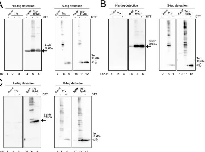
In the case of Rre28 (Fig. 4A), Rre37 (Fig. 4B) and SphR (Fig. 4C), the expression level of the TF protein in the TF strain (lane 4) was much lower than that in the Trx-TF strain (lane 5),
Fig 3. Examination of the interaction of Rre3, CopR, NrsR and CcaR with Trx inE.colicells.Soluble proteins from theE.coliOrigami2 strain (Control), the strain expressing only TrxMC35S(Trx), the strain expressing only a TF and the strain expressing both TrxMC35Sand a TF were separated by non-reducing 12% SDS-PAGE and immunoblot analysis was performed. (A) Rre3, (B) CopR, (C) NrsR and (D) CcaR were detected using a His-tag antibody (left panel) and Trx was detected with S-protein (right panel).±indicates with or without 100 mM DTT treatment. Black and white arrows indicate the TF monomer and the Trx monomer, respectively.
which made comparisons of the banding patterns difficult. Although the monomeric TF (black arrow) and high-order oligomers were detected in each Trx-TF strain (lane 5), we could not identify a Trx-TF complex among these bands due to the lack of the information regarding the banding pattern of the strain expressing only the recombinant TF. As shown inS3 Figs. E–G, the TFs mainly accumulated in the P fraction of the TF and TF-Trx strains and the
abundance of Trx was much lower in the Trx-TF strain than in the Trx strain. Since the inter-action of Rre28, Rre37 and SphR with Trx could not be evaluated by immunoblot analysis of the S fraction ofE.coli, we performed a co-purification analysis of extracts from the Trx-TF strains by nickel affinity chromatography. We determined that Trx co-purified with His-PedR from the Trx-His-PedR strain (S4 Fig. A), whereas co-purification of Trx was not apparent in extracts from the Trx-Rre28 (S4 Fig.C), Trx-Rre37 (S4 Fig. D) and Trx-SphR (S4 Fig. E) strains. We therefore concluded that Rre28, Rre37 and SphR do not interact with Trx inE.
colicells.
Fig 4. Examination of the interaction of Rre28, Rre37 and SphR with Trx inE.colicells.Soluble proteins from theE.coliOrigami2 strain (Control), the strain expressing only TrxMC35S(Trx), the strain expressing only a TF and the strain expressing both TrxMC35Sand a TF were separated by non-reducing 12% SDS-PAGE and immunoblot analysis was performed. (A) Rre28, (B) Rre37 and (C) SphR were detected using a His-tag antibody (left panel) and Trx was detected using S-protein (right panel).±indicates with or without 100 mM DTT treatment. Black and white arrows indicate the TF monomer and the Trx monomer, respectively.
Analysis of the Trx-RpaA interaction using purified proteins
To confirm the Trx-TF interaction detected inE.colicells, RpaA, RpaB and ManR were puri-fied under reducing conditions from RpaA, Trx-RpaB and ManR strains, respectively, by nickel affinity chromatography. The purified recombinant TFs were incubated with wild-type TrxM protein, alkylated using NEM or PEG-maleimide, and changes in oligomerization state and redox state of the constituent cysteine residues were examined by non-reducing SDS-PAGE.
RpaA has three cysteine residues, C64, C168 and C228 (Table 2), which are highly con-served among RpaA orthologs in cyanobacteria. The RpaA protein purified under reducing condition was a monomeric protein with an apparentmolecular massof approximately 30kDa
(Fig. 5A, lane 1, grey arrowhead). When incubated with 10 mM H2O2, a faint band
correspond-ing to a presumed RpaA dimer (white arrowhead) was detected (lane 2). The apparent amount of dimer formed by H2O2treatment was decreased by incubation with 100 mM DTT (lane 3),
but not affected by incubation with 0.5 or 5μM TrxM (lanes 5 and 6).
In order to establish whether the redox state of three cysteine residues in the monomeric RpaA is affected by the redox treatment, the same sample set shown inFig. 5Awas treated with the thiol-modifying agent PEG-maleimide (average molecular mass: 5 kDa), and then sub-jected to non-reducing SDS-PAGE (Fig. 5B). When RpaA treated with 100 mM DTT was fur-ther incubated with PEG-maleimide, a significant mobility shift was observed (lane 2) compared to the unmodified RpaA monomer (lane 1), indicating the attachment of PEG-mal-eimide to the three cysteine residues of RpaA. In contrast, when RpaA treated with 10 mM H2O2was further incubated with PEG-maleimide, the band of the reduced RpaA (termed Red)
was decreased in the strength and two new two bands (Ox1 and Ox2) appeared (lane 3). The electrophoretic mobility of Ox1 was greater than that of the Red band, indicating that one or two cysteine residues were not modified with PEG-maleimide. Ox2 showed the same electro-phoretic mobility as that of the unmodified RpaA monomer, indicating that no cysteine residue of Ox2 was modified with PEG-maleimide. Incubation with 100 mM DTT after treatment with 10 mM H2O2resulted in disappearance of Ox2 and an increase in the strength of the Red and
Ox1 bands (lane 4). This suggests that the three cysteine residues may be reversibly oxidized to sulfenic acid or involved in the formation of intramolecular disulfide bonds in Ox2. On the other hand, the insensitivity of Ox1 to treatment with reducing agents indicates that the cyste-ine residues of Ox1 may be oxidized to sulfinic or sulfonic acid. Incubation with 0.1 mM DTT after treatment with 10 mM H2O2(lane 5) did not change the banding pattern compared with
the sample treated with H2O2alone (lane 3), whereas addition of 0.5μM or 5μM Trx
concomi-tantly with 0.1 mM DTT resulted in the disappearance of the Ox2 band and an increase in the strength of the Red band (lanes 6 and 7). This indicates that the cysteine residue(s) of Ox2 were reduced by TrxM.
Analysis of the Trx-RpaB interaction using purified proteins
RpaB has only one cysteine residue, C59. Under reducing conditions, the RpaB protein was pu-rified as a monomer with an apparentmolecular massof approximately 34kD(Fig. 5C, lane 1, grey arrowhead). When incubated with 1 mM H2O2, an approximately 80 kDa band was
To determine whether the redox state of C59 in the monomeric RpaB is affected by redox treatments, the same sample set shown inFig. 5Cwas treated with PEG-maleimide, and then subjected to non-reducing SDS-PAGE (Fig. 5D). When RpaB that had been treated with 100 mM DTT was further incubated with PEG-maleimide, a mobility shift was observed (lane 2) compared with the unmodified RpaB monomer (lane 1), indicating the attachment of PEG-mal-eimide to C59 of RpaB. When incubated with 1 mM H2O2, the band corresponding to reduced
RpaB (Red) disappeared and two bands (Ox1 and Ox2) appeared (lane 3). Ox1 had apparent molecular mass of approximately 80 kDa, which was similar to that of the RpaB dimer (Fig. 5C), whereas Ox2 had the same molecular mass as the unmodified RpaB monomer (lane 1). Incuba-tion with 100 mM DTT after treatment with 1 mM H2O2resulted in detection of the Red band
instead of the Ox1 and Ox2 bands (lane 4). This reversible change between Red and Ox1/Ox2 suggests that Ox1 corresponds to an RpaB dimer of two monomers connected by an intermolec-ular disulfide bond, and that Ox2 corresponds to an RpaB monomer with an oxidized C59 resi-due that cannot be modified with PEG-maleimide. We propose that the C59 resiresi-due of Ox1 is oxidized to sulfenic acid. Although incubation with 0.1 mM DTT after treatment with 1 mM H2O2(lane 5) did not change the banding pattern resulting from the H2O2treatment alone
(lane 3), addition of 0.5μM or 5μM Trx concomitantly with 0.1 mM DTT resulted in a signifi-cant decrease in the strength of the Ox1 and Ox2 bands and an increase in the strength of the Red band (lanes 6 and 7). We interpret this result to suggest that dimeric RpaB and the oxidized form of monomeric RpaB can be reduced by TrxM. These results also suggested the possibility that TrxM can reduce the sulfenic acid of Ox1, which is consistent with reports that Trx can di-rectly reduce the cysteine sulfenic acid of 1-cys methionine sulfoxide reductase B [19,20].
Analysis of Trx-ManR interaction using purified proteins
ManR has three cysteine residues, (C64, C154 and C230;Table 2) and, of these, only C64, which is located in the receiver domain, is highly conserved among cyanobacterial species. Under reducing condition the ManR protein was purified as a monomer with an apparent mo-lecular massof approximately 28kDa,as determinedbySDS-PAGE(Fig. 5E, lane 1). We found that cysteine residues of the monomeric ManR could be fully oxidized using 100μM aldrithiol but not by H2O2(from 0.1 mM to 10 mM) (S5 Fig.B) and so we used aldrithiol as the oxidizing
agent for ManR. When ManR was incubated with 100μM aldrithiol, faint bands corresponding to dimeric and highly oligomeric forms were detected in addition to the monomeric form (lane 2). A decrease in the strength of the oligomer bands and an increase in the strength of the monomer band were observed following treatment with 100 mM DTT or 5μM Trx after treat-ment of 100μM aldrithiol (lanes 3 and 6).
To determine whether the redox state of three cysteine residues in the monomeric ManR is affected by redox treatments, the same sample set shown inFig. 5Ewas treated with PEG-malei-mide and then subjected to non-reducing SDS-PAGE (Fig. 5F). Modification of the fully re-duced form of ManR with this reagent resulted in a large mobility shift (Red, lane 2) indicating attachment of PEG-maleimide to the three cysteine residues of ManR. When ManR was incu-bated with 100μM aldrithiol, the strength of the Red band decreased and the Ox2 band ap-peared (lane 3). Ox2 had the same apparent molecular mass as the unmodified ManR
monomer, indicating that the cysteine residues were oxidized and were not modified with PEG-maleimide. Incubation with 100 mM DTT after oxidization with 100μM aldrithiol resulted in
Oxidized TFs were reduced by 100 mM DTT or TrxM (0.5 or 5μM) with indicated concentration of DTT. After
precipitation with 10% (w/v) trichloroacetic acid, TFs were subjected to the thiol modification using NEM or PEG-maleimide. Ox and Red indicate the oxidized and reduced forms of a TF, respectively.
the disappearance of the Ox2 band and an increase in the strength of the Red band (lane 4). In-cubation with 25μM DTT after treatment of 100μM aldrithiol caused a slight decrease in the strength of the Ox2 band (lane 5), whereas incubation with 0.5μM TrxM together with 25μM DTT resulted in a decrease in Ox2 and appearance of Ox1 (lane 6). We suggest that a reduction of one or two cysteine residues in Ox2 may have resulted in the attachment of the additional PEG-maleimide and the band shift to the Ox1 position. When the concentration of Trx was in-creased to 5μM, both Ox1 and Ox2 bands disappeared and the strength of the Red band was significantly increased (lane 7), indicating that all three cysteine residues were reduced by TrxM.
Discussion
Evaluation of the newly established screening system
In this study, we established a screening system using anE.colico-expression strain to detect in-teractions between Trx and TFs, and we used this system to test whether ten OmpR family TFs from S. 6803 interact with Trx. As a result, we identified three TFs, RpaA, RpaB and ManR, as potential Trx targets. The advantage of this screening system over other systems to detect pro-tein-protein interactions is that it can specifically detect the thiol-disulfide-exchange reaction with Trx, and is applicable to low-abundance proteins or proteins whose overexpression and pu-rification are technically challenging. For example, RpaB and ManR accumulated mostly in the insoluble pellet (P) fraction when overexpressed inE.coli(Figs. 2C and 2E). Nevertheless, we were able to detect the Trx-RpaB and Trx-ManR interactions in the soluble (S) fraction of theE.
colico-expression strain (Figs. 2D and 2F). This system is thus applicable if only a small amount of protein is expressed in the S fraction, which is expedient for the first screening of Trx-inter-acting proteins. Once the protein complex between a co-expressed TF and TrxMC35Sis detected,
the candidate TF can be purified fromE.coliand the interaction with Trx can be further evaluat-ed using the corresponding purifievaluat-ed proteins. Analysis of the revaluat-edox state of cysteine residues by modification with PEG-maleimide revealed that cysteine residues of RpaA, RpaB and ManR can be reduced by a thiol-disulfide-exchange reaction with Trx (Figs. 5B, 5D and 5F) and these data demonstrated the effectiveness of the screening system.
The potential interaction of Rre28, Rre3, CopR, NrsR, Rre37, SphR and CcaR with Trx was not confirmed in this study (Figs.3,4andS4 Fig.). However, it may be that post-translational modifications, or interaction with other proteins that are present in S. 6803 cells but not those of
E.colicells are required for a particular TF-Trx interaction. Consequently, we cannot exclude the possibility that these TFs do in fact interact with Trx in S. 6803 cells. To test this possibility, further studies are required to examine the interactions between Trx and these TFsin vivo.
The role of cysteine residues in OmpR family response regulators
Several studies have reported that OmpR family TFs form a dimer through a dimer interface comprised ofα4-β5-α5 structures within the receiver domain [22,23]. The conserved cysteine residue is located in theα3-helix and its involvement in dimerization has been unclear. We ob-served that the abundance of the RpaB dimer decreased after treatment with DTT or Trx (Figs. 5C and 5D), which suggests that the C59 of RpaB is involved in dimerization through the formation of intermolecular disulfide bonds. To date, as far as we are aware, there has been no report describing the oligomerization state of RpaBin vivo, as examined by non-reducing SDS-PAGE. Our immunoblotting analysis using an anti-RpaB antibody indicated that there is no RpaB dimer formation in LL-grown S.6803 cells (not shown). Further analysis will be required to test the possibility of dimer formation under specific growth conditions. In the case of RpaA and ManR, dimeric bands were detected, but the strength of these bands was significantly lower than those of the monomeric bands (Figs. 5A and 5E). The existence of dimeric forms of RpaA and ManR should thus be carefully assessed bothin vitroandin vivo.
Physiological significance of interaction of TFs with Trx
Our results provide the first evidence that RpaA, RpaB and ManR can interact with Trxin vitro. Further studies will be required to confirm the interaction of these TFs with Trxin vivo
and to elucidate the physiological significance of their interaction. However, the physiological significance of Trx-TF interaction is suggested by earlier studies. RpaA and RpaB (regulator of phycobilisome association) response regulators were initially identified as regulators involved in the distribution of phycobilisome-absorbed light energy between the two photosystems [24]. Subsequently, RpaA was reported to be involved in responses to salt and hyperosmotic stresses and HL exposure in S. 6803 [25,26], as well as to be a part of the circadian-clock system in S.7942 [27]. In S.6803, the Hik33-RpaA two-component pair controls the expression of differ-ent sets of genes under hyperosmotic and salt stress conditions [25]. Since a pair of histidine ki-nase and its cognate response regulator controls a set of genes in the accepted model of two-component systems, there must be unidentified regulatory mechanisms that enable the same pair of histidine kinase and response regulator to control different sets of genes. Our result sug-gests the possibility that the interaction between RpaA and Trx may provide another signal input under hyperosmotic or salt stress conditions.
phospho-accepting aspartate residue (D47), we propose that the redox state of C59 may affect the rate of phospho-transfer from Hik33 to RpaB, or the stability of the phosphorylated state of RpaB. Recently, a proteomic analysis ofSynechococcussp. PCC 7002 during light to dark transi-tions identified RpaB as a redox-sensitive protein [33]. A cell-permeable thiol-specific reactive probe was applied toSynechococcuscells under light or dark condition to label the free cysteine residues, and modified cysteine residues were then identified and quantified by liquid chroma-tography mass-spectrometry (LC-MS). The abundance of the labeled cysteine in RpaB was about 3-fold greater under light conditions than under dark conditions, indicating that a light-dependent reduction of the cysteine residue of RpaB. We suggest that the reduction of the cyste-ine residue during the dark to light transition may result from an interaction with Trx.
ManR has been reported to be a repressor of themntCABoperon encoding manganese trans-porter [34]. ManR is phosphorylated by the histidine kinase ManS and binds to themntCAB pro-moter under non-stress conditions, given sufficient amounts of Mn2+in the growth medium. Under Mn2+depleted conditions, there is no phosphotransfer from ManS and, consequently, de-phosphorylated ManR cannot bind to themntCABpromoter, leading to the induction of the
mntCABoperon [35]. Our results suggest a possible interaction between ManR with Trx to me-diate the redox regulation ofmntCABoperon. A recent proteomic study of the light to dark tran-sition in S.6803 resulted in the identification of ManR as a redox-sensitive protein and C154 was identified as a target of oxidation [21]. Furthermore, the amount of oxidized C154 of ManR was observed to be about 1.5-fold higher in cells exposed to dark conditions than those exposed to the light. When 3-(3,4-dichlorophenyl)-1,1-dimethylurea (DCMU), an inhibitor of photosyn-thetic electron transport, was added to the culture under light conditions, the amount of oxidized C154 increased approximately 2-fold compared with a culture without DCMU. Thus, C154 of ManR may be reduced by reducing equivalents from the photosynthetic electron transfer chain via Trx upon the shift from dark to light conditions.
The activity of response regulators has been thought to be under the control of their cognate histidine kinases. However, several recent reports indicate the importance of cysteine residues in the regulation of the activity of response regulators. Salmonella pathogenicity island 2 (SPI2) response regulator SsrB, which belongs to the NarL/FixJ family, was reported to sense reactive nitrogen species with modification of C203 in the C-terminal dimerization domain [36]. On the other hand, C45 located inα-helix 2 in the receiver domain, and not in the typicalα4-β5-α5 dimer interface, seems to be important for the activity of SsrB, based on the observation that a C45A and C45S substitutions cause impaired function [37]. We interpret the results of this cur-rent study to suggest, for the first time, that environmental signals transmitted from histidine kinases to response regulators may be attenuated by the reduction of cysteine residues by Trx. Furthermore there is a report that the OmpR protein co-purified with Trx fromE.colicells [38], so the interaction with Trx may be a universal mechanism to modulate the activity of OmpR-type response regulators. As a first step to characterize this novel regulatory mecha-nism, we are now characterizing the interaction of TFs with Trx in S.6803 cells.
Supporting Information
S1 Fig. Amino acid sequences of the receiver domain of OmpR family TFs in S. 6803.
Amino acid sequences of the response regulator receiver domain (PF00072 in the Pfam data-base (http://pfam.xfam.org/) of CopR, pCopR, NrsR, CcaR, ManR, Rre28, SphR, RpaA, RpaB, Rre3 and Rre37 were aligned using the CLUSTAL W 2.1 program. The solid and dashed arrows indicate a conserved cysteine residue found in OmpR family TF of S. 6803 and a putative phos-phor-accepting aspartate residue, respectively.
S2 Fig. Formation of TrxMC35S-TF complexes between recombinant proteins and in the
soluble fraction ofE.colico-expression strains.5μM of His-tagged TFs, S-tagged TrxMC35S
and a mixture of 5μM each of His-tagged TF and S-tagged TrxMC35Swere incubated with
H2O2(for RpaB, 1 mM) or aldrithiol (for ManR, 100μM), fractionated by non-reducing 12%
SDS-PAGE and stained with CBB (A). The oxidized recombinant proteins mentioned above and the soluble protein fractions (lysates) of the strain expressing both TrxMC35Sand TF were
separated by non-reducing 12% SDS-PAGE and immunoblot analysis was performed. (B) RpaB and (C) ManR were detected using a His-tag antibody (left panel) and Trx was de-tected using S-protein (right panel). The black arrow, white arrow and arrow head indicate the TF monomer, the Trx monomer and the Trx-TF complex, respectively. In both RpaB and ManR, the bands of the same size were present in the soluble fraction of the Trx-TF strains (lanes 1 and 7) and the mixture of purified proteins (lanes 3 and 9), showing that the bands de-tected specifically in the co-expression strains indeed correspond to Trx-TF complexes. On the other hand, when the His-tagged RpaA and S-tagged TrxMC35Swere mixed, the Trx-RpaA
complex was not formed. However, this does not mean that Trx does not interact with RpaA, since wild-type TrxM can reduce RpaA, as shown inFig. 5. The efficiency of the interaction may be lowered when TrxM has a C35S mutation.
(TIF)
S3 Fig. Expression levels of Rre3, CopR, NrsR, CcaR, Rre28, Rre37, SphR and Trx inE.coli cells.The wholeE.colicell extract (C), the soluble fraction (S) and the insoluble pellet fraction (P) of the control Origami2 strain (Control), the strain expressing only TrxMC35S(Trx), the
strain expressing only TF and the strain expressing both TrxMC35Sand TF were separated by
15% SDS-PAGE and stained with CBB. (TIF)
S4 Fig. Co-purification assay using the Trx-TF strains to assess the interaction of Rre3, CopR, NrsR and CcaR with Trx.Co-purification assays using (A) theE.colistrain expressing Trx and PedR (positive control), (B) the strain expressing Trx alone (negative control),(C)the Trx-Rre28 strain,(D)the Trx-Rre37 strain and(E)the Trx-SphR strain. The solubleE.coli
proteins were alkylated with NEM and each His-TF was purified using nickel resin under dena-turing condition. Soluble proteins alkylated with NEM (Sup) and the eluted fraction from nick-el resin (Elu) were separated by non-reducing SDS-PAGE, stained with CBB (left pennick-el) and detected by immunoblot analysis using a His-tag antibody and S-protein (right panel). ± indi-cates with or without 100 mM DTT treatment before electrophoresis. The black arrow, white arrow and arrow head indicate the TF monomer, the Trx monomer, and the Trx-TF complex, respectively. The 30 kDa Trx-PedR complex was detected in the eluate of the Trx-PedR strain using either an anti-His-tag antibody or S-protein, but not in the eluate from the Trx strain. The monomeric Rre28, Rre37, SphR proteins and high-order oligomer bands were detected with an ant-His-tag antibody in the eluate, but not using S-protein.
(TIF)
Acknowledgments
We thank Dr. Koji Kojima for purification of the wild-type TrxM. This work was supported by Network Joint Research Center for Materials and Devices (to Y.H.), Grant-in-Aid for Scientific Research (C) from the Ministry of Education, Culture, Sports, Science and Technology (MEXT) (to Y.H.), and CREST program from the Japan Science and Technology Agency (JST) (to T.H and Y.H.).
Author Contributions
Conceived and designed the experiments: TK YH. Performed the experiments: TK. Analyzed the data: TK. Contributed reagents/materials/analysis tools: YN TH. Wrote the paper: TK YH.
References
1. Lawlor DW (2009) Musings about the effects of environment on photosynthesis. Ann Bot 103: 543–
549. PMID:19205084
2. Muramatsu M, Hihara Y (2012) Acclimation to high-light conditions in cyanobacteria: from gene expres-sion to physiological responses. J Plant Res 125: 11–39. doi:10.1007/s10265-011-0454-6PMID: 22006212
3. Fey V, Wagner R, Bräutigam K, Pfannschmidt T (2005) Photosynthetic redox control of nuclear gene expression. J Exp Bot 56: 1491–1498. PMID:15863445
4. Pfannschmidt T (2003) Chloroplast redox signals: how photosynthesis controls its own genes. Trend Plant Sci 8: 33–41. PMID:12523998
5. Oelze ML, Kandlbinder A, Dietz KJ (2008) Redox regulation and overreduction control in the photosyn-thesizing cell: complexity in redox regulatory networks. Biochim Biophys Acta 1780: 1261–1272. doi: 10.1016/j.bbagen.2008.03.015PMID:18439433
6. Hihara Y, Sonoike K, Kanehisa M, Ikeuchi M (2003) DNA microarray analysis of the cyanobacterium
Synechocystissp. strain PCC 6803. J Bacteriol 185: 1719–1725. PMID:12591891
7. Sippola K, Aro EM (1999) Thiol redox state regulates expression ofpsbAgenes inSynechococcussp. PCC 7942. Plant Mol Biol 41: 425–433. PMID:10598108
8. Nakamura K, Hihara Y (2006) Photon flux density-dependent gene expression inSynechocystissp. PCC 6803 is regulated by a small, redox-responsive, LuxR-type regulator. J Biol Chem 281: 36758–36766. PMID:17035238
9. Horiuchi M, Nakamura K, Kojima K, Nishiyama Y, Hatakeyama W, Hisabori T, et al. (2010) The PedR transcriptional regulator interacts with thioredoxin to connect photosynthesis with gene expression in cyanobacteria. Biochem J 431: 135–140. doi:10.1042/BJ20100789PMID:20662766
10. Collet JF, Messens J (2010) Structure, function, and mechanism of thioredoxin proteins. Antioxid Redox Signal 13: 1205–1216. doi:10.1089/ars.2010.3114PMID:20136512
11. Lu J, Holmgren A (2014) The thioredoxin antioxidant system. Free Radic Biol Med 66: 75–87. doi:10. 1016/j.freeradbiomed.2013.07.036PMID:23899494
12. Hisabori T, Motohashi K, Hosoya-Matsuda N, Ueoka-Nakanishi H, Romano PG. (2007) Towards a functional dissection of thioredoxin networks in plant cells. Photochem Photobiol 83: 145–151. PMID: 16706599
13. Hishiya S, Hatakeyama W, Mizota Y, Hosoya-Matsuda N, Motohashi K, Ikeuchi M, et al. (2008) Binary reducing equivalent pathways using NADPH-thioredoxin reductase and ferredoxin-thioredoxin reduc-tase in the cyanobacteriumSynechocystissp. strain PCC 6803. Plant Cell Physiol 49: 11–18. PMID: 18003670
14. Lindahl M, Florencio FJ (2003) Thioredoxin-linked processes in cyanobacteria are as numerous as in chloroplasts, but targets are different. Proc Natl Acad Sci U S A 100: 16107–16112. PMID:14673118
15. Perez-Perez ME, Florencio FJ, Lindahl M (2006) Selecting thioredoxins for disulphide proteomics: tar-get proteomes of three thioredoxins from the cyanobacteriumSynechocystissp. PCC 6803. Proteo-mics 6: 186–195.
17. Seino Y, Takahashi T, Hihara Y (2009) The response regulator RpaB binds to the upstream element of photosystem I genes to work for positive regulation under low-light conditions inSynechocystissp. Strain PCC 6803. J Bacteriol 191: 1581–1586. doi:10.1128/JB.01588-08PMID:19074384
18. Itou H, Tanaka I (2001) The OmpR-family of proteins: insight into the tertiary structure and functions of two-component regulator proteins. J Biochem 129: 343–350. PMID:11226872
19. Tarrago L, Laugier E, Zaffagnini M, Marchand CH, Le Maréchal P, Lemaire SD, et al. (2010) Plant thior-edoxin CDSP32 regenerates 1-cys methionine sulfoxide reductase B activity through the direct reduc-tion of sulfenic acid. J Biol Chem 285: 14964–14972. doi:10.1074/jbc.M110.108373PMID:20236937
20. Kim HY, Kim JR (2008) Thioredoxin as a reducing agent for mammalian methionine sulfoxide reduc-tases B lacking resolving cysteine. Biochem Biophys Res Commun. 371:490–494. doi:10.1016/j.bbrc. 2008.04.101PMID:18452709
21. Guo J, Nguyen AY, Dai Z, Su D, Gaffrey MJ, Moore RJ, et al. (2014) Proteome-wide Light/Dark Modula-tion of Thiol OxidaModula-tion in Cyanobacteria Revealed by Quantitative Site-Specific Redox Proteomics. Mol Cell Proteomics mcp.M114.041160.
22. Menon S, Wang S (2001) Structure of the response regulator PhoP fromMycobacterium tuberculosis
reveals a dimer through the receiver domain. Biochemistry 50: 5948–5957.
23. Toro-Roman A, Wu T, Stock AM (2005) A common dimerization interface in bacterial response regula-tors KdpE and TorR. Prot Sci 14: 3077–3088.
24. Ashby MK, Mullineaux CW (1999) Cyanobacterialycf27gene products regulate energy transfer from phycobilisomes to photosystems I and II. FEMS Microbiol Lett 181: 253–260. PMID:10585546
25. Shoumskaya MA, Paithoonrangsarid K, Kanesaki Y, Los DA, Zinchenko VV, Tanticharoen M, et al. (2005) Identical Hik-Rre systems are involved in perception and transduction of salt signals and hyper-osmotic signals but regulate the expression of individual genes to different extents inSynechocystis. J Biol Chem 280: 21531–21538. PMID:15805106
26. Majeed W, Zhang Y, Xue Y, Ranade S, Blue RN, Wang Q, et al. (2012) RpaA regulates the accumula-tion of monomeric photosystem I and PsbA under high light condiaccumula-tions inSynechocystissp. PCC 6803. PLoS One 7: e45139. PMID:23024802
27. Takai N, Nakajima M, Oyama T, Kito R, Sugita C, Sugita M, et al. (2006) A KaiC-associating SasA-RpaA two-component regulatory system as a major circadian timing mediator in cyanobacteria. Proc Natl Acad Sci U S A 103: 12109–12114. PMID:16882723
28. Hanaoka M, Takai N, Hosokawa N, Fujiwara M, Akimoto Y, Kobori N, et al. (2012) RpaB, another re-sponse regulator operating circadian clock-dependent transcriptional regulation inSynechococcus elongatusPCC 7942. J Biol Chem 287: 26321–26327. doi:10.1074/jbc.M111.338251PMID: 22665493
29. Kappell AD, van Waasbergen LG (2007) The response regulator RpaB binds the high light regulatory 1 sequence upstream of the high-light-induciblehliBgene from the cyanobacteriumSynechocystisPCC 6803. Arch Microbiol 187: 337–342. PMID:17294172
30. Seki A, Hanaoka M, Akimoto Y, Masuda S, Iwasaki H, Tanaka K (2007) Induction of a group 2 sigma factor, RPOD3, by high light and the underlying mechanism inSynechococcus elongatusPCC 7942. J Biol Chem 282: 36887–36894. PMID:17977831
31. Hanaoka M, Tanaka K (2008) Dynamics of RpaB-promoter interaction during high light stress, revealed by chromatin immunoprecipitation (ChIP) analysis inSynechococcus elongatusPCC 7942. Plant J 56: 327–335. doi:10.1111/j.1365-313X.2008.03600.xPMID:18643976
32. Moronta-Barrios F, Espinosa J, Contreras A (2012)In vivofeatures of signal transduction by the essen-tial response regulator RpaB fromSynechococcus elongatusPCC 7942. Microbiology 158: 1229–
1237. doi:10.1099/mic.0.057679-0PMID:22322959
33. Ansong C, Sadler NC, Hill EA, Lewis MP, Zink EM, Smith RD, et al. (2014) Characterization of protein redox dynamics induced during light-to-dark transitions and nutrient limitation in cyanobacteria. Front Microbiol 5: 325. doi:10.3389/fmicb.2014.00325PMID:25071738
34. Yamaguchi K, Suzuki I, Yamamoto H, Lyukevich A, Bodrova I, Los DA, et al. (2002) A two-component Mn2+-sensing system negatively regulates expression of themntCABoperon inSynechocystis. Plant Cell 14: 2901–2913. PMID:12417709
35. Ogawa T, Bao DH, Katoh H, Shibata M, Pakrasi HB, Bhattacharyya-Pakrasi M (2002) A two-compo-nent signal transduction pathway regulates manganese homeostasis inSynechocystis6803, a photo-synthetic organism. J Biol Chem 277: 28981–28986. PMID:12039966
37. Carroll RK, Liao X, Morgan LK, Cicirelli EM, Li Y, Sheng W, et al. (2009) Structural and functional analy-sis of the C-terminal DNA binding domain of the Salmonella typhimurium SPI-2 response regulator SsrB. J Biol Chem 284: 12008–12019. doi:10.1074/jbc.M806261200PMID:19126546
38. Kumar JK, Tabor S, Richardson CC (2004) Proteomic analysis of thioredoxin-targeted proteins in