Identification of a Novel Bacterial Outer Membrane
Interleukin-1Β-Binding Protein from
Aggregatibacter
actinomycetemcomitans
Annamari Paino
1, Tuuli Ahlstrand
1, Jari Nuutila
1, Indre Navickaite
1, Maria Lahti
1, Heidi Tuominen
1,
Hannamari Välimaa
2,3, Urpo Lamminmäki
1, Marja T. Pöllänen
4, Riikka Ihalin
1*1 Department of Biochemistry and Food Chemistry, University of Turku, Turku, Finland, 2 Haartman Institute, Department of Virology, University of Helsinki, Helsinki, Finland, 3 Helsinki University Hospital Laboratory (HUSLAB), Helsinki University Hospital, Helsinki, Finland, 4 Institute of Dentistry, University of Turku, Turku, Finland
Abstract
Aggregatibacter actinomycetemcomitans is a gram-negative opportunistic oral pathogen. It is frequently associated with subgingival biofilms of both chronic and aggressive periodontitis, and the diseased sites of the periodontium exhibit increased levels of the proinflammatory mediator interleukin (IL)-1 . Some bacterial species can alter their physiological properties as a result of sensing IL-1 . We have recently shown that this cytokine localizes to the cytoplasm of A. actinomycetemcomitans in co-cultures with organotypic gingival mucosa. However, current knowledge about the mechanism underlying bacterial IL-1 sensing is still limited. In this study, we characterized the interaction of A. actinomycetemcomitans total membrane protein with IL-1 through electrophoretic mobility shift assays. The interacting protein, which we have designated bacterial interleukin receptor I (BilRI), was identified through mass spectrometry and was found to be Pasteurellaceae specific. Based on the results obtained using protein function prediction tools, this protein localizes to the outer membrane and contains a typical lipoprotein signal sequence. All six tested biofilm cultures of clinical A. actinomycetemcomitans strains expressed the protein according to phage display-derived antibody detection. Moreover, proteinase K treatment of whole A. actinomycetemcomitans
cells eliminated BilRI forms that were outer membrane specific, as determined through immunoblotting. The protein was overexpressed in Escherichia coli in both the outer membrane-associated form and a soluble cytoplasmic form. When assessed using flow cytometry, the BilRI-overexpressing E. coli cells were observed to bind β.5 times more biotinylated-IL-1 than the control cells, as detected with avidin-FITC. Overexpression of BilRI did not cause binding of a biotinylated negative control protein. In a microplate assay, soluble BilRI bound to IL-1 , but this binding was not specific, as a control protein for IL-1 also interacted with BilRI. Our findings suggest that A. actinomycetemcomitans
expresses an IL-1 -binding surface-exposed lipoprotein that may be part of the bacterial IL-1 -sensing system.
Citation: Paino A, Ahlstrand T, Nuutila J, Navickaite I, Lahti M, et al. (β01γ) Identification of a Novel Bacterial Outer Membrane Interleukin-1Β-Binding Protein from Aggregatibacter actinomycetemcomitans. PLoS ONE 8(7): e70509. doi:10.1γ71/journal.pone.0070509
Editor: José A. Bengoechea, Fundación Investigación Sanitaria Illes Balears, Spain
Received May γ1, β01γ; Accepted June β4, β01γ; Published July γ1, β01γ
Copyright: © β01γ Paino et al. This is an open-access article distributed under the terms of the Creative Commons Attribution License, which permits unrestricted use, distribution, and reproduction in any medium, provided the original author and source are credited.
Funding: This work was supported by the grants form the Academy of Finland (http://www.aka.fi/en-GB/A/), the Paulo Foundation (http://www.paulo.fi/in-english), The Ella and Georg Ehrnrooth Foundation (http://www.ellageorg.fi/en/about), and the Finnish Dental Society Apollonia (http://www.apollonia.fi/ Apollonia/www5.nsf/sp?open&cid=home) to RI, Biocenter Finland (http://www.biocenter.fi/) to UL and ML, and Erasmus to IN. The funders had no role in study design, data collection and analysis, decision to publish, or preparation of the manuscript.
Competing interests: The authors have declared that no competing interests exist. * E-mail: riikka.ihalin@utu.fi
Introduction
Aggregatibacter actinomycetemcomitans is a Gram-negative opportunistic human pathogen that causes chronic and aggressive forms of periodontitis [1-γ]. This pathogen is also associated with systemic diseases, such as cardiovascular diseases [4,5], and it possesses variety of virulence properties (reviewed in 6), which enhances its resistance against human defense mechanisms.
pathogens A. actinomyctemcomitans and P. gingivalis has been shown to cause changes in the gene expression profiles
of the commensal strains in the biofilm [15]. A.
actinomycetemcomitans employs several specific strategies for enhancing its survival in the host: it can secrete a leukotoxin that directly targets human phagocyte cells, monocytes and macrophages [16-18], while the other toxin produced by the species, cytolethal-distending toxin, indirectly modulates periodontal bone resorption [19] as well as periodontal keratinocyte and fibroblast proliferation [β0,β1]. However, little is known about the ability of the species to adjust its virulence properties after sensing inflammation-related environmental factors.
High levels of the human proinflammatory mediator interleukin (IL)-1 are typical of periodontal inflammation sites in tooth-supporting tissues [ββ]. IL-1 is a key inflammatory mediator of the human innate immune system. This cytokine is secreted primarily by human macrophages and monocytes after sensing microbial danger signals. IL-1 stimulates its own secretion [βγ], and the bursts of IL-1 that are released from human cells are highly controlled (reviewed in β4). Human mononuclear leukocytes detect A. actinomycetemcomitans as a pathogen via NLRPγ inflammasome [β5], which induces the maturation of pro-IL-1 to its biologically active form. Dysregulation of IL-1 activity can result in chronic diseases such as periodontitis (reviewed in β4,β6). In vitro, virulent bacteria may sense the inflammatory levels of this signal by binding it and altering their growth properties [β7-β9]. Furthermore, IL-1 is known to modulate virulence gene expression in Staphylococcus aureus [γ0]. In previous studies,
we observed that A. actinomycetemcomitans biofilm
sequestered and then took up human IL-1 [γ1]. In addition, A. actinomycetemcomitans increases the biofilm mass as a physiological response to IL-1 [γβ], similar to the biofilms formed by Gram-positive S. aureus [β8,β9]. As a secondary response, IL-1 binding temporarily decreases the metabolic activity of this species [γβ].
The findings of the studies described above strongly suggest that bacteria may use IL-1 as an indicator of the host inflammatory state. However, the mechanisms underlying the uptake and subsequent regulatory pathway of IL-1 in bacteria are not known. Currently, the only bacterial IL-1 -binding outer membrane receptor has been characterized from the
Gram-negative bacterium Yersinia pestis [γγ]. Gram-negative
Pseudomonas aeruginosa has been shown to specifically sense interferon (IFN)- via its outer membrane protein OprF [γ4]. The interleukin receptor of Yersinia is known as capsule antigen F1 assembly (Caf1A) protein. Caf1A is required for the outer membrane localization of capsule antigen F1 protein, which shows significant sequence similarity to a human IL-1α, receptor antagonist [γ5]. According to BLAST searches, the
A. actinomycetemcomitans genome does not show significant sequence similarity with genes encoding Caf1A, Caf1 or the human interleukin-1 receptor. Thus, we sought to specifically study the mechanism of IL-1 transfer inside bacterial cells using the oral pathogen A. actinomycetemcomitans as a model species.
In this paper, we describe the interaction of human IL-1 with
a hypothetical lipoprotein extracted from A.
actinomycetemcomitans from a dissolved total membrane protein fraction. Based on the observed interaction, we designated this putative protein bacterial interleukin-1 receptor I (BilRI). The interacting protein was identified via mass spectrometry (MS) analysis, and its subcellular location was analyzed using subcellular localization prediction tools for bacterial proteins. The putative bacterial outer membrane IL-1 receptor was overexpressed in an Escherichia coli strain designed for membrane protein overexpression [γ6] and was also produced in a soluble form in E. coli, without the typical lipoprotein signal sequence. The E. coli cells containing BilRI in their outer membrane showed an increased IL-1 -binding capacity, and the soluble form of the protein interacted with IL-1 in a microplate assay. Additionally, all of the tested clinical A. actinomycetemcomitans strains expressed BilRI, and the localization of the protein in the outer membrane was verified through proteinase K treatment. Knowledge of bacterial mechanisms for the binding and uptake of the central inflammatory cytokine IL-1 is essential for understanding the behavior and properties of the opportunistic pathogen under inflammatory conditions.
Results
Identification of an IL-1β-binding membrane protein in
A. actinomycetemcomitans
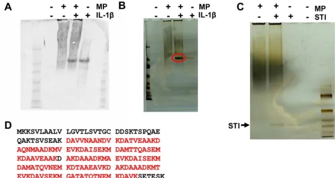
The protein band that interacted with the anti-IL-1 antibody was only visible in lanes containing either IL1 or the IL1 -membrane protein (MP) combination (Figure 1A). The intensity of this band was the same in both of these lanes (Figure 1A). However, in a silver-stained gel, the band was much more intense in the lane that contained both IL-1 and MP than in the lane containing only IL-1 (Figure 1B). The band was missing from the lane containing pure MP (Figure 1B). This phenomenon was not observed when IL-1 was replaced with the control protein soybean trypsin inhibitor (STI) (Figure 1C). Thus, it was suggested that in the IL-1 -MP lane, the intense band also contained proteins other than IL-1 and that those proteins most likely interacted with IL-1 . The intense protein band was extracted following non-denaturing-PAGE and identified as putative uncharacterized protein GγZCI1 of A. actinomycetemcomitans D17P-γ through LC-MS/MS analysis. The identified peptides covered 7β% of the total protein
sequence (Figure 1D). In addition to A.
actinomycetemcomitans protein GγZCI1, IL-1 was detected in the band, confirming the potential interaction of the novel protein with IL-1 . As the bacterial protein had no previously known function, we designated it bacterial interleukin receptor I (BilRI).
The amino acid sequence of BilRI was subjected to a BLAST
search against the genome sequence of A.
but not in E. coli. Its first 19 amino acids likely form the lipoprotein signal sequence, which directs the protein to the outer membrane in Gram-negative species (Figure βB).
Recombinant BilRI localized to the outer membrane of
E. coli
When BilRI was expressed in E. coli with its native signal sequence, the outer membrane protein fraction contained the recombinant protein (Figure 4A), which was identified from a silver-stained gel via LC-MS/MS. The purity of the outer membrane protein fraction was confirmed through the detection of heme proteins, which are only present in the inner membrane fraction [γ7] (Figure 4B).
Overexpression of BilRI in the E. coli outer membrane increased membrane frailty and caused
self-aggregation in the presence of calcium
The production of large amounts of recombinant BilRI in the outer membrane of E. coli led to increased frailty of E. coli cells (Figure 4C–E). When a bacterial pellet was frozen at -β0°C and
resuspended in PBS, the suspension was more viscous when it contained IPTG-induced cells than non-induced cells. Additionally, after centrifugation, the pellet was looser (Figure
4C), and the supernatant contained a large amount of DNA (Figure 4D), indicating that the outer membrane BilRI-producing E. coli cells broke down during the freezing and thawing cycle. However, 50% glycerol protected the cells from breakdown (Figure 4E).
When the E. coli cells producing native BilRI were incubated in the presence of 50 mM calcium, they began to aggregate. This phenomenon was not observed in non-induced E. coli
cells (Figure 4F).
Recombinant BilRI increased the binding of IL-1β to E. coli
The E. coli cells producing the recombinant full-length BilRI protein bound as much IL-1 as the control cells in which BilRI production was not induced based on estimation of the amount of binding as the number of positively stained cells (Figure 5A). However, when the mean fluorescence intensity was measured in the positively stained cells, it was observed that the recombinant BilRI-producing E. coli cells bound IL-1 more efficiently per cell compared to the control cells (Figure 5B). Neither the BilRI-producing E. coli cells nor the control cells showed any significant binding of the control protein STI (Figure 5A).
Figure 1. The uncharacterized putative membrane protein from A. actinomycetemcomitans interacted with IL-1β. Membrane proteins (MP) were isolated from A. actinomycetemcomitans and dissolved in 4% CHAPS, after which they were incubated with recombinant human IL-1 . The samples were subjected to native-PAGE and either blotted with anti-IL-1 (A) or silver stained (B). Anti-IL-1 detected similar amounts of IL-1 in samples containing MP+IL-1 and a sample of pure IL-1 (A). In the silver-stained gel, the equivalent band was more intense in the MP+IL-1 sample (B; red circle) than in the IL-1 sample, indicating the presence of proteins other than IL-1 . This phenomenon was not observed in the control experiment using the soybean trypsin inhibitor (STI) protein as a similar size control (C). The intense protein band (B; red circle) was cut out of the
silver-stained gel and identified as the putative uncharacterized GγZCI1 protein from A. actinomycetemcomitans D17P-γ via LC-MS/MS
analysis. The identified peptides (D; bold red) covered 7β% of the total protein sequence. doi: 10.1γ71/journal.pone.0070509.g001
The soluble recombinant form of BilRI bound IL-1β
The recombinant form of BilRI without the signal sequence, expressed and purified from E. coli cytoplasm as a soluble protein, was observed to bind IL-1 in a microplate assay (Figure 5C). Moreover, the level of binding to IL-1 was greater than to the blocking protein BSA (Figure 5C). However, no significant difference was found between IL-1 binding and STI binding (5C). Finally, IL-1 -coated wells bound the negative control protein from A. actinomycetemcomitans (N-terminal portion of RcpA) more weakly than BilRI (Figure 5C).
Different clinical isolates of A. actinomycetemcomitans
expressed BilRI in the outer membrane facing the extracellular space
The mature BilRI protein traveled to an extracellular position in A. actinomycetemcomitans D7S, as proteinase K treatment of whole cells digested BilRI (Figure 6A). The outer membrane protein fraction of strain D7S contained two forms of BilRI, with sizes of approximately γ5 kDa and 70 kDa (Figure 6A). The larger of the two was also found in the inner membrane preparate (Figure 6A). We hypothesized that the larger form was a lipidated immature form of BilRI, as large amounts of the smaller, γ5 kDa form were only detected in the outer membrane protein fraction (Figure 6A). However, both of these
Figure 2. The bacterial IL-1β-binding protein consisted of a lipoprotein signal sequence and four repeated sequences. Sequence analysis using the SignalP 4.1 Server [54] revealed that the bacterial IL-1 -binding protein, which we designated bacterial interleukin receptor I (BilRI), contained a putative signal sequence of 19 amino acids (A and B; bold red). The sequence also contained 4 different 10-amino acid-long sequences (A and B; bold green, blue, purple and grey) repeated in the same order three times. BLAST similarity searches [56] and Clustal W sequence alignment [57] revealed almost identical sequences in other A. actinomycetemcomitans (Aa) strains and A. aphrophilus (A. aphr.) (B).
doi: 10.1γ71/journal.pone.0070509.g00β
Figure 3. Only Pasteurellaceae species possessed proteins showing sequence similarity to BilRI. BLAST similarity searches [56] and Clustal W sequence alignment [57] only detected similar proteins in species belonging to the Pasteurellaceae
family. The proteins with the highest similarity were selected for further analysis. A bolded font indicates a similar amino acid to that seen in BilRI. Different colors were used to distinguish the signal sequence (red) and the repeating sequences (green, blue, purple and grey) from the other parts of the sequence (black).
doi: 10.1γ71/journal.pone.0070509.g00γ
forms are likely surface exposed in the outer membrane, as their respective bands could be eliminated by proteinase K treatment (Figure 6A). These bands could also be detected by another antibody clone, 16F7, in the same samples (Figure 6B). All four forms of BilRI (the proprotein, the unlipidated mature form and the 70 kDa and γ5 kDa forms) could be detected in in vitro biofilm cultures of all of the tested clinical isolates of A. actinomycetemcomitans, which were obtained from periodontitis patients (Figure 6C).
Discussion
The purpose of this study was to identify an IL-1 -binding receptor in the outer membrane of A. actinomycetemcomitans. Various bacterial species are known to display specific receptors for host cytokines [β7,β9,γ8], and two of such receptors have been identified [γγ,γ4]. Although we were able to demonstrate active binding and internalization of IL-1 in the cells of our model bacterium (A. actinomycetemcomitans) and to identify potential intracellular bacterial proteins that might interact with IL-1 in earlier works [γ1,γβ], we have not previously been able to identify the bacterial protein that
Figure 4. Recombinant E. coli expressing BilRI in the outer membrane showed membrane frailty and self-aggregation. The A. actinomycetemcomitans bilRI gene, including the signal sequence, was cloned into E. coli. The production of BilRI was induced using IPTG. The outer membrane fraction was isolated from both induced (IPTG+) and non-induced (IPTG-) cells, and different dilutions of the membrane samples were subjected to SDS-PAGE, followed by silver staining (A). The purity of the outer membrane (OM) fraction was confirmed by checking for the presence of inner membrane (IM) heme proteins using ECL [59]. Only the IM preparate and positive control cytochrome c generated a signal indicating the presence of heme (B). When IPTG-induced recombinant E. coli cells were frozen in the absence of protective agents, the cells began to break down, which was observed in the form of a spongy bacterial pellet (C) and high levels of DNA released in the cell supernatant (D). The presence of glycerol during freezing prevented the cells from breakdown, as they released less DNA during resuspension (E). When the IPTG-induced E. coli cells were incubated in the presence of 50 mM CaClβ at γ7°C for 1 hour, they started to self-aggregate (F).
doi: 10.1γ71/journal.pone.0070509.g004
interacts with IL-1 in the outer membrane of the bacterium. Because IL-1 sensing likely leads to the formation of a robust biofilm, discovering the outer membrane IL-1 receptor is crucial for future studies addressing the role of this sensory cascade in the virulence of this opportunistic pathogen.
We discovered that a previously uncharacterized outer
membrane lipoprotein of A. actinomycetemcomitans bound
IL-1 . Recombinant E. coli cells which overexpressed the mature protein bound more efficiently IL-1 than the control cells where the recombinant protein expression was not
induced. However, the finding that E. coli cells had intrinsic ability to bind IL-1 was expected, since E. coli has been shown to specifically bind IL-1 [β7]. Although the purified recombinant unlipidated form of the protein bound also the control protein STI in ELISA assays, STI did not bind significantly to the whole E. coli cells. This might be an indication that a membrane environment is needed for the protein to adapt to a form that binds specifically to IL-1 . The IL-1 binding protein was designated bacterial interleukin receptor I (BilRI) because this is the first identified bacterial
IL-Figure 5. Recombinant BilRI bound IL-1β both in the outer membrane of E. coli and as soluble protein. The IL-1 -binding capacity of recombinant E. coli cells containing BilRI in the outer membrane was studied using the Fluorokine™ assay (R&D Systems) and a flow cytometer. A similar number of the IPTG-induced cells bound IL-1 compared to non-induced cells (A). Neither group of cells showed significant binding of the control soybean trypsin inhibitor (STI) protein (A). However, the IPTG-induced recombinant E. coli cells bound IL-1 more efficiently than the non-induced cells, with the former showing a higher mean fluorescence intensity per positively stained cell than latter (B). When BilRI was expressed in E. coli without its signal sequence and purified from the cytoplasm, the obtained protein bound more efficiently to IL-1 than to BSA in a microplate assay (C). However, BilRI bound to IL-1 as efficiently as to STI (C). The negative outer membrane control protein from A. actinomycetemcomitans (the N-terminal portion of RcpA [γβ]) did not bind to IL-1 in the microplate assay (C). N=5 (A and B), and N=γ-8 (C). Statistically significant differences are indicated as follows: * p≤0.05, ** p<0.01, *** p<0.001 (Paired T-test).
doi: 10.1γ71/journal.pone.0070509.g005
binding protein with no other known function than the binding of IL-1 . The first bacterial IL-1 -binding protein to be described was the Caf1A usher protein of Yersinia pestis [γγ], but its main function is to take part to the production and construction of the F1 capsule [γ9]. An outer membrane protein A (OmpA) ortholog that senses IFN- and regulates virulence gene expression through quorum-sensing signaling, OprF of
Pseudomonas aeruginosa [γ4], is also involved in the adhesion of P. aeruginosa to various host cells [40,41]. The Caf1A usher protein and OprF are both porin proteins [4β,4γ] that form a channel through the outer membrane of Gram-negative bacteria. Bacterial lipoproteins linked to virulence traits can possess various functions, ranging from potential antigen activity to adhesion to host cells, and may play a role in antibiotic resistance (reviewed in 44). Additionally, some
bacterial lipoproteins function as receptors or components of more complex transport systems [44]. A recent study showed that the conserved lipoprotein Lpp found in Gram-negative species binds specifically to various cationic α-helical antimicrobial peptides and participates in the internalization of these antimicrobial peptides into the cytoplasm of the bacterium [45]. This demonstrates that bacterial lipoproteins may interact with components of the host innate defense system. Whether BilRI binds only IL-1 , or could it also interact with other host cytokines and chemokines, needs to be confirmed in further studies.
The results of the present study suggest that BilRI is a surface-exposed outer membrane lipoprotein that most likely attaches to the lipid bilayer through its lipid portion. This assumption is supported by the findings that most of the protein
Figure 6. Surface-exposed BilRI was expressed in various clinical isolates of A. actinomycetemcomitans grown in biofilms. The surface exposure of BilRI in A. actinomycetemcomitans was examined via proteinase K treatment. During proteinase K treatment, protein expression was inhibited with chloramphenicol, and the maturation of the proprotein was inhibited with globomycin. Intact A. actinomycetemcomitans D7S cells were incubated with proteinase K for various time periods ranging from 0 to β4 hours, and the cells were then lysed via sonication. The samples were subsequently subjected to SDS-PAGE and immunoblotted with an anti-BilRI antibody. Proteinase K treatment decreased the amount of both forms of BilRI (red quadrangles) detected in outer membrane (OM) protein samples with two different antibody clones, 16B8 (A) and 16F7 (B), suggesting that BilRI was surface exposed in A. actinomycetemcomitans. To examine BilRI expression in the clinical isolates, cell lysates were obtained from young (β1 hours) A. actinomycetemcomitans biofilm cultures through sonication. Samples containing approximately 0.γ×107
disrupted cells were subjected to SDS-PAGE and immunoblotted using an anti-BilRI antibody (B). The letters in parenthesis following the strain code indicate the serotypes (B).
doi: 10.1γ71/journal.pone.0070509.g006
forms that were dominant in the outer membrane protein fraction could be digested by proteinase K treatment of whole
A. actinomycetemcomitans cells and that the protein was highly soluble when produced as a cytosolic form without the signal sequence. The two forms that dominated the outer membrane protein fraction were approximately γ5 kDa and 70 kDa in size, which is larger than both the predicted proprotein (19 kDa) and mature unlipidated BilRI (17 kDa). The 70 kDa protein was also present in the inner membrane protein fraction. Thus, we hypothesized that the γ5 kDa form corresponded to mature lipidated BilRI, since it was only detected from the whole cells and the outer membrane fraction and not from the inner membrane faction. The level of the 19 kDa proprotein form remained constant throughout proteinase K treatment, as chloramphenicol inhibited protein synthesis, and globomycin impeded the processing of the pro-form to the unlipidated mature form. However, the lipidation and maturation process needs to be studied in more detail to confirm the exact composition of each protein form. It was unexpected to find that the recombinant BilRI attached to the extracellular side of the outer membrane in E. coli, as all of the lipoproteins previously identified in E. coli travel to the periplasmic side [46]. In summary, BilRI might be among the first proteins that interact with IL-1 during one of the most complex pathways through the cell wall of A. actinomycetemcomitans.
Sequence similarity searches revealed that protein sequences similar to BilRI were mainly found in the
Pasteurellaceae family, including different strains of A. actinomycetemcomitans and species such as Heamophilus influenzae, Haemophilus parainfluenzae, Haemophilus haemolyticus, Haemophilus somnus, Aggregatibacter aphrophilus, Aggregatibacter segnis and Pasteurella multocida. However, the function of the protein is unknown in majority of these strains. The only known function among these homologous proteins has been identified for Haemophilus ducrey fibrinogen binder A (FgbA), which binds to human fibrinogen and is an important virulence factor in humans [47]. However, we cannot claim that a similar protein cannot be found in other bacterial families, as similar binding structures can be formed from different amino acid sequences. Therefore, it is important to also determine the three-dimensional structure of BilRI to find similar receptors in other species.
Overexpression of BilRI in the outer membrane of E. coli
made the cells brittle and prone to lysis when they were frozen without a supplementary protective agent, such as glycerol. This finding was unexpected because we used an E. coli strain that was specifically designed for the production of outer membrane proteins. However, the vulnerability of the outer membrane of Gram-negative bacteria may restrain the recombinant production of outer membrane proteins containing an appropriate signal sequence, which has complicated the use of Gram-negative species as host strains in cell surface display applications [48]. We attempted to overcome this problem by shortening the IPTG induction time from three to two hours, in addition to freezing the cells in the presence of 50% glycerol. Further research is needed to decipher how the overexpression of BilRI affects the membrane integrity of A. actinomycetemcomitans.
In conclusion, we identified a potential first-line binder and receptor for the central human proinflammatory cytokine IL-1
from the opportunistic periodontal pathogen A.
actinomycetemcomitans. The interest in this finding is increased by the fact that no other functions have been described for this protein or its homologs in other bacterial species. Although specific binding of IL-1 by various Gram-negative and Gram-positive bacterial species has been reported [β7,β9,γγ], the identified BilRI protein could only be
found from the family Pasteurellaceae. Future studies
addressing the three-dimensional structure of the receptor will resolve whether the protein structure is entirely novel, or if it shared with other proteins present in different species. Since
human mononuclear leukocytes detect A.
actinomycetemcomitans as a pathogen leading to the production of biologically active IL-1 [β5], the findings reported here present new opportunities for studying the role of IL-1 uptake in the virulence of A. actinomycetemcomitans and in host–pathogen crosstalk.
Materials and Methods
Ethics Statement
Permission to collect and use clinical bacterial strains from A. actinomycetemcomitans positive patients was obtained from the Ethics committee of the Hospital District of Southwest Finland, Turku, Finland. Subgingival microbial samples from adult periodontitis patients were obtained with written informed consent.
Identification of an IL-1β-binding membrane protein
The total membrane protein fraction was isolated from A. actinomycetemcomitans D7S [49] using a previously described protocol [50] with some modification. Briefly, plate-grown (tryptic soy agar (TSA), 5% defibrinated sheep blood) A. actinomycetemcomitans cells (8 plates/extraction) were suspended in PBS1, pH 7.4 (10 mM NaβHPO4, 1.8 mM KHβPO4,
140 mM NaCl, β.7 mM KCl), in a total volume of γ0 ml. The cells were then centrifuged (4,000×g, β0 min, 4°C) and
resuspended in 8 ml of PBS1-saccharose (PBS1, 150 mM
saccharose, 1 mM Pefablock SC [Roche Diagnostics, Indianapolis, IN, USA]), disrupted via sonication (4×1 min on ice, separated by 1 min cooling periods), and whole cells and cell debris were removed via centrifugation (1 700×g, β0 min, 4°C). The supernatant was subsequently divided into two 6.5 ml
ultracentrifugation tubes (#γ55645, Beckman Instruments Inc., Palo Alto, CA, USA), and 500 µl of a 900 mM saccharose solution in PBS1 was pipetted below the supernatant using a
capillary pipette. The tubes were then centrifuged (150,000×g, β hours 45 min, 4°C) to separate the membrane proteins from
the soluble ones. The membrane proteins were located in the pellets, which were stored at -β0°C for further use following
removal of the supernatant.
One membrane protein pellet was dispersed in 400 µl of
PBS1 containing 4% CHAPS (Sigma) and 1 mM
phenylmethylsulfonyl fluoride (PMSF; Sigma), first by careful pipetting and then through slow rotation for γ0 min at RT and β.5 hours at γ7°C. The remaining insoluble material was
removed by spinning briefly in a minifuge. The protein content in the supernatant was determined using the method described by Lowry et al. [51]. Membrane proteins (total amount of β µg) had been solubilized as described above were incubated with γ00 ng of recombinant IL-1 (ReliaTech, GmbH, Braunschweig, Germany) for 1 hour at RT, after which the samples were run in a non-denaturing 4-15% Tris-HCl precast gel (Criterion, Bio-Rad, USA) and transferred to nitrocellulose membranes (Protran®Whatman®, Dassel, Germany) in an Amersham Biosciences Semi-dry blotter. Two controls were included in each gel, one of which contained only the membrane proteins, while the other contained only IL-1 . The
membranes were blocked with 5% skimmed milk in PBS1
containing 0.05% Tween-β0 (PBS1-T) at RT for 1 hour, washed
twice with PBS1-T at RT for 10 min and incubated with a rabbit
anti-IL-1 antibody (NB600-6γγ; Novus β80 Biologicals, Littleton, CO), diluted 1:4,000 in PBS1-T containing 0.5%
skimmed milk at 4°C overnight. Following incubation with the
primary antibody, the membrane was washed four times with PBS1-T for 5 min each and incubated with IRDye® 800CW
Donkey Anti-Rabbit IgG (H+L) (#9β6-γββ1γ, LI-COR Biosciences, Lincoln, NE, USA), diluted 1:10,000 in PBS1-T, at
RT for 1 hour. Prior to detection with Odyssey Infrared Imaging System (LI-COR Biosciences), the membrane was washed six times with PBS1-T for 5 min and then twice with PBS1 for 5 min.
Identical sample series were run and visualized using silver staining [5β].
A silver-stained protein band that reacted with IL-1 was cut from a lane containing both MP and IL-1 . The protein sample that was cut from the gel was in-gel digested with trypsin, and the resultant peptides were analyzed with a nanoflow HPLC system (EasyNano, Thermo, Fisher Scientific, Bremen, Germany) coupled to an LTQ Orbitrap Velos Pro mass spectrometer (Thermo, Fisher Scientific) equipped with a nano-electrospray ionization source. The peptides were first loaded onto a trapping column and were subsequently separated inline on a 15 cm C18 column (75 µm × 15 cm, Magic 5 µm β00 Å C18, Microm BioResources Inc., Sacramento, CA, USA). MS
data were acquired automatically using Thermo Xcalibur software (Thermo, Fisher Scientific). An information-dependent acquisition method was employed, which consisted of a TOF MS survey scan with a mass range of γ00-β,000 m/z. The ten most intensive peaks were selected for fragmentation. The obtained data files were searched for protein identification using Proteome Discover (1.γ) connected to in-house Mascot (v β.4) software against the UniProt database (release β01β_06). Only proteins with at least one “bold red” peptide were included in further analyses, as “bold red” indicated a peptide that was a best match for the assigned protein. Protein hits against species other than A. actinomycetemcomitans or humans were filtered out. Protein hits of less than two peptides were also removed.
Bioinformatics
The SOSUI-GramN server [5γ] was employed to predict the sub-cellular location of the protein. The sequence was analyzed using the SignalP 4.1 Server [54] to predict the length of the signal sequence and the LipoP 1.0 Server [55] to detect
the presence of the lipoprotein signal sequence. Sequence similarity searches were performed with SIB using the BLAST network service. The SIB BLAST network service employs a server developed at SIB and NCBI BLAST β software [56]. Sequence alignments were performed using the Clustal W (1.8γ) program of the SIB T-Coffee multiple sequence alignment package [57].
Cloning and expression of the IL-1β-binding membrane protein
The cloning of bilRI was performed as described for other recombinant A. actinomycetemcomitans proteins [γβ], with slight modifications. NdeI and XhoI restriction sites were introduced into the forward and reverse primers, respectively, and are underlined in the primers. The forward primer for bilRI
was 5’-ATACATATGAAAAAATCAGTATTAGCC-γ’, and the
reverse primer was
5’-ATACTCGAGTTATTTGCTTTCAGTTTC-γ’ (Eurofins MWG Operon, Ebersburg, Germany). The Phusion™ High-Fidelity DNA polymerase (Finnzymes, Espoo, Finland) was used to amplify the bilRI gene from A. actinomycetemcomitans D7S DNA. An annealing temperature of 54°C was selected for the
amplification reaction. The obtained PCR products were digested with NdeI and XhoI (Fermentas, Sankt Leon-Rot, Germany) and cloned into the pETγ6b vector (Novagen, Darmstadt, Germany) using T4 Ligase (Fermentas). The plasmids were then transformed into E. coli TOP10 cells (Invitrogen) via electroporation. Potential plasmid constructs were sequenced in both directions using Eurofins MWG Operon.
A plasmid construct that was verified to contain the correct
bilRI sequence was transformed into the E. coli C41(DEγ) RIL strain (Lucigen, Middleton, WI, USA), which was designed specifically for the production of membrane proteins [γ6]. Various growth temperatures (room temperature [RT], γ0°C,
γ7°C), isopropyl -D-thiogalactoside (IPTG) concentrations
(0.1, 0.5, 1.0 mM) and induction times (γ hours, overnight) were tested to find the optimal conditions for the production of the outer membrane lipoprotein. Outer membrane proteins
were extracted from E. coli [58] and analyzed via SDS-PAGE
with silver staining [5β]. The recombinant protein was identified from a silver-stained gel via mass spectrometry, as described above. Based on the results of these analyses, the expression of BilRI was induced for γ hours with 0.1 mM IPTG when cells first reached an optical density of 0.6 at 600 nm in a special medium (10 g/l tryptone, β4 g/l yeast extract, β.5 g/l KCl, β.5 g/l NaCl, 0.6 g/l NaOH) containing γ0 µg/ml kanamycin and chloramphenicol at γ7°C.
Determining the location of BilRI expressed in E. coli
nitrocellulose membrane and the heme proteins were detected with ECL (Pierce®, Thermo Life science) [59].
Effect of overexpressed outer membrane BilRI on E. coli membrane frailty and the self-aggregation of the recombinant cells
Recombinant E. coli cells containing the complete BilRI sequence were induced with IPTG as described above. Non-induced cells were used as controls. The cells were harvested (β000×g, 10 min, 4°C) and washed twice with PBS
1. The cell
pellets were frozen to -β0°C and then resuspended in PBS 1.
Next, the cell suspensions were centrifuged (16,000×g, 15 min, 4°C), and 10 µl of the supernatant was run in a 0.8% agarose
gel containing the Midori Green DNA Stain (Nippon Genetics Europe, Düren, Germany). To determine the optimal freezing conditions for the recombinant E. coli cells, cell pellets were suspended in different concentrations (β0-50%) of glycerol before freezing, and cell breakage was studied as described above.
Recombinant E. coli cells that had been stored in 50% glycerol were washed twice with HEPES buffer (10 mM HEPES, pH 7.4) and suspended at an OD600nm=1 in HEPES
buffer supplemented with 50 mM CaClβ. E. coli cells in which
the production of BilRI was not induced were used as controls. The cells were incubated in the CaClβ-supplemented buffer for
1 hour at γ7°C, after which the self-aggregation of the cells was
estimated visually.
Determination of the IL-1β-binding capacity of E. coli
cells expressing BilRI
BilRI expression was induced in E. coli as described above, except that the IPTG induction time was shortened to β hours. Cells stored in 50% glycerol were washed three times with PBS1 (5,900×g, 10 min, 4°C) prior to being fixed in PBS1
containing 1% formaldehyde, 1% BSA and 0.01% EDTA for β hours at 4°C. We applied the same fixation conditions which
were in a previous study, and were found to preserve the IL1 -binding capacity of A. actinomycetemcomitans [γβ]. The number of cells in the E. coli samples was adjusted to 108
using the OD-specific cell concentration conversion, according to which an OD600=1 for E. coli cells cultured in LB medium is
equal to 7.8±0.8x10⁸ cell/ml [60]. Following the adjustment of
cell numbers, the cells were washed once with PBS1, collected
via centrifugation and resuspended in 1 ml of PBS1. For flow
cytometric assays, the reagents from a commercial Fluorokine® kit (NFLB0, R&D Systems) were used for cell staining. First, β.5x106 cells in a final volume of β5 µl were
mixed with 10 µl of biotinylated IL-1 or 10 µl of the biotinylated control protein STI prior to incubation at 4°C for 1 hour. Then,
10 µl of the avidin-FITC label, or 5 µl of Syto9 (LIVE/DEAD ® BacLight™ Bacterial Viability and Counting Kit, Lγ4856), diluted 1:5 in sterile water, was added to the reaction mixture, and incubation was continued at 4°C for γ0 min. Finally, the
avidin-FITC-labeled samples were washed twice with 1x RDF1 buffer prior to resuspension of the cell pellets in 1 ml of the buffer. The samples were analyzed with a Cell Lab Quanta SC flow cytometer (Beckman Coulter, Inc.). During a flow cytometric run, the bacterial cells were excited at 488 nm by an
argon ion laser. The green fluorescence of FITC-labeled avidin binding biotinylated IL-1 on the cell surface or Syto9-stained nucleic acids was detected through a 5β5 nm band pass filter. The fluorescence signals were amplified in logarithmic mode. Two parameters (the mean fluorescence intensity [MFI] and the percentage of fluorescence-positive bacterial cells) were determined separately from approximately 10,000 bacteria at a flow rate of β00–γ00 events/s by gating the bacterial population according to the green fluorescence/side scatter (SSC) bivariate histogram. To exclude disturbing debris in the green fluorescence/SSC histogram, the discriminant was set to the SSC channel. Additionally, Syto9, which was the dye used for staining nucleic acids in both live and dead bacteria, was used to determine the actual proportion of bacteria in the sample.
Expression and purification of the cytosolic soluble form of BilRI
According to the obtained amino acid sequence, BilRI was predicted to contain a 19 amino acid-long signal sequence typical of Gram-negative membrane lipoproteins. To produce the soluble cytosolic form of the protein, the gene was cloned without the signal sequence-coding region using the following
primers: forward,
ATTCATATGTGTGATGACAGCAAAACTTC-γ’; reverse, 5’-ATACTCGAGTTTGCTTTCAGTTTCGC-γ’. The gene was then cloned into the pETγ6b vector. However, during this assessment, the recombinant protein did not contain the translation stop codon, and the 8-histidine coding tag was translated from the plasmid to the C-terminal end of the recombinant protein. PCR amplification and plasmid construction were performed as described above, and the plasmids were electroporated into E. coli XL1 blue cells. The potential plasmid constructs were sequenced in both directions using the Eurofins MWG Operon.
The correct plasmid construct was transformed into BLβ1-CodonPlus (DEγ)-RIL cells (Stratagene, La Jolla, CA, USA). The expression of cytosolic BilRI was induced for γ hours with 0.1 mM IPTG when the cells reached an optical density at 600 nm of 1.γ in TB medium (1β g/l tryptone, β4 g/l yeast extract, 0.4% glycerol, βγ.1 g/l KHβPO4 and 1β5.4 g/l K βHPO4)
containing γ0 µg/ml kanamycin and chloramphenicol at γ7°C.
The cells (10 g) were harvested (5,000×g, 10 min, 4°C) and
dissolved in Buffer A (50 mM Na-phosphate, 800 mM NaCl, β0 mM imidazole, pH 7.5), to which a small amount of DNase I (Roche Diagnostics, Mannheim, Germany) and 0.β mM PMSF were added. The cells were disrupted via sonication (4×15 s, separated by 1 min of incubation on ice), and intact cells and cell debris were collected via centrifugation (γ6,000×g, γ0 min, 4°C). The supernatant, containing soluble BilRI, was applied to
a HisTrap™HP column (Amersham Biosciences), then washed with 10% Buffer B (50 mM Na-phosphate, 800 mM NaCl, 0.5 M imidazole, pH 7.5), and BilRI was eluted with 40% Buffer B. Fractions that contained recombinant BilRI (eluted with 40% and 100% Buffer B) were pooled and purified through size-exclusion chromatography (Superdex β00 β6/60 column; GE Healthcare) and equilibrated with PBSβ (10 mM NaβHPO4, 145
mM NaCl, pH 7.β). Finally, the fractions containing BilRI were pooled, concentrated and stored at −70°C prior to use.
Interaction of the cytosolic soluble form of BilRI with IL-1β
The IL-1 -binding capacity of purified recombinant BilRI was examined using a microplate assay, similar to the method we employed to study the interaction with the DNA-binding protein HU [γ1], with slight modifications. A total of 100 ng of recombinant IL-1 was bound to each well, and the applied concentration of BilRI was 100 µg/ml. Bound recombinant BilRI was detected with His-Probe™-HRP (Thermo Scientific) and ABTS. As controls, similar amounts of STI (Sigma) and BSA (Sigma) were immobilized. The recombinant N-terminal portion
of the outer membrane RcpA protein of A.
actinomycetemcomitans [γβ] was used as a negative control protein that did not show significant binding to IL-1 .
Selection and screening of BilRI-recognizing antibody fragments
The synthetic single-chain antibody fragment (scFv) phage libraries ScFvM and ScFvP were cloned into the pEBγβx phagemid. The methods used for M1γ phage display, the cloning of the scFvs into the screening vector and the expression of scFv-AP (AP = bacterial alkaline phosphatase) fusion proteins were described by Huovinen et al. [61]. The ScFvP library was originally reported by Brockmann et al. [6β]. Briefly, purified recombinant soluble BilRI was immobilized on Dynabeads® M-β70 Epoxy (Life Technologies Inc.) using 0.γ mg of antigen per mg of beads, according to the instructions of the Dynabeads® Antibody Coupling Kit. The two antibody phage libraries were mixed in a 1:1 ratio for selection. The total phage input was 5x101β colony-forming units in the first round
and 5x10¹⁰ in the second round. The mass of antigen-coupled
beads used in the selections was 1 mg or 0.1 mg, respectively. The phage were incubated with the beads in TBS1 (50 mM
Tris-HCl, 150 mM NaCl, pH 7.5) containing 0.05% Tween-β0 and either 1% milk (1st round) or 1% BSA (βnd round) for 1 hour
at RT with rotation. The unbound phage were removed by washing two (1st round) or three times (βnd round) with the
buffer used during binding, followed by one wash with TBS1 +
0.05% Tween-β0 and one wash with TBS1. Elution of the
bound phage was performed with trypsin.
For single-clone immunoactivity screening, scFvs were cloned from the phagemid (second-round output) into the pLK06H vector using the SfiI restriction enzyme and expressed as scFv-AP fusion proteins in XL1-Blue (Stratagene) in a 96-well format. To test the activity of antibody fragments in sandwich immunoassays, BilRI was immobilized on Maxisorb plates (Nunc A/S, Thermo Fisher Scientific) (100 ng/100 µl/well in PBSγ, pH 7.4 [10 mM NaβHPO4, β mM KHβPO4, γ7 mM
NaCl, β.7 mM KCl]) through incubation at 4°C, overnight. After removing the unbound antigen, the wells were blocked with TBS1 + 1% milk for β hours, followed by the addition of the
scFv-AP sample (bacterial cell lysate). pNPP (1 mg/ml 4-nitrophenyl phosphate disodium salt hexahydrate
[Sigma-Aldrich, UK] in 500 mM Tris-HCl, β00 mM NaCl, 10 mM MgClβ,
pH 9.0) was used as a substrate for detection. Color development was measured with a Victor Multilabel counter (PerkinElmer/Wallac, Finland) at 405 nm. Ten active clones were identified and produced in 50 ml volumes, then extracted
from the cells using the freeze-thaw method. A lysate was employed in the experiments after removing cell debris via centrifugation. The capability of the active clones to bind BilRI was verified through western blotting, and clone 16B8 was selected for use in further analyses.
Expression of BilRI in various clinical isolates of A. actinomycetemcomitans
Clinical isolates of A. actinomycetemcomitans were collected from periodontitis patients. Subgingival microbial samples from adult periodontitis patients were obtained, with written informed consent, at the Community Dental Health Care Center of Turku (Institute of Dentistry, University of Turku) by students or dentists as part of periodontal examinations and treatment. The samples were collected at baseline or at the treatment
evaluation appointment, if disease still existed. A.
actinomycetemcomitans was detected in the samples via either PCR or culturing. Both chronic and aggressive periodontitis patients were included in the study. Patients were excluded if they had been treated with antibiotics during the past three months, were pregnant, had severe health problems or were on immunosuppressive medications. Patient smoking was recorded. A. actinomycetemcomitans strains were cultured at the Helsinki University Hospital Laboratory (HUSLAB, Helsinki, Finland), and the strains were further PCR serotyped [6γ,64] at the Department of Biochemistry and Food Chemistry, University of Turku. Additionally, three clinical strains, D7S (serotype a), SA1γ98 (serotype b) and SA1151 (serotype c), were used, which have shown IL-1 -binding capacity in our earlier studies [γβ]. Biofilm cultures were generated as we described for previous IL-1 binding assays [γβ]. Briefly, biofilms were cultured in cell culture bottles using a total culture volume of 5 ml and an inoculum of 5×10⁸ cells. The biofilms
were first cultured in TSB medium supplemented with 0.6% yeast extract and 0.8% glucose in a candle jar at γ7°C for
approximately 18 hours, after which they were washed twice with 10 ml of PBS1. Biofilm growth was continued in
RPMI-1640 medium supplemented with 4.1 mM glutamine (Sigma) for another γ hours, prior to the collection of the biofilm cells in 1 ml of PBS1 with a cell scraper. The cells were
suspended in Laemmli SDS-PAGE sample buffer at a final concentration of 450 mg/ml, corresponding to approximately 9×10⁹ CFU/ml. Then, the cells were disrupted through
sonication on ice (8-1β microns, 4x1 min, one minute break between each sonication), samples were boiled for 5 min, and aliquots containing 0.γ×107 disrupted cells were run in
10.5-14% Tris-HCl precast gels (Criterion, Bio-Rad), after which they were transferred to nitrocellulose membranes in an Amersham Biosciences Semi-dry blotter. The membranes were
blocked with β.5% BSA in TBSβ-T (β5 mM Tris, 0.15 M NaCl,
0.05% Tween-β0, pH 7.6) at 4°C overnight, followed by
washing twice with TBSβ-T for 5 min. The membrane was then
incubated with the alkaline phosphatase-fused recombinant anti-BilRI antibody clone 16B8, diluted to 1:500 in 0.5% BSA in TBSβ-T, at RT for β hours and washed again twice as
described above. The bound anti-BilRI antibody was detected using a 1:1,000 dilution (in 0.5% BSA supplemented TBSβ-T) of
Novus Biologicals, Cambridge, UK) at RT for β hours, after which the secondary antibody was detected with HRP-labeled streptavidin (Sβ4γ8, Sigma), diluted to β50 ng/ml in 0.5% BSA in PBS1-T, at RT for β hours. Finally, the membrane was
washed with PBS1-T and detected using the ECL substrate
(Pierce®, Thermo Scientific) and Biomax Light film (Kodak,
Rochester, NY, USA).
Proteinase K treatment to examine the surface exposure of BilRI in A. actinomycetemcomitans
To examine the surface exposure of BilRI in A.
actinomycetemcomitans cells, a slightly modified version of a previously published proteinase K treatment protocol [65] was
employed. A. actinomycetemcomitans D7S cells that had been
cultured for γ days on TSA plates were suspended in PBS1 and
collected via centrifugation at γ,800×g. The pellets were resuspended in PBS1, and the suspensions were filtered
through a 100 mm Nylon Cell Strainer (BD FalconTM #βγ60). The cell density was adjusted with Proteinase K buffer (50 mM Tris-HCl pH 7.5, 5 mM CaClβ, 40 µg/ml chloramphenicol)
supplemented with 55 µg/ml globomycin (G14β4, Sigma) to 1.7x10⁸ cells/ml. Chloramphenicol was used to hamper protein
synthesis, and globomycin was used to inhibit SPII function [66]. Pre-treatment was performed by shaking at γ7°C for γ0
minutes. Then, proteinase K was added to 4.5x10⁷ treated
bacteria to a final concentration of β mg/ml. In the control samples, proteinase K was replaced with sterile water. The proteolysis reactions were performed through rotation at γ7°C
for 5 hours or β1 hours, before the reactions were stopped by the addition of PMSF at a 1 mM final concentration. The cells were then collected and washed with proteinase K buffer supplemented with 1 mM PMSF. Finally, the pellets were suspended in Laemmli buffer and lysed via sonication. To remove intact cells, the samples were centrifuged at 1,100×g for β0 minutes. The soluble fraction was boiled prior to loading the samples into 10.5-14% Precast Tris-HCl Gels (Criterion, Bio-Rad). Additionally, the inner and outer membrane fractions of A. actinomycetemcomitans were extracted as described by Paul-Satyaseela et al. [50] and were used as control samples.
The proteins in the gels were electroblotted onto nitrocellulose membranes. When the proteolysis of BilRI was investigated, the membrane was blocked with γ% BSA in TBSβ-T at 4°C overnight. All remaining steps were performed at
RT. First, the membrane was washed twice with TBSβ-T prior
to a β hour incubation with a 1:500 dilution of an anti-BilRI
alkaline phosphatase-conjugated antibody in TBSβ-T
supplemented with 0.5% BSA (BSA/TBSβ-T). The primary
antibody was custom made through the M1γ phage display procedure, as described above. The membrane was
subsequently washed twice with TBSβ-T and incubated with a
1:1,000 dilution of a polyclonal bacterial anti-alkaline phosphatase antibody conjugated to biotin (NB600-500, Novus biologicals) in BSA/TBSβ-T for β hours. Following washing, the
membrane was incubated with β50 ng/ml HRP-labeled streptavidin (Sβ4γ8, Sigma) in BSA/TBSβ-T for 1 hour and
washed several times with TBSβ-T prior to the addition of the
ECL Western blotting substrate (Pierce®, Thermo Scientific). Biomax Light film (Kodak) was used in the detection step. The outer membrane protein RcpA was used as a positive control in proteolysis analysis and was immunostained accordingly. The
membrane was blocked with 5% skimmed milk in PBS1
supplemented with 0.05% Tween-β0 (PBS1-T) at RT for 1 hour.
After washing twice with PBS1-T the membrane was incubated
with a rabbit polyclonal anti-RcpA antibody (0.8 µg/ml; Abcell, Tampere) in PBS1-T containing 0.5% skimmed milk at 4°C
overnight. The next day, the membrane was incubated with an ECL™Rabbit IgG, HRP-linked whole Ab (5.8 ng/ml; NA9γ4, GE Healthcare) at RT for β hours, after which detection was carried out described for the BilRI experiments.
Acknowledgements
Tiina Pettersson, BSc, is thanked for her skillful technical assistance in preparing the anti-BilRI antibodies. Dr. Jan Oscarsson (Umeå University, Umeå, Sweden) is thanked for
providing the outer membrane protein fraction of A.
actinomycetemcomitans. Prof. Sirkka Asikainen (Umeå University, Umeå, Sweden) and Prof. Casey Chen (University of Southern California, Los Angeles, USA) are thanked for providing some of the clinical bacterial strains. Protein identification through mass spectrometry was performed in the Turku Proteomics Facility (Turku Centre for Biotechnology, Turku, Finland).
Author Contributions
Conceived and designed the experiments: AP RI TA JN IN HT UL ML MTP HV. Performed the experiments: AP TA IN JN HV ML RI. Analyzed the data: AP RI JN TA IN HT HV ML UL MTP. Contributed reagents/materials/analysis tools: RI UL JN HV MTP. Wrote the manuscript: AP RI JN UL MTP TA IN HT.
References
1. Zambon JJ (1985) Actinobacillus actinomycetemcomitans in human periodontal disease. J Clin Periodontol 1β: 1-β0. doi:10.1111/j. 1600-051X.1985.tb01γ48.x. PubMed: γ88β766.
β. Haffajee AD, Socransky SS (1994) Microbial etiological agents of destructive periodontal diseases. Periodontol β000 5: 78-111. doi: 10.1111/j.1600-0757.1994.tb000β0.x. PubMed: 967γ164.
γ. Teles RP, Gursky LC, Faveri M, Rosa EA, Teles FR et al. (β010) Relationships between subgingival microbiota and GCF biomarkers in generalized aggressive periodontitis. J Clin Periodontol γ7: γ1γ-γβγ. doi:10.1111/j.1600-051X.β010.015γ4.x. PubMed: β0447β54.
4. Hyvärinen K, Mäntylä P, Buhlin K, Paju S, Nieminen MS et al. (β01β) A common periodontal pathogen has an adverse association with both acute and stable coronary artery disease. Atherosclerosis ββγ:
478-484. doi:10.1016/j.atherosclerosis.β01β.05.0β1. PubMed: ββ704805.
5. Kozarov EV, Dorn BR, Shelburne CE, Dunn WA Jr, Progulske-Fox A (β005) Human atherosclerotic plaque contains viable invasive
Actinobacillus actinomycetemcomitans and Porphyromonas gingivalis. Arterioscler Thromb Vasc Biol β5: e17-e18. doi:10.1161/01.ATV. 0000155018.678γ5.1a. PubMed: 1566β0β5.
6. Fine DH, Kaplan JB, Kachlany SC, Schreiner HC (β006) How we got attached to Actinobacillus actinomycetemcomitans: A model for infectious diseases. Periodontol β000 4β: 114-157. doi:10.1111/j. 1600-0757.β006.00189.x. PubMed: 169γ0γ09.
7. Rosen G, Nisimov I, Helcer M, Sela MN (β00γ) Actinobacillus actinomycetemcomitans serotype b lipopolysaccharide mediates
coaggregation with Fusobacterium nucleatum. Infect Immun 71: γ65β-γ656. doi:10.11β8/IAI.71.6.γ65β-γ656.β00γ. PubMed: 1β761156. 8. Weiss EI, Shaniztki B, Dotan M, Ganeshkumar N, Kolenbrander PE et
al. (β000) Attachment of Fusobacterium nucleatum PK1594 to mammalian cells and its coaggregation with periodontopathogenic bacteria are mediated by the same galactose-binding adhesin. Oral Microbiol Immunol 15: γ71-γ77. doi:10.10γ4/j.1γ99-γ0βx. β000.150606.x. PubMed: 111544γ4.
9. Kaplan JB, Meyenhofer MF, Fine DH (β00γ) Biofilm growth and detachment of Actinobacillus actinomycetemcomitans. J Bacteriol 185: 1γ99-1404. doi:10.11β8/JB.185.4.1γ99-1404.β00γ. PubMed: 1β56β811.
10. Sánchez MC, Llama-Palacios A, Blanc V, León R, Herrera D et al. (β011) Structure, viability and bacterial kinetics of an in vitro biofilm model using six bacteria from the subgingival microbiota. J Periodontal Res 46: β5β-β60. doi:10.1111/j.1600-0765.β010.01γ41.x. PubMed: β1β616ββ.
11. Izano EA, Sadovskaya I, Vinogradov E, Mulks MH, Velliyagounder K et al. (β007) Poly-N-acetylglucosamine mediates biofilm formation and antibiotic resistance in Actinobacillus pleuropneumoniae. Microb Pathog 4γ: 1-9. doi:10.1016/j.micpath.β007.0β.004. PubMed: 1741β55β.
1β. Izano EA, Sadovskaya I, Wang H, Vinogradov E, Ragunath C et al. (β008) Poly-N-acetylglucosamine mediates biofilm formation and detergent resistance in Aggregatibacter actinomycetemcomitans. Microb Pathog 44: 5β-60. doi:10.1016/j.micpath.β007.08.004. PubMed: 178510β9.
1γ. Yuan L, Hillman JD, Progulske-Fox A (β005) Microarray analysis of quorum-sensing-regulated genes in Porphyromonas gingivalis. Infect Immun 7γ: 4146-4154. doi:10.11β8/IAI.7γ.7.4146-4154.β005. PubMed: 1597β504.
14. Yuan L, Rodrigues PH, Bélanger M, Dunn WA Jr, Progulske-Fox A (β008) Porphyromonas gingivalis htrA is involved in cellular invasion and in vivo survival. Microbiology 154: 1161-1169. doi:10.1099/mic. 0.β007/0151γ1-0. PubMed: 18γ75808.
15. Frias-Lopez J, Duran-Pinedo A (β01β) Effect of periodontal pathogens on the metatranscriptome of a healthy multispecies biofilm model. J Bacteriol 194: β08β-β095. doi:10.11β8/JB.06γβ8-11. PubMed: ββγβ8675.
16. Taichman NS, Dean RT, Sanderson CJ (1980) Biochemical and morphological characterization of the killing of human monocytes by a leukotoxin derived from Actinobacillus actinomycetemcomitans. Infect Immun β8: β58-β68. PubMed: 6155γ47.
17. Kelk P, Johansson A, Claesson R, Hänström L, Kalfas S (β00γ) Caspase 1 involvement in human monocyte lysis induced by
Actinobacillus actinomycetemcomitans leukotoxin. Infect Immun 71: 4448-4455. doi:10.11β8/IAI.71.8.4448-4455.β00γ. PubMed: 1β874γβ4. 18. Kelk P, Abd H, Claesson R, Sandström G, Sjöstedt A et al. (β011)
Cellular and molecular response of human macrophages exposed to
Aggregatibacter actinomycetemcomitans leukotoxin. Cell Death Dis β: e1β6. doi:10.10γ8/cddis.β011.6. PubMed: β1γ90060.
19. Belibasakis GN, Johansson A, Wang Y, Chen C, Kalfas S et al. (β005) The cytolethal distending toxin induces receptor activator of NF-kappaB ligand expression in human gingival fibroblasts and periodontal ligament cells. Infect Immun 7γ: γ4β-γ51. doi:10.11β8/IAI. 7γ.1.γ4β-γ51.β005. PubMed: 15618171.
β0. Belibasakis GN, Mattsson A, Wang Y, Chen C, Johansson A (β004) Cell cycle arrest of human gingival fibroblasts and periodontal ligament cells by Actinobacillus actinomycetemcomitans: Involvement of the cytolethal distending toxin. APMIS 11β: 674-685. doi:10.1111/j. 1600-046γ.β004.apm11β1006.x. PubMed: 15601γ19.
β1. Belibasakis G, Johansson A, Wang Y, Claesson R, Chen C et al. (β00β) Inhibited proliferation of human periodontal ligament cells and gingival fibroblasts by Actinobacillus actinomycetemcomitans: Involvement of the cytolethal distending toxin. Eur J Oral Sci 110: γ66-γ7γ. doi:10.10γ4/j.1600-07ββ.β00β.β1γ50.x. PubMed: 1β664467. ββ. Stashenko P, Fujiyoshi P, Obernesser MS, Prostak L, Haffajee AD et
al. (1991) Levels of interleukin 1 beta in tissue from sites of active periodontal disease. J Clin Periodontol 18: 548-554. doi:10.1111/j. 1600-051X.1991.tb00088.x. PubMed: 1894750.
βγ. Dinarello CA, Ikejima T, Warner SJ, Orencole SF, Lonnemann G et al. (1987) Interleukin 1 induces interleukin 1. I. induction of circulating interleukin 1 in rabbits in vivo and in human mononuclear cells in vitro. J Immunol 1γ9: 190β-1910. PubMed: γ49798β.
β4. Dinarello CA (β011) A clinical perspective of IL-1beta as the gatekeeper of inflammation. Eur J Immunol 41: 1β0γ-1β17. doi: 10.100β/eji.β01141550. PubMed: β15βγ780.
β5. Belibasakis GN, Johansson A (β01β) Aggregatibacter actinomycetemcomitans targets NLRPγ and NLRP6 inflammasome
expression in human mononuclear leukocytes. Cytokine 59: 1β4-1γ0. doi:10.1016/j.cyto.β01β.0γ.016. PubMed: ββ50γ597.
β6. Graves D (β008) Cytokines that promote periodontal tissue destruction. J Periodontol 79: 1585-1591. doi:10.190β/jop.β008.08018γ. PubMed: 1867γ014.
β7. Porat R, Clark BD, Wolff SM, Dinarello CA (1991) Enhancement of growth of virulent strains of Escherichia coli by interleukin-1. Science β54: 4γ0-4γβ. doi:10.11β6/science.18γγ8β0. PubMed: 18γγ8β0. β8. Kanangat S, Bronze MS, Meduri GU, Postlethwaite A, Stentz F et al.
(β001) Enhanced extracellular growth of Staphylococcus aureus in the presence of selected linear peptide fragments of human interleukin (IL)-1beta and IL-1 receptor antagonist. J Infect Dis 18γ: 65-69. doi: 10.1086/γ17645. PubMed: 11076706.
β9. McLaughlin RA, Hoogewerf AJ (β006) Interleukin-1beta-induced growth enhancement of Staphylococcus aureus occurs in biofilm but not planktonic cultures. Microb Pathog 41: 67-79. doi:10.1016/j.micpath. β006.04.005. PubMed: 16769197.
γ0. Kanangat S, Postlethwaite A, Cholera S, Williams L, Schaberg D (β007) Modulation of virulence gene expression in Staphylococcus aureus by interleukin-1beta: Novel implications in bacterial pathogenesis. Microbes Infect 9: 408-415. doi:10.1016/j.micinf. β006.1β.018. PubMed: 17γ07γ79.
γ1. Paino A, Lohermaa E, Sormunen R, Tuominen H, Korhonen J et al. (β01β) Interleukin-1beta is internalised by viable Aggregatibacter actinomycetemcomitans biofilm and locates to the outer edges of nucleoids. Cytokine 60: 565-574. doi:10.1016/j.cyto.β01β.07.0β4. PubMed: ββ898γ94.
γβ. Paino A, Tuominen H, Jääskeläinen M, Alanko J, Nuutila J et al. (β011) Trimeric form of intracellular ATP synthase subunit beta of
Aggregatibacter actinomycetemcomitans binds human interleukin-1beta. PLOS ONE 6: e189β9. doi:10.1γ71/journal.pone. 00189β9. PubMed: β15γγ109.
γγ. Zav’yalov VP, Chernovskaya TV, Navolotskaya EV, Karlyshev AV, MacIntyre S et al. (1995) Specific high affinity binding of human interleukin 1 beta by Caf1A usher protein of Yersinia pestis. FEBS Lett γ71: 65-68. doi:10.1016/0014-579γ(95)00878-D. PubMed: 7664886. γ4. Wu L, Estrada O, Zaborina O, Bains M, Shen L et al. (β005)
Recognition of host immune activation by Pseudomonas aeruginosa. Science γ09: 774-777. doi:10.11β6/science.111β4ββ. PubMed: 16051797.
γ5. Zav’yalov V, Denesyuk A, Zav’yalova G, Korpela T (1995) Molecular modeling of the steric structure of the envelope F1 antigen of Yersinia pestis. Immunol Lett 45: 19-ββ. doi:10.1016/0165-β478(94)00194-V. PubMed: 754β6β6.
γ6. Miroux B, Walker JE (1996) Over-production of proteins in Escherichia coli: Mutant hosts that allow synthesis of some membrane proteins and globular proteins at high levels. J Mol Biol β60: β89-β98. doi:10.1006/ jmbi.1996.0γ99. PubMed: 875779β.
γ7. Shell DM, Chiles L, Judd RC, Seal S, Rest RF (β00β) The Neisseria
lipooligosaccharide-specific alpha-β,γ-sialyltransferase is a surface-exposed outer membrane protein. Infect Immun 70: γ744-γ751. doi: 10.11β8/IAI.70.7.γ744-γ751.β00β. PubMed: 1β065517.
γ8. Luo G, Niesel DW, Shaban RA, Grimm EA, Klimpel GR (199γ) Tumor necrosis factor alpha binding to bacteria: Evidence for a high-affinity receptor and alteration of bacterial virulence properties. Infect Immun 61: 8γ0-8γ5. PubMed: 8γ81771.
γ9. Karlyshev AV, Galyov EE, Smirnov OY, Guzayev AP, Abramov VM et al. (199β) A new gene of the f1 operon of Y. pestis involved in the capsule biogenesis. FEBS Lett β97: 77-80. doi: 10.1016/0014-579γ(9β)80γγ1-A. PubMed: 1551441.
40. Azghani AO, Idell S, Bains M, Hancock RE (β00β) Pseudomonas aeruginosa outer membrane protein F is an adhesin in bacterial binding to lung epithelial cells in culture. Microb Pathog γγ: 109-114. doi: 10.1006/mpat.β00β.0514. PubMed: 1βββ0987.
41. Krishnan S, Prasadarao NV (β01β) Outer membrane protein A and OprF: Versatile roles in gram-negative bacterial infections. FEBS J β79: 919-9γ1. doi:10.1111/j.174β-4658.β01β.0848β.x. PubMed: βββ4016β. 4β. Yu X, Visweswaran GR, Duck Z, Marupakula S, MacIntyre S et al.
(β009) Caf1A usher possesses a Caf1 subunit-like domain that is crucial for Caf1 fibre secretion. Biochem J 418: 541-551. doi:10.104β/ BJβ008099β. PubMed: 190γβ149.
4γ. Benz R, Hancock RE (1981) Properties of the large ion-permeable pores formed from protein F of Pseudomonas aeruginosa in lipid bilayer membranes. Biochim Biophys Acta 646: β98-γ08. doi: 10.1016/0005-β7γ6(81)90γγ6-9. PubMed: 6β71β0β.
44. Kovacs-Simon A, Titball RW, Michell SL (β011) Lipoproteins of bacterial pathogens. Infect Immun 79: 548-561. doi:10.11β8/IAI. 0068β-10. PubMed: β09748β8.