Ischemia/Reperfusion Injury in Rat Bile Duct during Liver
Transplantation
Biao Wang1, Qi Zhang2, Bili Zhu3, Zhonglin Cui1, Jie Zhou1*
1Department of Hepatobiliary Surgery, Nanfang Hospital, Southern Medical University, Guangzhou, People’s Republic of China,2Liver Transplantation Center, The Third Affiliated Hospital of Sun Yat-sen University, Guangzhou, People’s Republic of China,3Huiqiao Department, Nanfang Hospital, Southern Medical University, Guangzhou, People’s Republic of China
Abstract
Background:Activation of Kupffer cell (KC) is acknowledged as a key event in the initiation and perpetuation of bile duct warm ischemia/reperfusion injury. The inhibitory effect of gadolinium chloride (GdCl3) on KC activation shows potential as a
protective intervention in liver injury, but there is less research with regard to bile duct injury.
Methods:Sixty-five male Sprague-Dawley rats (200–250 g) were randomly divided into three experimental groups: a sham group (n= 15), a control group (n= 25), and a GdCl3group (n= 25). Specimen was collected at 0.5, 2, 6, 12 and 24 h after
operation. Alanine aminotransferase (ALT), alkaline phosphatase (ALP) and total bilirubin (TBIL) of serum were measured. Tumor necrosis factor-a(TNF-a), Capase-3 activity and soluble Fas (sFas) were detected. The pathologic changes of bile duct
were observed. Immunochemistry for bile duct Fas was performed. Apoptosis of bile duct cells was evaluated by the terminal UDP nick end labeling assay.
Results:GdCl3significantly decreased the levels of ALT, ALP and TBIL at 2, 6, 12, and 24 h, and increased serum sFas at 2, 6
and 12 h (P,0.05). TNF-a was lower in the GdCl3 group than in the control group at 2, 6, 12 and 24 h (P,0.05).
Preadministration of GdCl3significantly reduced the Caspase-3 activity and bile duct cell apoptosis at 2, 6, 12 and 24 h. After
operation for 2, 6 and 12 h, the expression of Fas protein was lower in the GdCl3group than in the control group (P,0.05).
Conclusions:GdCl3plays an important role in suppressing bile duct cell apoptosis, including decreasing ALT, ALP, TBIL and
TNF-a; suppressing Fas-FasL-Caspase signal transduction during transplantation.
Citation:Wang B, Zhang Q, Zhu B, Cui Z, Zhou J (2013) Protective Effect of Gadolinium Chloride on Early Warm Ischemia/Reperfusion Injury in Rat Bile Duct during Liver Transplantation. PLoS ONE 8(1): e52743. doi:10.1371/journal.pone.0052743
Editor:Song Guo Zheng, University of Southern California, United States of America
ReceivedAugust 7, 2012;AcceptedNovember 21, 2012;PublishedJanuary 14, 2013
Copyright:ß2013 Wang et al. This is an open-access article distributed under the terms of the Creative Commons Attribution License, which permits unrestricted use, distribution, and reproduction in any medium, provided the original author and source are credited.
Funding:The authors have no support or funding to report.
Competing Interests:The authors have declared that no competing interests exist.
* E-mail: zhoujiejacky@126.com
Introduction
Biliary complications remain a major cause of morbidity and mortality after orthotopic liver transplantation [1,2]. Hepatic artery thrombosis [3], prolonged cold injury time [1,2,4], warm ischemia/reperfusion injury [1,4,5], and immunological rejection [6,7] have all been variously associated with structural changes and functional lesions of the biliary tract following grafting. Among these pathogenic factors, warm ischemia/reperfusion injury is recognized as the major cause of the early phase of biliary lesion development [1,8].
The process of biliary tract warm ischemia/reperfusion injury is a cascade of inflammatory events involving multiple interconnect-ed factors [1,2,4,9]. Recent studies have indicatinterconnect-ed that activatinterconnect-ed Kupffer cell (KC) release a large amount of proinflammatory cytokines, such as reactive oxygen species, tumor necrosis factor-a
(TNF-a), and proteases during the initial phase of warm ischemia/
reperfusion injury [10,11,12]. The interaction of cytokines causing neutrophil adherence, disturbance of the biliary tract microcircu-lation, and bile duct cell apoptosis lead to the nonfunctioning
epithelium [9,11,13]. As a consequence, activation of KC has been identified as a critical event in the initiation and perpetuation of bile duct warm ischemia/reperfusion injury. To attenuate inflammation, substances that inhibit KC activity have been characterized. Gadolinium chloride (GdCl3), a rare earth metal
salt with marked similarity to calcium salts in regard to crystal radii, can potentially inhibit the phagocytic and proteolytic activation of KC by interfering with calcium uptake and calcium-dependent cellular processes [10,14]. Although GdCl3
suitable for investigation of the effect of GdCl3on rat bile duct
early warm ischemia/reperfusion injury during transplantation.
Materials and Methods
Materials
Sixty-five male Sprague-Dawley rats (200–250 g) were obtained from Southern Medical University Animal Center, Guangzhou, China. All animals received humane care in compliance with the European Convention on Animal Care. All surgical procedures were approved by the Southern Medical University Animal Care and Use Committee. GdCl3was purchased from Sigma-Aldrich
Co., LLC (USA). Serum TNF-a and soluble Fas (sFas) were
measured by ELISA kits supplied by R&D systems, Inc. (USA). Caspase-3 activity was measured using Caspase-3 Activity Assay kit (Beyotime Institute of Biotechnology, Nantong, Jiansu, China). The terminal UDP nick end labeling (TUNEL) assay kit was supplied by Roche Diagnostics Corporation (USA).
Experimental Groups
The rats were randomly divided into three experimental groups: (1) a sham group (n= 15), which underwent laparotomy and liver dissection without liver transplantation; (2) a GdCl3group (n= 25),
which was pretreated with GdCl3 before orthotopic autologous
liver transplantation; and (3) a control group (n= 25), which was administered an equivalent volume of 0.9% normal saline before orthotopic autologous liver transplantation. GdCl3 was injected
intravenously into the tail at a dose of 10 mg/kg body weight 48 and 24 h before surgery. The rats were kept under a 12 h/12 h light/dark cycle, had free access to rat chow and water (Animal Center, Southern Medical University, Guangzhou, China), and were fasted for 8 h before surgery. The rats were sacrificed at 0.5, 2, 6, 12 and 24 h after surgery for the measurement of serum transaminase (alanine aminotransferase [ALT], total bilirubin [TBIL] and alkaline phosphatase [ALP]), TNF-a, Caspase-3
activity and sFas levels, performance of the TUNEL assay and Pathological examination, and determination of the Fas protein expression.
Surgical Procedures
The orthotopic autologous liver transplantation model was induced according to methods previously described [18]. Under ether anesthesia, the abdomen was opened through an inverted T-incision, and the left diaphragmatic, hepatoesophageal ligament, and right adrenal veins were separated and ligated. The suprahepatic and infrahepatic inferior vena cava was anatomized, and the common bile duct, portal vein, and celiac trunk were separated over the margin of the duodenal bulb. After clipping the distal portal vein and celiac trunk with bulldog clamps, lactated Ringer’s solution (4uC, including 20 ml heparin 12.5 U/ml) was simultaneously injected into both the portal vein and coeliac trunk at the rate of 2.5 ml/min to perform cold perfusion with transfixion pins. Subsequently, the clamps were placed on the suprahepatic and infrahepatic inferior vena cava, and an incision was placed above the clipped area in the infrahepatic inferior vena cava as an outflow tract. The cold perfusion phase was terminated when the normal liver color faded. The portal vein, celiac trunk, and infrahepatic inferior vena cava outflow tract were then repaired. Blockages in the portal vein and inferior vena cava were relieved, followed by blockage of the hepatic artery for 2 h to induce warm ischemia/reperfusion injury. After this period of occlusion, the clamp was removed. Thus, the bile duct ischemia/ reperfusion injury model was established.
Serum Analysis
Blood was collected from the heart via the diaphragm for the measurement of ALT, ALP, DBIL, TNF-aand sFas levels. ALT,
ALP and DBIL were determined using commercial kits, with the automatic biochemical analyzer (Olympus-AU5400; Japan) pro-vided by the Laboratory of Southern Hospital of Southern Medical University. Serum TNF-aand sFas were measured using
ELISA according to the manufacturer’s instructions.
Caspase-3 Activity Assay
Protein of extrahepatic bile duct were prepared following manufacturer’s instruction by using Bradford Protein Assay kit (Beyotime Institute of Biotechnology, Nantong, Jiansu, China) in which tissue were mixed with Ac-DEVD-pNA substrate for 2 h at 37uC prior to colorimetric measurement ofp-nitroanilide product at 405 nm.
Pathological Examination
The tissue of extrahepatic bile duct preserved in 5% parafor-maldehyde was taken, cropped and made into wax pattern, thereafter sliced up, dyed with hematoxylin-eosin. Edema of bile duct cells, infiltration of inflammatory cells were observed with high power field microscope. With the modified method, the sections of bile duct were assessed for pyknosis, cell necrosis and cell shedding. Histological indicators were analyzed by the semiquantitative assessment of changes from score 0 to 3, in which zero represented normal tissue, and the score increased gradually from 1 (minimal changes) to 3 (severe alteration from normal tissue).
TUNEL Staining
The concentration of apoptotic cells was determined by TUNEL staining of extrahepatic bile duct sections according to the manufacturer’s instructions. Extrahepatic bile duct was cut into 4-mm section. Following TUNEL staining, cells with brown
nuclei were recognized as positive. Apoptotic cells were counted at magnification6400 in 5 randomly and blindly chosen fields per section. The apoptotic index (AI) was defined as the percentage of apoptotic cells among the total number of cells.
Immunohistochemistry
Immunohistochemical analysis of Fas was performed overnight using anti-Fas polyclonal rabbit antibody (Santa Cruz Biotechnol-ogy, USA) at a dilution of 1:100 at 4uC. A Polinl-2 Plus HRP rabbit/mouse kit (Zhongshan Goldenbridge Biotech Co., Ltd, China) was used according to the manufacturer’s instructions. Immunoperoxidase staining was performed with the diaminoben-zidine substrate kit (Zhongshan Goldenbrideg Biotech Co., Ltd). Negative control slides were prepared without the primary antibody while still performing all the other steps. Brown–yellow cytoplasmic granules were recognized as positive staining. The number of positive cells was counted in 5 random microscopic fields under the light microscope at a magnification of 6400.
Relative protein levels were presented as the percentage of positive cells among the total number of cells.
Statistical Analysis
All data are presented as mean6standard deviation. Statistical analysis was performed using analysis of variance when comparing three groups, followed by the Fisher’s least significant different test. Values were analyzed using the statistical package SPSS for Windows version 13.0 (SPSS Inc., Chicago, IL, USA).P values
Results
Serum Transaminases
Serum ALT, ALP and DBIL concentrations at different time points are shown in Fig. 1. Those for ALT, ALP and DBIL levels were significantly higher in the control group than in the sham group at all indicated time points (P,0.01); Bile duct function was evaluated with ATL, ALP and TBIL, respectively. In most case, the damage was most evident in control group. Pretreatment with GdCl3 significantly alleviated the damage at 2, 6, 12 and 24 h
after operation (P,0.05).
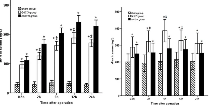
Serum TNF-aand sFas levels
As shown in Fig. 2, the serum level of TNF-a was markedly
increased during warm ischemia/reperfusion, reaching a plateau 12 h after operation. Pretreatment with GdCl3 significantly
decreased this level at 2, 6, 12 and 24 h after operation (P,0.05). In addition, the sFas level was increased in the animals induced with bile duct warm ischemia/reperfusion injury. Preadminstration of GdCl3 led to a significant increase in sFas
levels between 2 and 12 h after operation (P,0.05).
Caspase-3 Activity
At 2, 6, 12 and 24 h after operation, the Caspase-3 activity in GdCl3group was lower than in the control group (P,0.05), but
there was no significant difference between the GdCl3and control
groups at 0.5 hour. The Caspase-3 activity of both groups gradually increased and reached a peak between 6 and 12 hours (Table 1).
Apoptosis of Bile Duct Cell
The bile duct sections were stained by the TUNEL method to evaluate cell apoptosis. The number of TUNEL-stained cells increased in the ischemia/reperfusion groups compared with sham group (P,0.05). Administration of GdCl3prevented the increase
in bile duct apoptosis at 2, 6, 12 and 24 h after surgery (P,0.05) (Table 2).
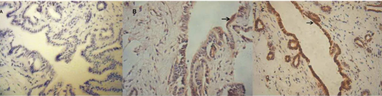
Expression of Fas Protein
To elucidate the mechanisms of apoptosis, the concentration of apoptotic Fas protein was measured by immunohistochemistry in
all three groups (Fig. 3). Warm ischemia/reperfusion injury induced an increase in the number of Fas-positive cells compared with the sham group at all indicated time points (P,0.05). Following pretreatment with GdCl3, the expression of Fas was
significantly lower than that in the control group at 2, 6 and 12 h after surgery (P,0.05), but was higher than that in the sham group (Fig. 4).
Pathological Examination
Statistical significance was found among groups. The bile duct showed a normal appearance in sham group, and more marked histological changes occurred in control group. The injury at 6 hour after operation was more serious than other time points (Fig. 5).
Discussion
Recent studies have demonstrated that GdCl3 can prevent
alcoholic liver injury, minimize ischemia/reperfusion injury, and prevent primary graft dysfuction after experimental liver trans-plantation [10,15,16]. However, the mechanism involved is still incompletely understood; it may be partly related to cell apoptosis. In this study, we demonstrated by the TUNEL method that the number of apoptotic bile duct cells increased markedly in the early stage of warm ischemia/reperfusion injury, and these number decreased markedly after GdCl3treatment. These findings suggest
that GdCl3 plays an important role in suppressing bile duct cell
apoptosis during the early stage of warm ischemia/reperfusion injury.
Serum enzymes are key indicators with regard to bile duct cells injury. In the present study, ALT, ALP and TBIL were markedly elevated during the first 12 h, and then slowly decreased, and the levels of ALT, ALP and TBIL were significantly increased in the control group. Likewise, in biliary pathomorphologic changes, the biliary tract injury in GdCl3group was less serious than that in the
control group. The analysis of serum enzymes and pathomorpho-logic changes showed that the bile duct ischemia/reperfusion injury took place in the early stage, but recovery occurred in the middle and late stage. Meanwhile, the administration of GdCl3
resulted in a significant decrease in serum enzyme activity. As we known, KC participates in many pathophysiologic responses after reperfusion, including the release of activation factors and free
Figure 1. Serum transaminases concentrations in different groups at different time points.Serum levels of ALT, ALP and TBIL were dramatic increased at the early stage of warm ischemia/reperfusion injury. Serum transaminases concentrations were decreased by preadminstration of GdCl3.*Significant increase compared with the sham group (P,0.05);{
statistically significant difference between the GdCl3and control groups (P,0.05). Error bars represent standard deviations.
radicals. The activation factors and free radicals may interaction to decrease the prostaglandin production at that stage. These changes then activated phospholipase A2, resulting in endothelial
cells degeneration and blood coagulation in the biliary tract microcirculation [19]. In addition, since the blood supply to the extrahepatic bile duct is derived solely from the hepatic artery [20], bile duct injury was aggravated.
Likewise, in biliary pathomorphologic changes, the bile duct injury in GdCl3 group was less serious than that in the control
group. To further elucidate the precise mechanism of GdCl3, we
investigated the regulators of apoptosis. Raised TNF-a levels
strongly induced apoptosis and necrosis [10,13], while TNF-a
triggered leukocyte chemotaxis and activated many of the proteins involved in apoptosis, such as the proteases 3 and caspase-8 [21]. Activated caspase-3, along with mitochondria cytochrome-C released into the cytoplasm, cleaved cellular substrates to
morphological changes in cells and nuclei during apoptosis [22]. Our data demonstrated that TNF-awas overproduced during the
early phase of warm ischemia/reperfusion injury while pretreat-ment with GdCl3 significantly down-regulated its production,
roughly in accordance with the findings from the TUNEL assay. With regard to liver transplantation, KC have been identified as the critical source of TNF-a [23]. GdCl3 competitively binded
with the KC calcium receptor to block the activity of nuclear factor-kappa B (NF-kB), consequently inhibiting the transcription
of TNF-a[10,13,14,24].
The Fas-Fas ligand is another pathway associated with bile duct cell apoptosis during warm ischemia/reperfusion. An increase in sFas inhibits apoptosis in Fas-expressing cells by binding either the Fas ligand (FasL) or membrane-bound Fas or by interacting with other proteins expressed on these cells [25]. In this study, our data Figure 2. Time course of changes in the TNF-aand sFas levels.TNF-awas significantly increased and reached a peak at 12 h in both ischemia/ reperfusion groups. TNF-awas significantly lower in the GdCl3group than in the control group except at 0.5 h, while serum sFas level was increased in animals with ischemia/reperfusion injury. Preadminstration of GdCl3led to a significant increase in sFas level between 2 and 12 h after operation. doi:10.1371/journal.pone.0052743.g002
Table 1.The Caspase-3 activity in the three experimental groups at different time points.
Group Time point (h)
0.5 2 6 12 24
Sham 5.8061.37 6.4560.53 5.8460.93 5.6761.01 6.3461.12
GdCl3 7.1261.78 8.6962.33{
10.1662.30*{
11.6961.99*{
11.3163.14*{
Control 9.5461.98* 11.98
62.21* 14.12
61.21* 15.92
62.66* 15.03 62.59*
F 4.50 7.38 22.73 21.39 10.34
P 0.04 0.01 ,0.01 ,0.01 ,0.01
P,0.05 was considered significant.
*Significant increase compared with the sham group;
{
significant reduction compared with the control group. doi:10.1371/journal.pone.0052743.t001
Table 2.Apoptosis of bile duct cells in the three experimental groups at different time points.
Group Time point (h)
0.5 2 6 12 24
Sham 2.0660.78 2.3760.27 2.2960.29 2.3260.21 2.3060.20
GdCl3 14.6861.06* 16.99 61.39*{
18.2861.68*{
17.3161.23*{
16.5461.11*{
Control 16.4861.70* 21.83
61.93* 25.46
62.16* 24.98
62.06* 24.56 61.78*
F 124.23 159.98 168.59 208.99 261.79
P ,0.01 ,0.01 ,0.01 ,0.01 ,0.01
Quantitative analysis of TUNEL-stained cell was conducted by apoptosis index (AI).
*Significant increase compared with the sham group;
{significant reduction compared with the control group.
showed that pretreatment with GdCl3clearly increased sFas levels
in rats with ischemia/reperfusion injury. A correlation was also found between TUNEL assay and serum sFas levels. The results demonstrated that GdCl3 may activate a negative feedback
mechanism during warm ischemia/reperfusion injury by increas-ing sFas, aimed at prevention of additional cell loss.
To further explore the roles of GdCl3in bile duct apoptosis, we
measured Fas protein by immunohistochemistry. Fas-mediated apoptosis is regarded as an important effector process in progressive bile duct loss [26]. Binding of Fas to its ligand results in receptor cross-linking and apoptosis of Fas-positive cells via cellular pathways, including receptor oligomerization and recruit-ment of the Fas-associated protein with the death domain, which mediates the activation of the proteolytic activity of caspase-8 and other downstream caspases, such as caspase-3 [7,27]. In the present study, Fas immunoreactivity was seen in both ischemia/
reperfusion groups, and its expression was more markedly enhanced in the control group than in the GdCl3 group. The
caspase-3 results conformed roughly with the apoptosis from the TUNEL method. After the operation for 2, 6, 12 and 24 hours, the Caspase-3 activity in GdCl3 group was lower than in the
control group. GdCl3suppressed the expression of Fas in bile duct
cells during ischemia/reperfusion; this may be related to the suppression of cytokine-induced KC activation. As mentioned above, the apoptotic process involves various genes, proteins, and activation factors. In the study, pretreatment with GdCl3 may
largely decrease the caspase-3 activity, and some researchers have implied that caspase-3 cleavage is strongly augmented soon after reperfusion of liver grafts [10]. So we conjecture GdCl3 could
depress the expression of Fas protein and caspase-3 activity to suppress bile duct apoptosis.
In summary, the present study demonstrated that GdCl3
regulates bile duct apoptosis after warm ischemia/reperfusion injury. These effects may be related to the suppression of the Fas-Figure 3. Immunohistochemical detection of Fas in the sham and ischemia/reperfusion groups.Paraffin-embedded sections from the sham (A), GdCl3(B), and control groups (C) at 6 h following ischemia/reperfusion were reacted with anti-Fas serum. Arrows indicate Fas-positive cells (magnification6400).
doi:10.1371/journal.pone.0052743.g003
Figure 4. Distribution of Fas-positive cells in serial bile duct samples.The percentage of positive cells in GdCl3and control groups increased slowly for the first 6 hours and reached a peak at 6 hour, but the rate of increase in GdCl3 group was slower than in the control group.
doi:10.1371/journal.pone.0052743.g004
Figure 5. Comparison of pathomorphology in different groups.
In control and GdCl3groups, the main injury of bile duct was at 6 hour after surgery. Pretreatment with GdCl3may attenuate the bile duct injury during the ischemia/reperfusion.
FasL pathway and the inhibition of the caspase-3. These findings provide strong evidence that GdCl3 protects the rat bile duct
against early warm ischemia/reperfusion injury during liver transplantation.
Author Contributions
Conceived and designed the experiments: JZ. Performed the experiments: BW BLZ ZLC. Analyzed the data: BW QZ JZ. Contributed reagents/ materials/analysis tools: BLZ. Wrote the paper: BW QZ.
References
1. Wang MF, Jin ZK, Chen DZ, Li XL, Zhao X, et al. (2011) Risk factors of severe ischemic biliary complications after liver transplantation. Hepatobiliary Pancreat Dis Int 10: 374–379.
2. Heidenhain C, Pratschke J, Puhl G, Neumann U, Pascher A, et al. (2010) Incidence of and risk factors for ischemic-type biliary lesions following orthotopic liver transplantation. Transpl Int 23: 14–22.
3. Singhal A, Stokes K, Sebastian A, Wright HI, Kohli V (2010) Endovascular treatment of hepatic artery thrombosis following liver transplantation. Transpl Int 23: 245–256.
4. de Vera ME, Lopez-Solis R, Dvorchik I, Campos S, Morris W, et al. (2009) Liver transplantation using donation after cardiac death donors: long-term follow-up from a single center. Am J Transplant 9: 773–781.
5. Monbaliu D, Vekemans K, Hoekstra H, Vaahtera L, Libbrecht L, et al. (2009) Multifactorial biological modulation of warm ischemia reperfusion injury in liver transplantation from non-heart-beating donors eliminates primary nonfunction and reduces bile salt toxicity. Ann Surg 250: 808–817.
6. Paulsen M, Janssen O (2011) Pro- and anti-apoptotic CD95 signaling in T cells. Cell Commun Signal 9: 7.
7. Strasser A, Jost PJ, Nagata S (2009) The many roles of FAS receptor signaling in the immune system. Immunity 30: 180–192.
8. Cag M, Audet M, Saouli AC, Panaro F, Piardi T, et al. (2010) Does arterialisation time influence biliary tract complications after orthotopic liver transplantation? Transplant Proc 42: 3630–3633.
9. Ban D, Kudo A, Sui S, Tanaka S, Nakamura N, et al. (2009) Decreased Mrp2-dependent bile flow in the post-warm ischemic rat liver. J Surg Res 153: 310– 316.
10. Rentsch M, Puellmann K, Sirek S, Iesalnieks I, Kienle K, et al. (2005) Benefit of Kupffer cell modulation with glycine versus Kupffer cell depletion after liver transplantation in the rat: effects on postischemic reperfusion injury, apoptotic cell death graft regeneration and survival. Transpl Int 18: 1079–1089. 11. Tian Y, Jochum W, Georgiev P, Moritz W, Graf R, et al. (2006) Kupffer
cell-dependent TNF-alpha signaling mediates injury in the arterialized small-for-size liver transplantation in the mouse. Proc Natl Acad Sci U S A 103: 4598–4603. 12. Liu P, McGuire GM, Fisher MA, Farhood A, Smith CW, et al. (1995) Activation of Kupffer cells and neutrophils for reactive oxygen formation is responsible for endotoxin-enhanced liver injury after hepatic ischemia. Shock 3: 56–62. 13. Esposito E, Cuzzocrea S (2009) TNF-alpha as a therapeutic target in
inflammatory diseases, ischemia-reperfusion injury and trauma. Curr Med Chem 16: 3152–3167.
14. Lee CM, Yeoh GC, Olynyk JK (2004) Differential effects of gadolinium chloride on Kupffer cells in vivo and in vitro. Int J Biochem Cell Biol 36: 481–488. 15. Schneider L, Hackert T, Longerich T, Hartwig W, Fritz S, et al. (2010) Effects of
gadolinium chloride and glycine on hepatic and pancreatic tissue damage in alcoholic pancreatitis. Pancreas 39: 502–509.
16. Li JY, Gu X, Zhang WH, Jia S, Zhou Y (2009) GdCl3 abates hepatic ischemia-reperfusion injury by inhibiting apoptosis in rats. Hepatobiliary Pancreat Dis Int 8: 518–523.
17. Henrich D, Lehnert M, Herzog C, Niederlaender S, Relja B, et al. (2008) Differential effects of GdCl3- or MDP treatment on rat liver microcirculation and gene expression in the hepatic non-parenchymal cell fraction in LPS shock. Microcirculation 15: 427–439.
18. Zhao HF, Zhang GW, Zhou J, Lin JH, Cui ZL, et al. (2009) Biliary tract injury caused by different relative warm ischemia time in liver transplantation in rats. Hepatobiliary Pancreat Dis Int 8: 247–254.
19. Ohkohchi N (2001) Suppression of Kupffer cell function is a key for liver transplantation from the non-heart-beating donor. Transplant Proc 33: 3728– 3731.
20. Guichelaar MM, Benson JT, Malinchoc M, Krom RA, Wiesner RH, et al. (2003) Risk factors for and clinical course of non-anastomotic biliary strictures after liver transplantation. Am J Transplant 3: 885–890.
21. Montalvo-Jave EE, Escalante-Tattersfield T, Ortega-Salgado JA, Pina E, Geller DA (2008) Factors in the pathophysiology of the liver ischemia-reperfusion injury. J Surg Res 147: 153–159.
22. Nagata S (1999) Fas ligand-induced apoptosis. Annu Rev Genet 33: 29–55. 23. Giakoustidis D, Papageorgiou G, Iliadis S, Giakoustidis A, Kostopoulou E, et al.
(2006) The protective effect of alpha-tocopherol and GdCl3 against hepatic ischemia/reperfusion injury. Surg Today 36: 450–456.
24. Crescenzi E, Pacifico F, Lavorgna A, De Palma R, D’Aiuto E, et al. (2011) NF-kappaB-dependent cytokine secretion controls Fas expression on chemotherapy-induced premature senescent tumor cells. Oncogene 30: 2707–2717. 25. Cardinal H, Brophy JM, Bogaty P, Joseph L, Hebert MJ, et al. (2010) Usefulness
of soluble fas levels for improving diagnostic accuracy and prognosis for acute coronary syndromes. Am J Cardiol 105: 797–803.
26. Iwata M, Harada K, Katayanagi K, Saito T, Kaneko S, et al. (2003) Apoptosis of murine cultured biliary epithelial cells induced by glycochenodeoxycholic acid involves Fas receptor and its ligand. Hepatol Res 25: 329–342.