VEGF Expression after Warm Ischemia and Reperfusion in
Rat Liver
Mei Zhang1., Wujun Li2., Liang Yu1
, Shengli Wu1*
1Department of Hepatobiliary Surgery, the First Affiliated Hospital of Xi’an Jiaotong University, Xi’an, P.R. China,2Department of General Surgery, the First Affiliated Hospital of Xi’an Medical University, Xi’an, P.R. China
Abstract
Background:Hypoxia-inducible factor-1a (HIF-1a) is overexpressed in many human tumors and their metastases, and is closely associated with a more aggressive tumor phenotype. The aim of the present study was to investigate the effect of resveratrol (RES) on the expression of ischemic-induced HIF-1aand vascular endothelial growth factor (VEGF) in rat liver.
Methods:Twenty-four rats were randomized into Sham, ischemia/reperfusion (I/R), and RES preconditioning groups. I/R was induced by portal pedicle clamping for 60 minutes followed by reperfusion for 60 minutes. The rats in RES group underwent the same surgical procedure as I/R group, and received 20 mg/kg resveratrol intravenously 30 min prior to ischemia. Blood and liver tissue samples were collected and subjected to biochemical assays, RT-PCR, and Western blot assays.
Results:I/R resulted in a significant (P,0.05) increase in liver HIF-1aand VEGF at both mRNA and protein levels 60 minutes after reperfusion. The mRNA and protein expressions of HIF-1a and VEGF decreased significantly in RES group when compared to I/R group (P,0.05).
Conclusion:The inhibiting effect of RES on the expressions of HIF-1aand VEGF induced by I/R in rat liver suggested that HIF-1a/VEGF could be a promising drug target for RES in the development of an effective anticancer therapy for the prevention of hepatic tumor growth and metastasis.
Citation:Zhang M, Li W, Yu L, Wu S (2014) The Suppressive Effect of Resveratrol on HIF-1aand VEGF Expression after Warm Ischemia and Reperfusion in Rat Liver. PLoS ONE 9(10): e109589. doi:10.1371/journal.pone.0109589
Editor:Aditya Bhushan Pant, Indian Institute of Toxicology Reserach, India
ReceivedMay 26, 2014;AcceptedSeptember 2, 2014;PublishedOctober 8, 2014
Copyright:ß2014 Zhang et al. This is an open-access article distributed under the terms of the Creative Commons Attribution License, which permits unrestricted use, distribution, and reproduction in any medium, provided the original author and source are credited.
Data Availability:The authors confirm that all data underlying the findings are fully available without restriction. All relevant data are within the paper.
Funding:The authors have no support or funding to report.
Competing Interests:The authors have declared that no competing interests exist.
* Email: [email protected]
.These authors contributed equally to this work.
Introduction
Hepatocellular carcinoma (HCC) is one of the most common malignancies in the world [1]. Surgical resection and liver transplantation are conventional treatment modalities that can offer long-term survival for patients with HCC. However, the high incidence of tumor recurrence and metastasis after liver surgery remains a major problem [2]. Hepatic ischemia/reperfusion (I/R) injury is a phenomenon inevitable during liver surgery and promotes liver tumor growth and metastases through activation of cell adhesion, invasion, and angiogenesis pathways [3]. Hypoxia-inducible factor-1 alpha (HIF-1a) is one of the key regulators of hypoxia/ischemia [4]. Accumulating evidence indicated that the outgrowth of hepatic micrometastases is stimulated by I/R injuries during surgery and may at least in part, be stimulated by an increased HIF-1astabilization [5,6]. HIF-1astimulates transcrip-tion of multiple genes, including angiogenic vascular endothelial growth factor (VEGF) [7], an important growth factor involved in
tumor angiogenesis [8], and HIF-1a/VEGF pathway have been implicated in the development of multiple tumors [9–11].
Resveratrol (trans-3,49,5-trihydroxystilbene, RES) is a natural polyphenolic phytoalexin found in various plant species [12]. Numerous studies have demonstrated its diverse pharmacological activities, including antitumor and chemopreventive properties [13]. Studies in animal models have demonstrated that RES exerts potent anticarcinogenic effects via affecting diverse cellular events associated with tumor initiation, promotion, and progression [14]. Recently, RES has been found to inhibit angiogenesis and its antiangiogenic effects had been investigated in the setting of in vitro hypoxia, but the underlying mechanism of its antiangiogenic activity remains unclear [15].
Materials and Methods
Animals
Male Sprague-Dawley (SD) rats 9–10 weeks old weighing 190– 210 g were purchased from the Animal Center of Xi’an Jiaotong University (Xi’an, China). All rats were allowed free access to water and standard laboratory chow. Before operation the rats were fasted for 12 h and only allowed free access to water. Care was provided in accordance with the ‘‘Guide for the care and use of laboratory animals’’ (NIH publication No. 85–23, revised in 1996). The study was approved by the Xi’an Jiaotong University Institutional Animal Care and Use Committee.
Reagents
Resveratrol and dimethyl sulfoxide (DMSO) were purchased from Sigma Chemical Co., USA. RPMI-1640 was from Gibco-BRL, USA. The RES was dissolved and sterilized in DMSO and then diluted in RPMI-1640 to 4 mg/mL.
Experimental design
Rats were anesthetized with an intraperitoneal injection of pentobarbital sodium (50 mg/kg; Nembutal, Abbott Laboratories, North Chicago, IL). Twenty-four rats were randomly divided into three experimental groups (eight rats in each group) as follows: Sham operation group, a 6-cm midline abdominal incision was made to expose the liver and laparotomy was carried out for 60 min with no hepatic ischemia; I/R group: I/R was induced by portal pedicle clamping with an atraumatic microvascular clip for 60 minutes followed by removal of the clip for 60 minutes, and rats received an equivalent volume of placebo solution (RPMI-1640); and RES preconditioning group: rats in this group underwent the same surgical procedure as I/R group, and received one-shot injection of RES (4 mg/mL) at a dose of 20 mg/kg body weight through vena dorsalis penis 30 min prior to ischemia. All rats were euthanized with an overdose of pentobarbital (100 mg/kg IV) followed by exsanguinations at 60 min after clip removal, while Sham operation group animals were killed at the same time points after surgery. The liver was removed, and the inferior vena cava was cannulated so that blood samples could be taken from its suprahepatic segment. Blood samples were centrifuged at 4000 r/min at 4uC for 3 min and serum was taken and immediately processed. Liver tissues were snap-frozen in liquid nitrogen and stored at 280uC for further analysis.
Measurement of Serum Liver Enzymes
In all three groups serum alanine aminotransaminase (ALT), alkaline phosphates (ALP), and total bilirubin (TBIL) concentra-tions were measured using Hitachi AU5400 automatic biochem-ical analyzer (Hitachi Corp., Japan) and Roche Diagnostics kit (Roche, USA) at 60 min after reperfusion.
Real-time RT-PCR
After homogenization of liver tissue by the use of a MM301 Mixer Mill (Retsch, Haan, Germany), total cellular RNA was extracted from the liver tissue by using TriPure Reagent Isolation Reagent (Roche). RNA concentration was determined using UV spectrophotometer. Five hundred nanograms of RNA were reverse-transcribed and amplified to cDNA using real time RT-PCR with iScript One-Step RT-RT-PCR Kit with SYBR Green (BioRad, USA). b-actin gene was used as an internal control. Primer sequences used in this study were designed using NCBI Primer-Blast (http://www.ncbi.nlm.nih.gov/tools/primer-blast) as follows: for the HIF-1a, sense 59
-ACTGCACAGGCCACATT-CAT-39 and antisense 59-CGAGGCTGTGTCGACTGAGA-39; for the VEGF, sense 59-AGGCGAGGCAGCTTGAGTTA-39
and antisense 59-CTGTCGACGGTGACGATGGT-39; for theb -actin, sense 59-CCTAGGCACCAGGGTGTGAT-39 and anti-sense 59-TTGGTGACAATGCCGTGTTC-39. The initial dena-turation phase was 3 min at 95uC followed by 39 cycles of denaturation at 95uC for 10 s and annealing at 55uC for 30 s. Relative quantification of PCR products was performed after normalization tob-actin.
Western blot analysis
After homogenization of liver tissue by the use of a MM301 Mixer Mill (Retsch, Haan, Germany), total cellular protein was extracted from the liver tissue by using tissue protein extraction buffer (Pierce, Rockford, IL, USA) containing protease inhibitors (Protease Inhibitor Cocktail 100X, Pierce). Protein concentrations were determined and the samples were subjected to sodium dodecyl sulfate/polyacrylamide gel electrophoresis and transferred to a nitrocellulose membrane (ECL, Amersham, Buckingham-shire, UK). The membranes were then blocked for 60 min and subsequently incubated with primary antibodies (1:3000) overnight at 4uC prior to incubation with anti-mouse IgG conjugated to horseradish peroxidase (1:6000) for 120 min at room temperature. Finally, the signals were detected using an enhanced chemilumi-nescence detection kit (Amersham, Piscataway, NJ, USA). The chemiluminescent signal was captured by a UVP BioSpectrum500 imaging system (UVP, Upland, CA, USA). Protein expression was quantified by densitometry and normalized tob-actin expression. Anti-HIF-1a, anti-VEGF, and anti-b-actin antibodies were obtained from Santa Cruz Biotechnology, Inc. (Santa Cruz, CA, United States).
Statistical analysis
All data are presented as the means6standard deviation of the mean. Statistical analysis was performed using SPSS 16.0 software. Differences among groups were tested by one-way analysis of variance (ANOVA) with post-hoc Student-Newman-Keuls meth-od. A P-value ,0.05 was considered to indicate a statistically significant result.
Results
Liver function
Serum ALT, ALP and TBIL concentrations in different groups are shown in Table. 1. At 60 min post reperfusion, serum ALT, ALP and TBIL levels were significantly higher in I/R group than in the sham operation group (allP,0.05). Pretreatment with RES (20 mg/kg) showed a significant decrease in levels of serum ALT, ALP and TBIL than in I/R group (allp,0.05).
HIF-1aexpression
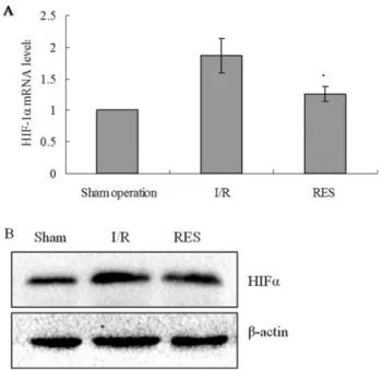
The expression of HIF-1a in livers of experimental rats was examined by real-time RT-PCR and western blotting methods. Compared to sham operation group, both mRNA and protein expressions of HIF-1awere significantly increased in the livers of rats in I/R group (all P,0.05). Compared to I/R group, a significant reduction in HIF-1a mRNA and protein expression levels in RES preconditioning group was observed (allP,0.05; Fig. 1A and B).
VEGF expression
expressions of VEGF were significantly increased in the livers of rats in I/R group (all P,0.05). Compared to I/R group, a significant reduction in VEGF mRNA and protein expression levels in RES preconditioning group was observed (all P,0.05; Fig. 2A and B).
Discussion
Hepatic I/R injury may cause metabolic and structural hepatic damage [16] and has been proposed as a key clinical problem associated with liver transplantation and major liver surgery [17]. It involves a complex series of events, such as mitochondrial deenergization, adenosine-59-triphosphate depletion, alterations of electrolyte homeostasis, as well as Kupffer cell activation, oxidative stress changes and upregulation of proinflammatory cytokine signaling [18]. At the same time, cellular response to low tissue oxygen concentrations is mediated by HIF-1 to protect liver from I/R injury.
HIF-1 is composed of HIF-1aand HIF-1bsubunits [19]. HIF-1awas firstly described by Semenza in 1992 and the expression of which is tightly regulated by low oxygen tension [20], whereas
HIF-1b is constitutively expressed. Under normoxic conditions, HIF-1a protein is induced and continuously degraded by the ubiquitin-proteasome pathway in the cytoplasmic cellular com-partment. However, under hypoxic conditions the blockade of degradation lead to the remarkable accumulation and transloca-tion of HIF-1a protein to the nucleus, where it heterodimerizes with HIF-1b. This HIF-1 complex then initiates transcriptional activation via binding with hypoxia responsive elements in the promoter regions of target genes [21], including VEGF, erythro-poietin, glycolytic enzymes, transferrin and a variety of other proteins that are important for adaptation and survival under hypoxic stress [22].
VEGF, an immediate downstream target gene of HIF-1a, plays a pivotal role in tumor angiogenesis [23], especially under conditions of intratumoral hypoxia. It promotes the proliferation of vessel endothelial cells, inhibits the apoptosis of vessel endothelial cells, and stimulates the formation of blood vessels [20]. Furthermore, it stimulates the production of hepatocyte growth factor (HGF), which is regarded as an initiator of liver
Table 1.Serum biochemical parameters in different groups (mean6SD).
Groups n ALT (U/L) ALP(U/L) TBIL (umol/L)
Sham 8 43.85615.64 85.39621.65 6.5761.12
I/R 8 1465.506316.37* 415.71668.43* 35.4165.87*
RES 8 837.656205.53** 297.42623.44** 21.4763.69**
*p,0.05vsSham operation group; **p,0.05vsI/R group.
doi:10.1371/journal.pone.0109589.t001
Figure 1. Expression of HIFamRNA in rat livers.(A) HIFamRNA levels were determined by real-time RT-PCR. Relative fold induction for HIFamRNA (means6SD) in I/R and RES group rat livers is presented relative to the expression in Sham operation group rat livers (*P,0.05 compared with I/R group). (B) Western blot analysis for HIFaprotein expression in the indicated groups. b-actin was used as a loading control.
doi:10.1371/journal.pone.0109589.g001
Figure 2. Expression of VEGF mRNA in rat livers.(A) VEGF mRNA levels were determined by real-time RT-PCR. Relative fold induction for VEGF mRNA (means6SD) in I/R and RES group rat livers is presented relative to the expression in Sham operation group rat livers (*P,0.05 compared with I/R group). (B) Western blot analysis for VEGF protein expression in the indicated groups. b-actin was used as a loading control.
regeneration [24]. Therefore, a stimulation of HIF-1a via liver ischemia, could be a double-edged sword; i.e., it protects the liver against I/R injuries, but a side effect could be the promotion of recurrence and metastasis of HCC through angiogenesis.
RES has been reported to have several biologic effects such as a potent antioxidative effect via prevention of lipid peroxidation, anti-platelet activity, an estrogenic activity, and anti-inflammatory activity attributed to cyclooxgenase inhibition [25]. Previous study had reported the hepatoprotective effects of RES in hepatic I/R and the protective effects of RES may be associated with its antioxidant activity and free radical scavenging activity which are released during the reperfusion period [26]. In recent years, RES has been found to inhibit tumor angiogenesis [27], but the mechanism of its antiangiogenic activity remains to be elucidated. Yu et al. reported that RES inhibits VEGF expression of HepG2 cells through a NF-kappa B-mediated mechanism [28]. Cao et al. reported that RES may inhibit human ovarian cancer progression and angiogenesis by inhibiting HIF-1a and VEGF expression through multiple mechanisms, including the inhibition of AKT and mitogen-activated protein kinase activation, the inhibition of several protein translational regulators, and inducing HIF-1a protein degradation through the proteasome pathway [29]. Zhang et al. showed that RES directly inhibits hypoxia-mediated HIF-1a protein accumulation by inhibiting its degradation via the proteasomal pathway in both SCC-9 and HepG2 cells [30]. In the present study, as expected, serum ALT, ALP and TBIL levels were significantly higher in I/R group than in the sham operation group at 60 min post reperfusion, while pretreatment with RES (20 mg/kg) showed a significant decrease in levels of serum ALT,
ALP and TBIL than in I/R group. Moreover, we further showed that the mRNA and protein expressions of HIF-1a and VEGF were increased significantly in rats subjected to 60 minutes of warm liver ischemia and 60 minutes of reperfusion compared to the control group, while the mRNA and protein expressions of HIF-1a and VEGF decreased significantly in RES group when compared to I/R group. These findings affirmed the results of previous studies showing the hepatoprotective effects of RES in hepatic I/R. More importantly, we provided the first evidence supporting the antiangiogenic effects of RES in the setting of in vivo hypoxia, which are consistent with previous findings, suggesting that RES inhibits angiogenesis at least partly through regulating the expressions of HIF-1a and VEGF. However, additional studies are needed to identify the detailed mechanisms by which RES regulated the expressions of HIF-1aand VEGF.
Taken together, our present study has provided evidence that RES, exerts its antiangiogenic effects through inhibiting HIF-1a and its downstream target gene, VEGF, in a rat model of hepatic I/R injury. HIF-1a/VEGF axis, as a key regulator of tumor growth and metastasis, could be a promising drug target for RES in the development of an effective anticancer therapy for the prevention of hepatic tumor growth and metastasis.
Author Contributions
Conceived and designed the experiments: SW. Performed the experiments: MZ WL. Analyzed the data: LY. Contributed reagents/materials/analysis tools: MZ. Wrote the paper: SW.
References
1. Bosch FX, Ribes J, Dı´az M, Cle´ries R (2004) Primary liver cancer: worldwide incidence and trends. Gastroenterology 127: S5–S16.
2. Zhang Y, Shi ZL, Yang X, Yin ZF (2014) Targeting of circulating hepatocellular carcinoma cells to prevent postoperative recurrence and metastasis. World J -Gastroenterol 20: 142–147.
3. Li CX, Shao Y, Ng KT, Liu XB, Ling CC, et al. (2012) FTY720 suppresses liver tumor metastasis by reducing the population of circulating endothelial progenitor cells. PLoS One 7: e32380.
4. Cursio R, Miele C, Filippa N, Van Obberghen E, Gugenheim J (2008) Liver HIF-1 alpha induction precedes apoptosis following normothermic ischemia-reperfusion in rats. Transplant Proc 40: 2042–2045.
5. van der Bilt JD, Kranenburg O, Nijkamp MW, Smakman N, Veenendaal LM, et al. (2005) Ischemia/reperfusion accelerates the outgrowth of hepatic micrometastases in a highly standardized murine model. Hepatology 42: 165– 175.
6. Knudsen AR, Kannerup AS, Grønbæk H, Andersen KJ, Funch-Jensen P, et al. (2011) Effects of ischemic pre- and postconditioning on HIF-1a, VEGF and TGF-bexpression after warm ischemia and reperfusion in the rat liver. Comp Hepatol 10: 3.
7. Boros P, Tarcsafalvi A, Wang L, Megyesi J, Liu J, et al. (2001) Intrahepatic expression and release of vascular endothelial growth factor following orthotopic liver transplantation in the rat. Transplantation 72: 805–811.
8. Tamagawa K, Horiuchi T, Uchinami M, Doi K, Yoshida M, et al. (2008) Hepatic ischemia-reperfusion increases vascular endothelial growth factor and cancer growth in rats. J Surg Res 148: 158–163.
9. Chai ZT, Kong J, Zhu XD, Zhang YY, Lu L, et al. (2013) MicroRNA-26a inhibits angiogenesis by down-regulating VEGFA through the PIK3C2a/Akt/ HIF-1apathway in hepatocellular carcinoma. PLoS One 8: e77957. 10. An X, Xu G, Yang L, Wang Y, Li Y, et al. (2014) Expression of
hypoxia-inducible factor-1a, vascular endothelial growth factor and prolyl hydroxylase domain protein 2 in cutaneous squamous cell carcinoma and precursor lesions and their relationship with histological stages and clinical features. J Dermatol 41: 76–83.
11. Shi D, Xie F, Zhang Y, Tian Y, Chen W, et al. (2014) TFAP2A Regulates Nasopharyngeal Carcinoma Growth and Survival by Targeting HIF-1a Signaling Pathway. Cancer Prev Res (Phila) 7: 266–277.
12. Soleas GJ, Diamandis EP, Goldberg DM (1997) Wine as a biological fluid: history, production, and role in disease prevention. J Clin Lab Anal 11: 287– 313.
13. Singh CK, George J, Ahmad N (2013) Resveratrol-based combinatorial strategies for cancer management. Ann N Y Acad Sci 1290: 113–121.
14. Aziz MH, Kumar R, Ahmad N (2003) Cancer chemoprevention by resveratrol: in vitro and in vivo studies and the underlying mechanisms. Int J Oncol 23: 17– 28.
15. Wen D, Huang X, Zhang M, Zhang L, Chen J, et al. (2013) Resveratrol attenuates diabetic nephropathy via modulating angiogenesis. PLoS One 8: e82336.
16. Shin T, Kuboki S, Huber N, Eismann T, Galloway E, et al. (2008) Activation of peroxisome proliferator-activated receptor-gamma during hepatic ischemia is age-dependent. J Surg Res 147: 200–205.
17. Ito K, Ozasa H, Noda Y, Koike Y, Arii S, et al. (2008) Effect of non-essential amino acid glycine administration on the liver regeneration of partially hepatectomized rats with hepatic ischemia/reperfusion injury. Clin Nutr 27: 773–780.
18. Papadopoulos D, Siempis T, Theodorakou E, Tsoulfas G (2013) Hepatic ischemia and reperfusion injury and trauma: current concepts. Arch Trauma Res 2: 63–70.
19. Wang GL, Jiang BH, Rue EA, Semenza GL (1995) Hypoxia-inducible factor 1 is a basic-helix-loop-helix-PAS heterodimer regulated by cellular O2 tension. Proc Natl Acad Sci U S A 92: 5510–5514.
20. Xu LF, Ni JY, Sun HL, Chen YT, Wu YD (2013) Effects of hypoxia-inducible factor-1a silencing on the proliferation of CBRH-7919 hepatoma cells. World J Gastroenterol 19: 1749–1759.
21. Semenza GL, Jiang BH, Leung SW, Passantino R, Concordet JP, et al. (1996) Hypoxia response elements in the aldolase A, enolase 1, and lactate dehydrogenase A gene promoters contain essential binding sites for hypoxia-inducible factor-1. J Biol Chem 271: 32529–32537.
22. Vaupel P (2004) The role of hypoxia-induced factors in tumor progression. Oncologist 9: 10–17.
23. Semenza GL (2003) Targeting HIF-1 for cancer therapy. Nat Rev Cancer 3: 721–732.
24. Michalopoulos GK (2007) Liver regeneration. J Cell Physiol 213: 286–300. 25. Wu SL, Yu L, Jiao XY, Meng KW, Pan CE (2006) The suppressive effect of
resveratrol on protein kinase C theta in peripheral blood T lymphocytes in a rat liver transplantation model. Transplant Proc 38: 3052–3054.
26. Gedik E, Girgin S, Ozturk H, Obay BD, Ozturk H, et al. (2008) Resveratrol attenuates oxidative stress and histological alterations induced by liver ischemia/ reperfusion in rats. World J Gastroenterol 14: 7101–7106.
28. Yu HB, Zhang HF, Zhang X, Li DY, Xue HZ, et al. (2010) Resveratrol inhibits VEGF expression of human hepatocellular carcinoma cells through a NF-kappa B-mediated mechanism. Hepatogastroenterology 57: 1241–1246.
29. Cao ZX, Fang J, Xia C, Shi XL, Jiang BH (2004) Trans-3,4,59-trihydrox-ystibene inhibits hypoxia-inducible factor-1aand vascular endothelial growth
factor expression in human ovarian cancer cells. Clin Cancer Res 10: 5253– 5263.