PknE, a Serine/Threonine Protein Kinase of
Mycobacterium tuberculosis
Initiates Survival Crosstalk
That Also Impacts HIV Coinfection
Dinesh Kumar Parandhaman1, Luke Elizabeth Hanna2, Sujatha Narayanan1*
1Department of Immunology, National Institute for Research in Tuberculosis, Chennai, TamilNadu, India,2Department of Clinic Research, HIV division, National Institute for Research in Tuberculosis, Chennai, TamilNadu, India
Abstract
Serine threonine protein kinases (STPK) play a major role in the pathogenesis of Mycobacterium tuberculosis. Here, we examined the role of STPKpknE, using a deletion mutantDpknEin the modulation of intracellular signaling events that favor M. tuberculosis survival. Phosphorylation kinetics of MAPK (p38MAPK, ErkK and SAPK/JNK) was defective in DpknE compared to wild-type infected macrophages. This defective signaling dramatically delayed and reduced the phosphorylation kinetics of transcription factors ATF-2 and c-JUN inDpknEinfected macrophages. MAPK inhibitors instead of reducing the phosphorylation inDpknEinfected macrophages, revealed crosstalks with ErkKsignaling influenced by SAPK/JNK and p38 pathways independently. Modulations in intra cellular signaling altered the expression of coreceptors CCR5 and CXCR4 in DpknE infected macrophages. In conclusion, pknE plays a role in MAPK crosstalks that enables intracellular survival ofM. tuberculosis. This survival strategy also impacts HIV/TB coinfection.
Citation:Parandhaman DK, Hanna LE, Narayanan S (2014) PknE, a Serine/Threonine Protein Kinase ofMycobacterium tuberculosisInitiates Survival Crosstalk That Also Impacts HIV Coinfection. PLoS ONE 9(1): e83541. doi:10.1371/journal.pone.0083541
Editor:Volker Briken, University of Maryland, United States of America
ReceivedApril 4, 2013;AcceptedNovember 5, 2013;PublishedJanuary 8, 2014
Copyright:ß2014 Parandhaman et al. This is an open-access article distributed under the terms of the Creative Commons Attribution License, which permits unrestricted use, distribution, and reproduction in any medium, provided the original author and source are credited.
Funding:DKP was supported by ICMR Senior Research fellowship and the study was carried with intramural funds. The funders had no role in study design, data collection and analysis, decision to publish, or preparation of the manuscript.
Competing Interests:The authors have declared that no competing interests exist. * E-mail: sujatha.sujatha36@gmail.com
Introduction
The global incidence of tuberculosis (TB) caused by Mycobac-terium tuberculosis (MTB) has increased due to the emergence of drug resistant strains and HIV coinfection [1]. The adaptation of MTB in hostile environments is regulated by serine/threonine protein kinases (STPK). Among the 11 STPKs encoded by MTB,
pknE, pknG, pknH, pknI and pknK play a role in its intracellular survival [2–6].
STPKs are prime targets for new drug discovery and share only 30% homology with their human counterparts [7]. The likelihood of STPKs in mediating intracellular signaling events in the host remains elusive. However, two other MTB genes,eisandmPTPB
were reported to play a role in the modulation of host intracellular signaling [8,9].
Mitogen activated protein kinase (MAPK) cascades are evolu-tionarily conserved signaling pathways in eukaryotes that play a role in cell proliferation, cell differentiation, cell movement and cell death [10]. MAPK family is divided into four main subfamilies namely extracellular regulated kinases 1 and 2 (ErkK), Jun N-terminal kinases (JNKs), p38 MAPK and Erk5 [11]. MAPKs were reported to have cooperated signaling with shared substrates [12]. ErkKpathway activated by growth factors and mitogens plays a major role in regulating cell proliferation and differentiation. On the other hand, environmental stress, inflammatory cytokines and stress-dependent apoptosis stimulate p38 and SAPK/JNK path-ways [10]. Activated MAPKs signal the transcription factors to regulate the expression of cytokines and iNOS [10]. Studies on
Mycobacteriumhave shown the suppression of MAPK signaling as a mechanism to prevent macrophage activation [10].
TB predominates in HIV-infected individuals due to weakened immune functions that lead to reactivation of latent MTB. Disease progression in HIV/TB coinfected individuals is accelerated by both MTB and HIV [13]. Cellular components of MTB are known to regulate coreceptors CXCR4 and CCR5 involved in HIV entry [14], but the molecular mechanisms underlying this phenomenon are not well-understood. Previously, we reported that pknE expressed under nitric oxide (NO) stress suppresses multiple apoptotic pathways thereby supporting intracellular survival of MTB and that purified PknE cross-reacts with SAPK/JNK antibody [3].
In the present study, the influence of pknE on intracellular signaling that favors MTB survival and its impact on the outcome of HIV/TB coinfection were studied. Our data shows thatpknEof MTB influences the crosstalk between the MAPK pathways to regulate inflammation and HIV/TB coinfection.
Methods
Bacterial strains and culture conditions
Cell culture, infection, inhibitors and nitrate stress experiments
THP-1 cells were maintained, differentiated and infected as reported earlier [15]. Cells were pretreated for 1 h with inhibitors of Akt (Wortmannin, 100 nM), arginase (Nv -Hydroxy-nor-L-arginine diacetate, 100mM), caspase-8 (Z-IETD-FMK, 25mmol/L), caspase-9 (Z-LEHD-FMK, 25mmol/L), ErkK (PD98059, 20mM), p38 (SB203580, 10mM), SAPK/JNK (SP600125, 10mM) and TP53 (pifithrin-a, 5mmol/L) purchased from Calbiochem, USA, and infected with MTB strains. For nitrate stress experiments, post-infection with MTB, the cells were treated with 10 mM sodium nitroprusside (SNP) as reported earlier [15].
Western blotting
Cell lysates were prepared as reported earlier [15] and the immunoblots were probed with rabbit anti-human polyclonal antibodies (Cell Signaling Technologies) against phospho and non-phospho Akt, p38, ErkKand SAPK/JNK (1:1000) and detected using horseradish peroxidase-conjugated goat anti-rabbit antibody (1:300) (Amersham Biosciences).
Transcription factor ELISA
Nuclear factors were isolated using the procedure reported earlier [16]. Briefly, 16106THP-1 macrophages were lysed using 300ml of buffer A (10 mM HEPES–KOH, pH 7.9, 1.5 mM MgCl2, 10 mM KCl, 0.5 mM DTT, 0.2 mM PMSF), centrifuged for 10 s at 15,000 g and the supernatants were labeled as cytosolic fraction. The cell pellet was resuspended in 200ml of icecold buffer B (20 mM HEPES–KOH, pH 7.9, 25% glycerol, 420 nM NaCl, 1.5 mM MgCl2, 0.2 mM EDTA, 0.5 mM DTT, 0.2 mM PMSF), centrifuged at 15,000 g for 10 s and the supernatants were stored at280uC until use. The concentration of proteins was quantified using bicinchoninic acid method (Sigma).
Trans-AM ATF-2, c-JUN and NF-kB kit from Active Motif (Carlsbad, CA) were used to determine the levels of ATF-2, c-JUN and p65NF-kB in nuclear extract. 2mg of nuclear extract was added to wells coated with oligonucleotides containing the consensus binding site for the respective nuclear factors, followed by addition of primary antibody, horseradish peroxidase-conju-gated secondary antibody and the substrate. The absorbance was read at 450 nm (with a reference wavelength at 650 nm). The specificity of the assay was monitored using competitive binding of wild-type or mutated consensus oligonucleotides before the addition of nuclear extracts.
HIV/TB coinfection
24 h post-infection with MTB, THP-1 cells were infected with 500TCID50of CCR5 (92UG005) and CXCR4 (92UG024)-tropic HIV-1 virus for 2 h at 37uC. Post infection extracellular virus was removed by wash using serum-free RPMI and replenished with RPMI containing 10% FBS. The supernatant was harvested on day 4 to estimate the viral p24 levels by sandwich ELISA (Perkin Elmer). Similarly, monocyte derived macrophages (MDM) were isolated from the blood received from healthy volunteers (Jeevan blood bank, http://www.jeevan.org/blood/index.html) after writ-ten informed consent approved by institutional ethics committee review board (NIRT IEC protocol number 2006 006) and the coinfection experiment was carried out as described above.
Statistics
Statistical analysis was carried out using graph pad prism v5.0. One way and Two way ANOVA were used depending on the data and p value,0.05 was considered statistically significant.
Results
DpknEdecreases phosphorylation of MAPKs
The phosphorylation kinetics of ErkK, p38MAPK and SAPK/ JNK were compared in THP-1-derived macrophages infected with MTB strains Rv,DpknE and CDE (complemented DpknE). Control cells and Lipopolysaccharide (LPS) were used as appropriate controls.
Rv-infected macrophages had increased levels of phosphoryla-tion of ErkK, p38MAPK and SAPK/JNK from 30 min post-infection compared to controls (Figures 1A–C). In contrast,DpknE -infected macrophages abrogated the phosphorylation of ErkKat 240 min (p,0.0001, for all the time periods), reduced the phosphorylation of p38MAPK at 60 min (p,0.0001) and selectively inhibited the phosphorylation of p46 subunit of SAPK/JNK at 120 min (p,0.0001) post infection compared to Rv-infected macrophages (Figures 1A–C). These data reveal that
pknEmodulates the MAPK signaling thereby providing a survival niche for MTB.
DpknEdecreases phosphorylation of ATF-2 and c-JUN while NF-kB is unaltered
Decreased MAPK signaling in DpknE-infected macrophages prompted us to study the expression of transcription factors ATF-2, c-JUN and NF-kB, the final targets for cellular activation.
Phosphorylation of ATF-2 and c-JUN in Rv-infected macro-phages peaked at 60 min and returned to baseline at 240 min post-infection, while inDpknE-infected macrophages it peaked at 120 min and reached baseline levels at 240 min post-infection. Phosphorylation of ATF-2 and c-JUN in LPS-treated macrophag-es peaked at 120 min post-treatment and reached baseline valumacrophag-es at 240 min post-infection (Figures 2A,2B). Phosphorylation kinetics of NF-kB was similar in both Rv and DpknE-infected macrophages (Figure 2C). CDE was able to reverse the altered phosphorylation events observed inDpknE-infected macrophages. This clearly shows that deletion of pknE reduces cellular inflammation due to delayed and reduced activation of transcrip-tion factors and reconfirms our previous finding that pknE
contributes to inflammatory responses [15].
MAPK signaling inDpknE-infected macrophages is unaltered in the presence of pathway-specific inhibitors
The inhibition of MAPK signaling byDpknE-infected macro-phages was assessed using pathway-specific inhibitors. Surprising-ly, p38MAPK, ErkK, SAPK/JNK or Akt inhibitors were unable to suppress the phosphorylation inDpknE-infected macrophages as observed in macrophages infected with Rv (data not shown).
DpknEinduces crosstalk between ErkKand SAPK/JNK
signaling
The absence of inhibition in the presence of MAPK and Akt inhibitors observed in DpknE-infected macrophages suggests two plausible facts; either DpknE induces phosphatases of MAPK differentially or that it initiates a crosstalk response. We chose to analyze ErkKcrosstalk based on its reduced expression inDpknE -infected macrophages and its role in cellular survival.
inhibitor did not affect SAPK/JNK phosphorylation, SAPK/JNK inhibitor reduced phosphorylation of ErkK in DpknE-infected macrophages as compared to Rv-infected macrophages (Figure 3A). p38MAPK inhibitor also modestly reduced ErkK phosphorylation inDpknE-infected macrophages when compared to Rv-infected macrophages (Figure 3B). Thus MTB is able to initiate crosstalk modulations inside the host for its survival and
pknEcontributes to these responses.
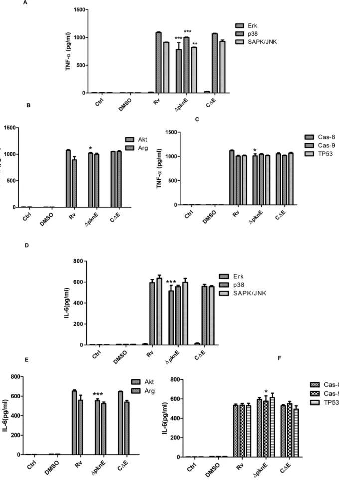
DpknEmodulates the secretion of cytokines TNF-aand IL-6 in response to intracellular signaling
The current observation on crosstalk and our previous observations [3,15] thatDpknE-infected macrophages are defective in producing pro and anti-inflammatory cytokines compelled us to assess production of TNF-aand IL-6 in the presence of pathway-specific inhibitors.
Three signaling pathways, MAPK (ErkK, p38MAPK, SAPK/ JNK), survival (Akt, arginase) and apoptosis (caspase-8, caspase-9, TP53) were studied. While ErkK inhibitor almost inhibited production of TNF-a and IL-6 in Rv-infected macrophages, increased production of these cytokines inDpknE-infected macro-phages was observed as compared to Rv infected macromacro-phages (p,0.0001, for both) (Figures 4A,4D).DpknE-infected macrophag-Figure 1.DpknEinfected macrophages are defective in MAPK signaling.Controls (Ctrl, LPS) and infected cells were lysed post infection at varied time points and subjected to western blotting. The blots were probed with phospho A) Erk1/2, E) p38, and I) SAPK/JNK and their respective non phospho (C, G and I) antibodies. The results are from three independent experiments. Figures B, D, F, H, J, K, and L depict the corresponding densitometry values of phospho and nonphospho antibody probed blots. *, *** denotes p,0.05 and p,0.0001, whenDpknEinfected macrophages compared with Rv (one – way ANOVA). The abbreviations ctrl denotes control and LPS denotes lipopolysaccharide.
doi:10.1371/journal.pone.0083541.g001
Figure 2. DpknEinfected macrophages have reduced
expres-sion of transcription factors. Nuclear fractions isolated post infection was subjected to DNA binding ELISA for A) ATF-2, B) c-JUN, and C) NF-kB. The results from three independent experiments are shown. The O.D values denote standard error of the means. *denote p,0.05 (Two way ANOVA) whenDpknEversus Rv infected macrophag-es were compared.
doi:10.1371/journal.pone.0083541.g002
Figure 3.DpknEinfected macrophages potentiates a crosstalk between SAPK/JNK and ErkK, and p38 and ErkKpathways. Cells to be infected were treated with A) SAPK/JNK and B) p38 inhibitors, for 1 h and lyzed 1 h post infection. The lysates were subjected to western blotting and probed with phospho and non phospho ErkK antibodies. The results from three independent experiments are shown. The abbreviations Ctrl denote control and LPS denotes lipopolysaccharide.
es had reduced secretion of TNF-ain the presence of p38MAPK and SAPK/JNK inhibitor (p,0.0001, p,0.001 respectively in DpknEversus Rv-infected macrophages) (Figure 4A). Secretion of IL-6 in both Rv andDpknE-infected macrophages was unaffected in the presence of p38 and SAPK/JNK inhibitors (Figure 4D).
In comparison with Rv-infected macrophages,DpknE-infected macrophages produced reduced amounts of TNF-aand IL-6 in the presence of Akt inhibitor (p,0.05 and p,0.0001 respectively), and arginase inhibitor had a reciprocal effect on their secretion (Figures 4B, 4E). In CDE-infected macrophages cytokine levels were restored to that observed in Rv-infected macrophages.
Apoptosis pathway (caspase-8, 9, TP53) inhibitors did not have any effect on the secretion of TNF-a or IL-6 secretion in Rv-infected macrophages (Figures 4C, 4F). However, in DpknE -infected macrophages secretion of TNF-a was decreased in the presence of caspase-8 inhibitor as compared to Rv infected macrophages (p,0.05). IL-6 was modestly increased in DpknE -infected macrophages in the presence of caspase-8/9 and TP53 inhibitors, of which only caspase-9 inhibition was significant (p,0.05) (Figure 4F). CDE-infected macrophages had cytokine levels comparable to that of Rv-infected macrophages. Collective-ly, these data show that secretion of TNF-aand IL-6 is influenced by the genes of MTB that enable crosstalk between intracellular pathways in the host and that pknE plays a significant role in crosstalk response thereby modulating the secretion of inflamma-tory cytokines.
Nitrate stress response
From our previous observations [3,15] we found thatpknEhas a role in nitrate stress response suppressing the host cell apoptosis. The role ofpknEin modulating intracellular signaling in response to nitrate stress was studied using an exogenous NO donor, sodium nitroprusside.
Phosphorylation of p38MAPK was higher in DpknE-infected macrophages while ErkK and SAPK/JNK were reduced in DpknE-infected macrophages, similar to that observed in the absence of NO donor (Figures 5A, 5B and 5C). Phosphorylation of ATF-2 was increased in DpknE-infected as compared to Rv-infected cells (p,0.05, Figure 5D), but phosphorylation of c-JUN was similar in both Rv andDpknE-infected macrophages (data not shown). Phosphorylation of NF-kB was reduced inDpknE-infected as compared to Rv-infected macrophages (p,0.05) (Figure 5E). CDE-infected macrophages had the restored phenotype of Rv-infected macrophages. Thus, in the presence of NO stressDpknE -infected macrophages reproduced events observed in the endog-enous NO host response. This clearly proves thatpknEresponds to NO stress in the host and by modulating signaling events enables the intracellular survival of MTB.
DpknE-infected macrophages modulate the expression of receptors for HIV entry
The role of pknE in the modulation of CCR5 and CXCR4 receptors involved in HIV entry was investigated based on our observation that DpknE has defective MAPK signaling, and the knowledge that MAPK signaling influences modulation of coreceptors.
The expression kinetics of CCR5 and CXCR4 was examined on days 1 and 2 post-infection. Expression of CCR5 was reduced in DpknE-infected macrophages as compared to Rv-infected
macrophages on both days (p,0.05; Figure 6A). Conversely, expression of CXCR4 was increased in macrophages infected with DpknE when compared to Rv-infected macrophages (p,0.05; Figure 6E). CDE-infected macrophages had comparable levels of coreceptor expression to that of Rv-infected macrophages.
Reduction in CCR5 byDpknE-infected macrophages is influenced by intracellular signaling cascades
Our previous [15] and present findings, persuaded us to examine the modulation of HIV receptors by MAPK, survival and apoptosis family of inhibitors. While MAPK inhibitors reduced the expression of CCR5 in DpknE-infected macrophages (p,0.05), SAPK/JNK inhibitors increased the expression of CCR5 in comparison with Rv-infected cells (Figure 6B). Akt inhibition did not have any effect on CCR5 expression (Figure 6C). DpknE -infected macrophages had increased expression of CCR5 in the presence of arginase inhibitor as compared to Rv-infected macrophages (p,0.001; Figure 6C). In the presence of TP53 inhibitor, both Rv andDpknE-infected macrophages had greater reduction in the expression of CCR5 (Figure 6D). CDE-infected macrophages were able to restore the expression levels similar to Rv-infected macrophages.
Increase in CXCR4 byDpknE-infected macrophages is influenced by intracellular signaling cascades
Modulation of CXCR4 expression was also assessed in the presence of MAPK, survival and apoptosis inhibitors. In general, MAPK and Akt inhibitors increased the expression of CXCR4 in Rv-infected macrophages. In contrast,DpknE-infected macrophag-es had significantly reduced CXCR4 exprmacrophag-ession in the prmacrophag-esence of ErkKinhibitor (p,0.0001, Figure 6F) and moderate reduction in the presence of p38MAPK and SAPK/JNK inhibitors, compared to Rv infected macrophages (Figure 6F). Akt inhibitor did not affect expression of CXCR4 (Figure 6G), but arginase inhibitor reduced the expression of CXCR4 inDpknE-infected macrophages (p,0.05 when compared to Rv-infected cells) (Figure 6G). TP53 inhibitor reduced the expression of CXCR4 in Rv, DpknE and CDE-infected macrophages (Figure 6H). CDE-infected macro-phages reversed the changes observed in DpknE-infected macro-phages.
DpknEmodulates coinfection of MTB-infected THP-1 cells and MDM with HIV
THP-1 macrophages and MDM were coinfected with an MTB strain (Rv,DpknEand CDE) and a CCR5 (R5) or CXCR4 (X4)-tropic HIV strain to examine the effect of coreceptor modulation on HIV entry and infection, by measuring HIV-1 p24 antigen levels in infected culture supernatants. While THP-1 macrophages coinfected withDpknEand R5-tropic virus had reduced p24 levels, cells coinfected with X4 virus had increased p24 levels (See Table S1). This validates our finding thatDpknE-infected macrophages had increased CXCR4 and decreased CCR5 expression.
To further confirm these findings, coinfection was performed in MDM obtained from normal healthy individuals. In MDM model of infection, p24 antigen levels were increased inDpknE-infected macrophages coinfected with R5 as well as X4-tropic viruses as compared to Rv-coinfected cells (p,0.05, Figures 7A,7C). Fur-ther, in the presence of SAPK/JNK inhibitor, DpknE-infected family, and F) caspase family ] was analyzed using ELISA on day 1. The error bars represent standard error of the means. Data is from three independent experiments. The symbols *, **, *** denotes p,0.05, p,0.001 and p,0.0001(one way ANOVA) respectively, whenDpknEwas compared to Rv infected macrophages.
macrophages coinfected with R5 as well as X4-tropic viruses had higher p24 antigen levels as compared to Rv-coinfected macro-phages (p,0.05, Figures 7B,7D).
The observations of increased p24 levels in macrophages coinfected with R5 tropic virus andDpknEcorroborates increased CCR5 expression observed inDpknEinfected macrophages treated with SAPK/JNK inhibitor. Further, decreased p24 levels in R5 tropic virus and Rv coinfected macrophages confirms decreased CCR5 expression observed in RV infected macrophages treated with SAPK/JNK inhibitor. This data for the first time shows that
pknE contributes to the co-pathogenesis of HIV by modulating intracellular signaling in the host.
Discussion
Virulence and infectivity of MTB modulates various apoptotic paradigms thereby reducing immunity of the host [18,19].pknEof MTB suppresses cell death of the host by inhibiting intrinsic pathway of apoptosis and arginase2 dependent mechanisms [15]. MAPK signaling in eukaryotes plays an important role in cytokine and apoptosis regulation [10,20]. The present study investigates the role ofpknEin modulating MAPK cascades and its impact on HIV/TB coinfection.
Analysis of MAPK signaling showed DpknE-infected macro-phages to have decreased ErkK phosphorylation. This observa-tion corresponds with our previous finding that DpknE-infected macrophages had reduced phosphorylation of Akt, an upstream activator of ErkK[15], correlating the findings of Yang et al [21]. In addition, selective inhibition of p46SAPK/JNK was observed in DpknE infected macrophages. These inferences prompted us to examine the phosphorylation kinetics of transcription factors c-JUN, ATF-2 and NF-kB that are regulated by MAPK cascades. Phosphorylation of c-JUN and ATF-2 were dramatically delayed and reduced inDpknEas compared to Rv-infected macrophages. This defective MAPK signaling could be a reason for theDpknE
infected macrophages to have dampened cytokine secretion and execute apoptosis independent of extrinsic pathway (TNF-a) and iNOS [15]. This concurs with previous findings that c-JUN and ATF-2 induces the secretion of TNF-a, activating p46SAPK/JNK and iNOS [22,23]. The current data suggest that deletion ofpknE
results in deactivation of survival pathways inside the host. This underlines the role for pknE in modulating host intracellular cascades.
Use of pathway specific inhibitors to confirm the defective MAPK signaling inDpknE-infected macrophages did not reverse the effects. However, the secretion of TNF-a and IL-6 were modulated by inhibitors to various intracellular pathways. Observations from the inhibitor studies suggested the probability of crosstalk responses andDpknEinfected macrophages had cross talk responses between ErkK and SAPK/JNK, and p38MAPK and ErkK pathways. This is in concordance with earlier studies where crosstalks within MAPK signaling were reported [17]. Our study for the first time demonstrates the role ofpknEin crosstalk responses essential for the intracellular survival of MTB.
In our earlier observationspknEwas found to respond NO stress that results in suppression of apoptosis [3,15]. In the present study,
the function of pknE in modulating intracellular signaling in response to the NO stress of the host was examined using SNP as NO donor that mimics in vivo situations of NO stress [24]. As expected,DpknE-infected macrophages had reduced phosphoryla-tion of MAPKs that confirms pknE in modulating intracellular signaling during NO stress of the host.
Our findings suggest thatpknEincreases ErkKsignaling thereby suppressing apoptosis which favors the survival of MTB. This is analogous with a previous report where ErkK was shown to suppress apoptotic signals [25].
In the next part of the study we endeavored to analyze the significance of pknE in the co-pathogenesis of HIV. This was investigated since the data about mycobacterial genes involved in HIV coinfection remains unexplored. Nevertheless earlier reports have shown MAPKs, Akt, chemokines, apoptosis, IL-12 and MTB to modulate CCR5 and CXCR4 coreceptors involved in HIV entry [26–32].
Here we found that THP-1 macrophages infected with DpknE
suppressed CCR5 but increased CXCR4 expression as compared to the wild-type strain. This finding was further confirmed by coinfection studies with MTB and HIV-1 tropic strains. Next we examined various intracellular pathways that could influence this modulation. MAPK and arginase signaling were found to play an important role in the expression of CCR5 and CXCR4 in macrophages infected withDpknE.
For the first time, we show that DpknEinduces apoptosis and down modulates intracellular events that suppress CCR5 expres-sion. This concurs with a previous study where CCR5 was shown to induce antiapoptotic signals via Akt and ErkK [32]. The modulations of coreceptor expression were further investigated in MDM derived from normal healthy individuals. In contrast to THP-1 model of coinfection,DpknE increased the levels of p24 antigen upon coinfection with either R5 or X4 tropic HIV-1 strains. Among the MAPK and arginase signaling, SAPK/JNK was chosen for further validation. Inhibition of the SAPK/JNK signaling and coinfection with either R5 or X4 tropic HIV-1 strains increased the p24 antigen levels in DpknE coinfected macrophages. However inhibition of SAPK/JNK signaling markedly reduced the p24 levels in macrophages coinfected with either R5 or X4 tropic HIV-1 strains and Rv. These data suggests SAPK/JNK signaling as one among the cascade that regulates CCR5/CXCR4 expression. This is in concordance with an earlier report where inhibition of SAPK/JNK was shown to reduce CCR5 expression [28].The reasons for disparity in coinfection studies between THP-1 and MDM cells could be multifactorial including differences in CD4 receptor expression, genetic compo-sition of the host, etc [33]. In contrast to our study, p38 signaling was reported to regulate the expression of coreceptors upon infection with MTB [34]. Our study using various pathway specific inhibitors and HIV/TB model of coinfection authenticate the significance of SAPK/JNK pathway in regulating the coreceptor expression.
In conclusion, our previous [3,15] and the current findings show that pknE contributes to the intracellular survival of MTB by initiating crosstalks within the intracellular signaling of the host. Figure 5.DpknEinfected macrophages show defective MAPK signaling in the presence of NO stress.Controls (ctrl, LPS) and infected cells were lysed post NO stress and subjected to western blotting. The blots were probed with phospho A) Erk1/2, B) p38, and C) SAPK/JNK with their respective non phospho antibodies and the results of three independent experiments are given. The corresponding densitometry values of phospho and nonphospho are given at the end of antibody probed blots. *** denotes p,0.0001, whenDpknEinfected macrophages compared with Rv (one – way ANOVA). The abbreviations ctrl denotes control, LPS denotes lipopolysaccharide, and NO denotes nitric oxide. Nuclear fractions isolated post infection was subjected to DNA binding ELISA D) ATF-2 and E) NF-kB. The results are from three independent experiments. The error bars denote standard error of the means. * denotes p,0.05 (one way ANOVA) whenDpknE+NO was compared to Rv+NO treated macrophages.
This protective strategy employed by MTB provides a favorable niche for HIV infection.
Supporting Information
Table S1 DpknE coinfected with CCR5 has reduced while with CXCR4 have increased p24 levels in THP-1 model of coinfection. THP-1 derived macrophages were
infected withM. tuberculosisstrains followed by coinfection with a CCR5 and CXCR4 tropic virus. The p24 antigen levels were estimated using ELISA on day 4. *, ** denotes p,0.05 and p,0.001 (one way – Anova) whenDpknE was compared to Rv infected macrophages.
(DOC)
Figure 6.DpknEinfected macrophages modulate the expression of coreceptors CCR5 and CXCR4 by intracellular cascades.Cells post infection were stained with CCR5 (A) and CXCR4 (E) antibody and the expression was analyzed in a time dependent manner using FACS, * denotes p,0.05 (Twoway – Anova) whenDpknEwas compared to Rv infected macrophages. Cells post infection in the presence of inhibitors CCR5 [B) MAPK family, C) survival family and D) TP53] and CXCR4 [F) MAPK family, G) survival family, and H) caspase family] expression was analyzed on day1 post infection using FACS. The symbols *, **, ***denotes p,0.05, p,0.001 and p,0.0001 respectively (one way – Anova) whenDpknEwas compared to Rv infected macrophages.
doi:10.1371/journal.pone.0083541.g006
Acknowledgments
We would like to acknowledge Dr.P.R. Narayanan, former director, NIRT for his constant support and encouragement. We thank the technical assistance rendered by Ms. Suganthi and Mr. Senthilnathan. We thank Mr. Anbalagan for his help in FACS analysis. We thank National Institutes of Health, USA for providing CCR5 and CXCR4 tropic HIV strains. We, also thank Mr. John, radiographer for his help in developing the western
blots. We sincerely thank Jeevan blood bank for their co-operation in collecting blood samples from normal healthy subjects.
Author Contributions
Conceived and designed the experiments: DKP SN. Performed the experiments: DKP LEH. Analyzed the data: DKP LEHSN. Contributed reagents/materials/analysis tools: SN. Wrote the paper: DKP SN LEH.
References
1. Chao J, Wong D, Zheng X, Poirier V, Bach H, et al. (2010) Protein kinase and phosphatase signaling in Mycobacterium tuberculosis physiology and patho-genesis. Biochim Biophys Acta 1804: 620–627.
2. Gopalaswamy R, Narayanan S, Chen B, Jacobs WR, Av-Gay Y (2009) The serine/threonine protein kinase PknI controls the growth of Mycobacterium tuberculosis upon infection. FEMS Microbiol Lett 295: 23–29.
3. Jayakumar D, Jacobs WR, Jr., Narayanan S (2008) Protein kinase E of Mycobacterium tuberculosis has a role in the nitric oxide stress response and apoptosis in a human macrophage model of infection. Cell Microbiol 10: 365– 374.
4. Malhotra V, Arteaga-Cortes LT, Clay G, Clark-Curtiss JE (2010) Mycobacte-rium tuberculosis protein kinase K confers survival advantage during early infection in mice and regulates growth in culture and during persistent infection: implications for immune modulation. Microbiology 156: 2829–2841. 5. Papavinasasundaram KG, Chan B, Chung JH, Colston MJ, Davis EO, et al.
(2005) Deletion of the Mycobacterium tuberculosis pknH gene confers a higher bacillary load during the chronic phase of infection in BALB/c mice. J Bacteriol 187: 5751–5760.
6. Walburger A, Koul A, Ferrari G, Nguyen L, Prescianotto-Baschong C, et al. (2004) Protein kinase G from pathogenic mycobacteria promotes survival within macrophages. Science 304: 1800–1804.
7. Meszaros B, Toth J, Vertessy BG, Dosztanyi Z, Simon I (2011) Proteins with complex architecture as potential targets for drug design: a case study of Mycobacterium tuberculosis. PLoS Comput Biol 7: e1002118.
8. Shin DM, Jeon BY, Lee HM, Jin HS, Yuk JM, et al. (2010) Mycobacterium tuberculosis eis regulates autophagy, inflammation, and cell death through redox-dependent signaling. PLoS Pathog 6: e1001230.
9. Zhou B, He Y, Zhang X, Xu J, Luo Y, et al. (2010) Targeting mycobacterium protein tyrosine phosphatase B for antituberculosis agents. Proc Natl Acad Sci U S A 107: 4573–4578.
10. Schorey JS, Cooper AM (2003) Macrophage signalling upon mycobacterial infection: the MAP kinases lead the way. Cell Microbiol 5: 133–142. 11. Gui T, Sun Y, Shimokado A, Muragaki Y (2012) The Roles of
Mitogen-Activated Protein Kinase Pathways in TGF-beta-Induced Epithelial-Mesenchy-mal Transition. J Signal Transduct 2012: 289243.
12. Blumenthal A, Ehlers S, Ernst M, Flad HD, Reiling N (2002) Control of mycobacterial replication in human macrophages: roles of extracellular signal-regulated kinases 1 and 2 and p38 mitogen-activated protein kinase pathways. Infect Immun 70: 4961–4967.
13. Pawlowski A, Jansson M, Skold M, Rottenberg ME, Kallenius G (2012) Tuberculosis and HIV co-infection. PLoS Pathog 8: e1002464.
14. Rosas-Taraco AG, Arce-Mendoza AY, Caballero-Olin G, Salinas-Carmona MC (2006) Mycobacterium tuberculosis upregulates coreceptors CCR5 and CXCR4 while HIV modulates CD14 favoring concurrent infection. AIDS Res Hum Retroviruses 22: 45–51.
15. Kumar D, Narayanan S (2012) pknE, a serine/threonine kinase of Mycobac-terium tuberculosis modulates multiple apoptotic paradigms. Infect Genet Evol 12: 737–747.
16. Rojas M, Olivier M, Garcia LF (2002) Activation of JAK2/STAT1-alpha-dependent signaling events during Mycobacterium tuberculosis-induced macro-phage apoptosis. Cell Immunol 217: 58–66.
17. Frost JA, Steen H, Shapiro P, Lewis T, Ahn N, et al. (1997) Cross-cascade activation of ERKs and ternary complex factors by Rho family proteins. EMBO J 16: 6426–6438.
18. Behar SM, Martin CJ, Booty MG, Nishimura T, Zhao X, et al. (2011) Apoptosis is an innate defense function of macrophages against Mycobacterium tuberculosis. Mucosal Immunol 4: 279–287.
19. Lee J, Repasy T, Papavinasasundaram K, Sassetti C, Kornfeld H (2011) Mycobacterium tuberculosis induces an atypical cell death mode to escape from infected macrophages. PLoS One 6: e18367.
20. Zhou HR, Islam Z, Pestka JJ (2005) Induction of competing apoptotic and survival signaling pathways in the macrophage by the ribotoxic trichothecene deoxynivalenol. Toxicol Sci 87: 113–122.
21. Yang CS, Lee JS, Jung SB, Oh JH, Song CH, et al. (2006) Differential regulation of interleukin-12 and tumour necrosis factor-alpha by phosphatidylinositol 3-kinase and ERK 1/2 pathways during Mycobacterium tuberculosis infection. Clin Exp Immunol 143: 150–160.
22. Barthel R, Tsytsykova AV, Barczak AK, Tsai EY, Dascher CC, et al. (2003) Regulation of tumor necrosis factor alpha gene expression by mycobacteria involves the assembly of a unique enhanceosome dependent on the coactivator proteins CBP/p300. Mol Cell Biol 23: 526–533.
23. Lahti A, Jalonen U, Kankaanranta H, Moilanen E (2003) c-Jun NH2-terminal kinase inhibitor anthra(1,9-cd)pyrazol-6(2H)-one reduces inducible nitric-oxide synthase expression by destabilizing mRNA in activated macrophages. Mol Pharmacol 64: 308–315.
24. Blond D, Raoul H, Le Grand R, Dormont D (2000) Nitric oxide synthesis enhances human immunodeficiency virus replication in primary human macrophages. J Virol 74: 8904–8912.
25. Kim SJ, Ju JW, Oh CD, Yoon YM, Song WK, et al. (2002) ERK-1/2 and p38 kinase oppositely regulate nitric oxide-induced apoptosis of chondrocytes in association with p53, caspase-3, and differentiation status. J Biol Chem 277: 1332–1339.
26. Dasgupta B, Roychoudhury K, Ganguly S, Akbar MA, Das P, et al. (2003) Infection of human mononuclear phagocytes and macrophage-like THP1 cells with Leishmania donovani results in modulation of expression of a subset of chemokines and a chemokine receptor. Scand J Immunol 57: 366–374. 27. Fraziano M, Cappelli G, Santucci M, Mariani F, Amicosante M, et al. (1999)
Expression of CCR5 is increased in human monocyte-derived macrophages and alveolar macrophages in the course of in vivo and in vitro Mycobacterium tuberculosis infection. AIDS Res Hum Retroviruses 15: 869–874.
28. Giri RK, Rajagopal V, Shahi S, Zlokovic BV, Kalra VK (2005) Mechanism of amyloid peptide induced CCR5 expression in monocytes and its inhibition by siRNA for Egr-1. Am J Physiol Cell Physiol 289: C264–276.
29. Hoshino Y, Tse DB, Rochford G, Prabhakar S, Hoshino S, et al. (2004) Mycobacterium tuberculosis-induced CXCR4 and chemokine expression leads to preferential X4 HIV-1 replication in human macrophages. J Immunol 172: 6251–6258.
30. Lande R, Giacomini E, Grassi T, Remoli ME, Iona E, et al. (2003) IFN-alpha beta released by Mycobacterium tuberculosis-infected human dendritic cells induces the expression of CXCL10: selective recruitment of NK and activated T cells. J Immunol 170: 1174–1182.
31. Mukai T, Iwasaki M, Gao P, Tomura M, Yashiro-Ohtani Y, et al. (2001) IL-12 plays a pivotal role in LFA-1-mediated T cell adhesiveness by up-regulation of CCR5 expression. J Leukoc Biol 70: 422–430.
32. Tyner JW, Uchida O, Kajiwara N, Kim EY, Patel AC, et al. (2005) CCL5-CCR5 interaction provides antiapoptotic signals for macrophage survival during viral infection. Nat Med 11: 1180–1187.
33. Cassol E, Alfano M, Biswas P, Poli G (2006) Monocyte-derived macrophages and myeloid cell lines as targets of HIV-1 replication and persistence. J Leukoc Biol 80: 1018–1030.
34. Lei J, Wu C, Wang X, Wang H (2005) p38 MAPK-dependent and YY1-mediated chemokine receptors CCR5 and CXCR4 up-regulation in U937 cell line infected by Mycobacterium tuberculosis or Actinobacillus actinomycetem-comitans. Biochem Biophys Res Commun 329: 610–615.