Expre ssio n o f p5 3 pro te in in pituitary
ade no m as
Departamentos de 1Endocrinologia, 2Patologia and 3Neurocirurgia,
Fundação Faculdade Federal de Ciências Médicas de
Porto Alegre e Irmandade Santa Casa de Misericórdia de Porto Alegre, Porto Alegre, RS, Brasil
M.C. O liveira1, C.P. Marroni1,
C.B. Pizarro1, J.F. Pereira-Lima1,
L.M. Barbosa-Coutinho2
and N.P. Ferreira3
Abstract
Inactivating mutations of TP53, a tumor suppressor gene, are associ-ated with abnormal cell proliferation. Although p53 expression is common in many human malignancies, p53 protein has seldom been evaluated in pituitary tumors. When detected, the percentage of p53-positive cells is low, and, in general, it is exclusive for invasive lesions. The aim of the present study was to use immunohistochemis-try to determine the presence of p53 protein in pituitary adenomas from tumor samples of 163 surgeries performed in 148 patients (40% male, 60% female). In 35% of the cases the adenoma was nonfunc-tional, while in the others it was associated with PRL, GH and/or ACTH endocrine hypersecretion syndrome. Macroadenomas were observed in 83.2% of the cases with available neuroimage evaluation, of which 28% invaded the cavernous, sphenoid and/or ethmoidal sinus, bone, third ventricle or subfrontal lobe. p53 protein was de-tected in 2/148 patients (1.3%). Immunohistochemistry was positive for PRL and GH in these cases. Due to the high percentage of invasive pituitary adenomas found in our study, the low frequency of p53 detection suggests that it is inadequate as a routine marker for aggres-siveness and as a predictive factor of tumor behavior.
Co rre spo nde nce
M.C. O liveira Dona Mimi Moro, 40 90480-050 Porto Alegre, RS Brasil
Fax: + 51-328-6761 E-mail: mco@ portoweb.com.br
Received April 27, 2001 Accepted March 8, 2002
Ke y wo rds
·Pituitary
·Pituitary adenoma ·Invasive adenoma ·p53
·Immunohistochemistry
Pituitary adenomas are frequent, being diagnosed in about 27% of glands examined in autopsies (1) and incidentally in about 20% of imaging scans of the sellar area (2). More uncommon are the adenomas diag-nosed clinically because of either their asso-ciation with endocrine clinical syndromes or symptoms resulting from the presence of a tumor mass.
Pituitary tumorigenesis has classically in-volved the hypothalamic theory and the pitu-itary intrinsic defect, the latter involving clonality, activation of oncogenes and inacti-vation of tumor suppressor genes. Both theo-ries are possibly operant, the process starting
from transformed cells, whose progression depends on hormonal stimuli and other growth factors (3).
immu-nohistochemistry (4). Inactivating mutations of TP53 are among the most frequent alter-ations in human neoplasias, but they seldom have been evaluated in pituitary tumors. While some investigators did not detect p53 expression in pituitary adenomas (5,6), oth-ers reported it in invasive (7,8) and noninva-sive adenomas (9).
The present investigation was designed to determine the presence of p53 in human pituitary adenomas of different hormonal phenotypes.
Tissue from surgeries performed on 148 patients was used. Patients had undergone resection of pituitary tumors at a reference neurosurgery hospital in southern Brazil. The medical records of these patients were re-viewed for clinical presentation, hormonal evaluation, pituitary image and surgical reintervention. Tumors were considered as microadenomas when they were less than 10 mm in size, and as macroadenomas when they were 10 mm or larger, regardless of the presence of suprasellar extension. Invasive adenomas were those which showed inva-sion of the cavernous, sphenoid or ethmoi-dal sinus, bone, third ventricle or subfrontal lobe. Recurrent tumors were those in which a new tumor growth resulted in another
sur-gical resection.
Samples embedded in paraffin, stored in a bank of adenomatous pituitary tissue, were cut and sections were mounted on glass slides prepared with organosilane. The immuno-histochemical study was based on the strepta-vidin-biotin immunoperoxidase method (Strept ABComplex/HRP, Dako A/S, Glos-trup, Denmark). Briefly, the sections were submitted to deparaffinization, rehydration, blockade of endogenous peroxidase activity, antigen retrieval, and incubation with so-dium citrate, and were then placed in a mi-crowave oven for 10 min at maximum po-tency and for 10 min at medium popo-tency. The primary antibody used for detection of p53, at 1:50 dilution, was the mouse mono-clonal antibody (Dako) that recognizes an epitope at the N-terminus of the human p53 protein and reacts with both the wild type and mutant type of the protein. The second-ary antibody was the anti-mouse biotinyl-ated antibody (Vectastain®
, Vector Labora-tories, Inc., Burlingame, CA, USA). Anti-gen-antibody complexes were detected with 3,3-diaminobenzidine. In all assays, posi-tive (carcinoma of the esophagus) and nega-tive (exclusion of primary antibody in reac-tion) controls were used.
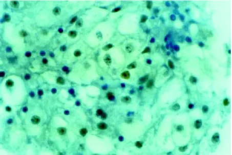
Glass slides were examined under the light microscope. The criterion for a positive p53 immunoreaction was the presence of at least a group of immunostained cell nuclei (see Figure 1).
A total of 167 surgeries were performed on the 148 patients studied here (60% fe-males and 40% fe-males, ranging in age from 16 to 76 years, mean 44.4 years). Fifteen patients underwent two surgeries and two patients were submitted to three surgeries. Nonfunctioning adenomas made up 35% of cases, and the others were associated with endocrine hypersecretion syndrome of ACTH, GH and/or PRL. Sellar scans (com-puted tomography or magnetic resonance) were available for 125 patients. Of these, 104 (83.2% of cases) presented
adenomas, 35 of which (28% of total) with invasive characteristics.
p53 protein was positive in 2/148 pa-tients (1.3%). Immunohistochemistry re-vealed that both adenomas were GH- and PRL-producing. One of the patients was a 35-year-old male with clinical acromegaly and PRL levels above 20,000 ng/ml. He presented a giant adenoma with invasion of the sphenoid and nasal fossae. The Ki-67 index of this adenoma (Oliveira MC, Pizarro CB, Barbosa-Coutinho LM and Ferreira NP, unpublished results) was 0.20%. The patient died within a few months. The other patient was a 71-year-old female also with clinical acromegaly. The pituitary scan showed an intrasellar lesion with roof erosion. The Ki-67 index of this adenoma was 0.36%. Six years after surgery, the patient remains healthy.
Mean patient age and sex distribution were in agreement with the literature (10,11), as also was the frequency of the different types of hormonal secretion (11). Most of the adenomas found were macroadenomas, and 28% of these were of the invasive type. The incidence of invasion by pituitary ad-enomas reported in the literature is high, ranging from 10 to 85% depending on the criterion used to define whether an adenoma is invasive or not. The index is much higher when microscopic invasion of the dura mater is considered.
Expression of p53 protein has seldom been studied in pituitary adenomas and dif-ferent results were obtained in the available reports. A number of studies failed to detect p53 in adenomas and even in pituitary carci-nomas (5,6,10,12), while others detected the presence of the mutated protein (7-9,13). The reasons for these discrepancies are un-clear, but must be related to difficulties in the various steps of the immunohistochemi-cal technique used, criteria for positive reac-tion, and characteristics of the material stud-ied, which include small samples, coexist-ence of normal tissue in the middle of
neo-plastic tissue, and heterogeneity concerning the function and characteristics of cell repli-cation in each adenoma.
Sumi et al. (5) did not detect p53 protein in 40 adenomas of several hormonal pheno-types, of which 17 were recurrent and 3 were invasive. Gandour-Edwards et al. (6) did not detect the presence of p53 in 5 normal pitu-itary tissues and in 20 invasive and noninva-sive adenomas. Lübke et al. (12), in a study of 19 ACTH-secreting adenomas only, ob-tained negative results, as also did Müller et al. (10). The latter group studied patients with acromegaly, 27 with plurihormonal ad-enomas and 21 with GH- and PRL-secreting adenomas. Immunohistochemistry for p53 was negative in all cases studied.
Buckley et al. (7) used three different antibodies to evaluate 121 adenomas of all functional classifications, of which 95 were benign and 26 presented local invasion or invasion over distance. The presence of p53 was detected in 16% of invasive tumors, but only in ACTH-secreting tumors (2/4) and in nonfunctioning tumors (4/15).
An analysis of the above data shows that findings are inconsistent and that positive reaction for p53, when detected, is restricted either to a given type of hormonal secretion (or to its absence) or to aggressiveness char-acteristics. The highest frequency of p53 immunoreaction in ACTH-secreting ad-enomas has not been clarified (14). More-over, the percentage of positive results, even if it was significant within the unique subset in which it was found, was too low in rela-tion to the whole sample studied.
The present series is the largest among those reviewed. Here p53 was detected in a very low percentage (1.3%) of the total sample of 148 and in 5.7% of invasive ad-enomas. It is remarkable that in both cases of positive immunoreaction, concomitant se-cretion of GH and PRL by the tumor was observed. There is one study in the literature reporting a PRL-secreting adenoma, a GH-secreting adenoma, and a stem cell adenoma among five p53-positive adenomas found (8), and another study reporting a PRL-se-creting adenoma and another one sePRL-se-creting GH + PRL + ACTH (9), but such findings are rare. Unlike Buckleys report (7), p53 was not detected in any of the nonfunction-ing or ACTH-secretnonfunction-ing adenomas, though Thapar et al. (8) suggested that there is no link between positive reaction for p53 and tumor functional profile.
The possible association between p53 detection and cell replication indices has not been explored in the literature. The Ki-67 index is a nuclear antigen expressed in pro-liferating cells during the G1, S, G2 and M phases of the cell cycle (15). Pituitary ad-enomas in general are associated with either absence of a positive reaction for Ki-67 anti-gen or low levels of it, usually under 1%. However, a significant difference has been established between invasive and noninva-sive tumors. The mean Ki-67 index (in
stud-ies using MIB-1 for detection of Ki-67 anti-gen in material embedded in paraffin) ranges from 0.15 (16) to 2.01 for noninvasive ad-enomas (11) and from 1.27 (17) to 4.66 for invasive adenomas (18). Our figures are 0.86 for microadenomas, 1.12 for macroadenomas with or without suprasellar extension, and 2.01 for invasive adenomas (Oliveira MC, Pizarro CB, Barbosa-Coutinho LM and Fer-reira NP, unpublished data). Both patients with a positive reaction for p53 in this series were evaluated for Ki-67 index, showing low positive values of 0.2 and 0.36. The small number of cases precludes correlating p53 with the Ki-67 index.
Re fe re nce s
1. Burrow GN, Wortzman G, Rew castle NB, Holgate RC & Kovacs K (1981). M icro-adenomas of the pituitary and abnormal sellar tomograms in an unselected au-topsy series. New England Journal of M edicine, 304: 156-158.
2. Elster AD (1993). M odern imaging of the pituitary. Radiology, 187: 1-14.
3. Asa SL & Ezzat S (1998). The cytogenesis and pathogenesis of pituitary adenomas. Endocrine Review s, 19: 798-827. 4. Harris C & Hollstein M (1993). Clinical
im-plications of the p53 tumor-suppressor gene. New England Journal of M edicine, 329: 1318-1327.
5. Sumi T, Stefaneanu L, Kovacs K, Asa S & Rindi G (1993). Im m unohistochem ical study of p53 protein in human and animal pituitary tumors. Endocrine Pathology, 4: 95-99.
6. Gandour-Edw ards R, Kapadia SB, Janecka IP, M artinez AJ & Barnes L (1995). Bio-logic markers of invasive pituitary ad-enomas involving the sphenoid sinus. M odern Pathology, 8: 160-164.
7. Buckley N, Bates AS, Broome JC, Strange RC, Perrett CW, Burke CW & Clayton RN (1994). p53 protein accumulates in Cush-ing’s adenomas and invasive non-func-tional adenomas. Journal of Clinical Endo-crinology and M etabolism, 79: 1513-1516. 8. Thapar K, Scheithauer BW, Kovacs K, Pernicone PJ & Law s Jr ER (1996). p53 expression in pituitary adenomas and car-cinomas: correlation w ith invasiveness
and tumor grow th fractions. Neurosur-gery, 38: 765-771.
9. Levy A, Hall L, Yeudall WA & Lightman SL (1994). p53 gene mutations in pituitary adenomas: rare events. Clinical Endocri-nology, 41: 809-814.
10. M üller W , Saeger W , W ellhausen L, Derw ahl KM , Hamacher C & Ludecke DK (1999). M arkers of function and prolifera-tion in non-invasive and invasive bi- and plurihormonal adenomas of patients w ith acrom egaly: an im m unohistochem ical study. Pathology, Research and Practice, 195: 595-603.
11. M astronardi L, Guiducci A, Spera C, Puzzilli F, Liberati F & M aira G (1999). Ki-67 labelling index and invasiveness among anterior pituitary adenomas: anal-ysis of 103 cases using the M IB-1 mono-clonal antibody. Journal of Clinical Pathol-ogy, 52: 107-111.
12. Lübke D, Saeger W & Lüdecke DK (1995). Proliferation markers and EGF in ACTH-secreting adenomas and carcinomas of the pituitary. Endocrine Pathology, 5: 45-55.
13. Suliman M , Royds J, Cullen D, Timperley W, Pow ell T, Battersby R & Jones TH (2000). M arkers of pituitary adenoma in-vasiveness. Pituitary, 3: 19 (Abstract OC11).
14. Clayton RN, Boggild M , Bates AS, Bicknell J, Simpson D & Farrell W (1997). Tumour suppressor genes in the pathogenesis of human pituitary tumours. Hormone
Re-search, 47: 185-193.
15. Gerdes J, Schw ab U, Lemke H & Stein H (1983). Production of a mouse monoclon-al antibody reactive w ith a human nuclear antigen associated w ith cell proliferation. International Journal of Cancer, 31: 13-20. 16. Atkin SL, Green VL, Hipkin LJ, Landolt AM , Foy PM , Jeffreys RV & White M C (1997). A comparison of proliferation indi-ces in human anterior pituitary adenomas using formalin-fixed tissue and in vitro cell culture. Journal of Neurosurgery, 87: 85-88.
17. M izoue T, Kaw amoto H, Arita K, Kurisu K, Tominaga A & Uozumi T (1997). M IB1 immunopositivity is associated w ith rapid regrow th of pituitary adenomas. Acta Neurochirurgica, 139: 426-432.
18. Thapar K, Kovacs K, Scheithauer BW, Stefaneanu L, Horvath E, Pernicone PJ, M urray D & Law s ER (1996). Proliferative activity and invasiveness among pituitary adenomas and carcinomas: an analysis using the M IB-1 antibody. Neurosurgery, 38: 99-107.
19. Copelli SB, Loza Coll M A & Bruno OD (1999). Absence of mutations in the p53 tumor suppressor gene in non-invasive Cushing adenomas. M edicine, 59: 459-462.