online | memorias.ioc.fiocruz.br
Control of
Mycobacterium fortuitum
and
Mycobacterium intracellulare
infections with respect to distinct granuloma formations
in livers of BALB/c mice
Tânia Regina Marques da Silva, Antonio Luis de Oliveira Almeida Petersen, Theo de Araújo Santos, Taís Fontoura de Almeida, Luiz Antônio Rodrigues de Freitas, Patrícia Sampaio Tavares Veras/+
Centro de Pesquisas Gonçalo Moniz, Fundação Oswaldo Cruz-Fiocruz, Rua Waldemar Falcão 121, 40296-710 Salvador, BA, Brasil
Mycobacterium fortuitum is a rapidly growing nontuberculous Mycobacterium that can cause a range of
dis-eases in humans. Complications from M. fortuitum infection have been associated with numerous surgical proce-dures. A protective immune response against pathogenic mycobacterial infections is dependent on the granuloma formation. Within the granuloma, the macrophage effector response can inhibit bacterial replication and mediate the intracellular killing of bacteria. The granulomatous responses of BALB/c mice to rapidly and slowly growing my-cobacteria were assessed in vivo and the bacterial loads in spleens and livers from M. fortuitum and Mycobacterium
intracellulare-infected mice, as well as the number and size of granulomas in liver sections, were quantified.
Bacte-rial loads were found to be approximately two times lower in M. fortuitum-infected mice than in M. intracellulare -infected mice and M. fortuitum-infected mice presented fewer granulomas compared to M. intracellulare-infected mice. These granulomas were characterized by the presence of Mac-1+ and CD4+ cells. Additionally, IFN-γ mRNA expression was higher in the livers of M. fortuitum-infected mice than in those of M. intracellulare-infected mice. These data clearly show that mice are more capable of controlling an infection with M. fortuitum than M.
intracel-lulare. This capacity is likely related to distinct granuloma formations in mice infected with M. fortuitum but not
with M. intracellulare.
Key words: Mycobacterium fortuitum - Mycobacterium intracellulare - granuloma - liver - control of infection
Nontuberculous mycobacteria (NTM) include
dif-ferent species of the genus Mycobacterium that do not
belong to the Mycobacterium tuberculosis complex.
These include both slowly growing [e.g., Mycobacterium
avium-intracellulare (MAI)] and rapidly growing (e.g.,
Mycobacterium fortuitum and Mycobacterium
absces-sus) species (Runyon 1959). NTM are human
opportu-nistic pathogens and are predominantly acquired from the environment. A large number of NTM species have been recovered from soil, household dust, water, dairy products, cold-blooded animals, vegetation and human faeces (Ho et al. 2006). These species can also colonize surgical equipment and materials, such as endoscopes and solutions (Brown-Elliott & Wallace 2005).
In humans, NTM are organisms that belong to a heterogeneous group in which each species of bacteria should be studied separately (Alvarez-Uria 2010). These pathogens can cause a range of diseases affecting a vari-ety of tissues, including the lungs, lymph nodes, skin and soft and skeletal tissue. These diseases can also affect the genitourinary systems and cause disseminated infec-tions (Ho et al. 2006, Griffith et al. 2007, Jarzembowski
Financial support: CNPq (306672/2008-1) + Corresponding author: pveras@bahia.fiocruz.br Received 14 January 2010
Accepted 15 June 2010
& Young 2008). MAI is primarily a pulmonary pathogen and is the NTM species most commonly associated with human disease (Griffith et al. 2007). Inhalation of this bacterium may cause pulmonary disease, whereas the in-gestion of contaminated water may cause a disseminated disease. A cutaneous manifestation can be attributed to direct inoculation, direct contact or disseminated dis-ease (Weitzul et al. 2000). Infections caused by rapidly
growing NTM including M. fortuitum can appear after
surgical procedures, such as liposuction, silicone injec-tion and breast implantainjec-tion, or after intravenous catheter insertion, exposure to prosthetic material and pacemaker placement (Sungkanuparph et al. 2003, Palwade et al. 2006, Uslan et al. 2006). There is still no defined optimal treatment for NTM infections because these organisms are resistant to the standard antituberculous agents. In addition, susceptibility to anti-mycobacterial agents var-ies across different NTM specvar-ies (ATS 1997).
develop compromised granulomas changes from chronic to acute, leading to high morbidity and mortality from mycobacterial infections (Ladel et al. 1995).
The static spatial localization of cells within
Myco-bacterium-induced granulomas has been extensively studied by standard histological and immunohistochem-ical methods (Bouley et al. 2001). Little is known, how-ever, about the events involving granuloma formation during infection by rapidly growing mycobacteria. Previous studies have shown that granuloma formation
could depend on the Mycobacterium properties (van der
Sar et al. 2004) and on the genetic background of the host (Orrell et al. 1992). Using a mammalian model, we compared the granulomatous response of BALB/c mice
infected with rapidly or slowly growing Mycobacterium.
We compared the bacterial load and the number and size
of granulomas from M. fortuitum and M. intracellulare
-infected mice. In addition, the cellular composition and mRNA expression of regulatory cytokines and induct-ible nitric oxide synthase (iNOS) at the site of infection were assessed. The two species of environmental
myco-bacteria induced distinct responses in BALB/c mice. M.
fortuitum-infected mice efficiently controlled the
bacte-ria while M. intracellulare-infected mice did not. The
bacterial load in the spleen and liver, as well, the number
of granulomas and the extent of IFN-γ mRNA expres
-sion in the liver were higher in M. fortuitum than in M.
intracellulare-infected mice.
SUBJECTS, MATERIALS AND METHODS
Animals - All animal experiments were performed ac-cording to the standards of the Oswaldo Cruz Foundation guidelines for animal experimentation and the Committee of Ethics on Animal Experimentation of Gonçalo Moniz Research Center (CPqGM-Fiocruz). Inbred 4-8-week-old female BALB/c mice were obtained from the Animal Fa-cilities Centre of CPqGM-Fiocruz and were maintained under specific pathogen-free conditions.
Bacterial strains and infection - M. intracellulare
(ATCC-13950) was obtained from the Prof. Hélio Fraga
Reference Center (Rio de Janeiro, Brazil) and M.
for-tuitum was isolated from naturally infected C57BL/6 mice, as previously described (Da Silva et al. 2002). A bacterial inoculum was prepared by thawing a frozen aliquot of a mycobacterium suspension and diluting it into sterile saline. Mice were injected intravenously via
the orbital plexus with a suspension of 107 bacteria per
mouse in a final volume of 50 µL. Mice were euthanized at three, seven, 14, 28 and 60 days after infection and the bacterial loads in the infected organs were determined. The liver and spleen were aseptically collected, weighed and homogenized in 7H9 agar medium (Difco, Detroit, MI, USA) and the bacterial load was determined by plat-ing serial 10-fold dilutions of tissue homogenates on Middlebrook 7H10 medium (Difco, Detroit, MI, USA) supplemented with OADC (oleic acid, albumin, dex-trose, catalase; Becton Dickinson, Germany). The plates
were incubated for 2-3 weeks at 37oC. Control mice of
the same sex and age were injected with sterile saline. The inoculum viability was confirmed by dilution
plat-ing of the suspension in duplicate on Middlebrook 7H10 medium (Difco, Detroit, MI, USA) supplemented with OADC. The viability was consistently 80%. Four to five mice per group were euthanized at each time point.
Histopathology - Histological sections from the liver and spleen tissues were collected three, seven, 14, 28 and 60 days after infection. Samples from the liver were fixed in 10% buffered formalin before paraffin embedding. Sections (4-5 µm-thick) were stained with haematoxy-lin and eosin (H&E) to evaluate pathological changes, as previously described (Flynn et al. 1998). Granulo-mas were quantified by counting 10 microscopic fields at 200x magnification per slide. Four to five mice were used per experimental time point. The number of experi-ments is indicated in the figure legend. Typical clusters of more than six mononuclear cells were characterized as a granuloma. All sections were examined by at least two of the authors in a blinded analysis.
Morphometry - The granuloma size was measured blindly in formalin-inflated livers that were paraffin-embedded, sectioned and stained with H&E. Four to five mice were used per experimental time point. At least 10 lesions per mouse were video-scanned at 400x magni-fication and the granuloma area was analyzed using the
ImagePro 6.0software after calibration.
Immunohistochemistry - Samples of liver were em-bedded in Tissue-Tek (Sakura, Zoeterwoude, The
Neth-erlands), immediately frozen on N2 and stored at -70oC.
The frozen tissues were sectioned (6 µm thick) on a cry-ostat (Leica, Nussloch, Germany), air dried and fixed
in acetone (10 min at 4oC). After a rinse in
phosphate-buffered saline, sections were pretreated for 10 min with
0.003% H2O2 and then with avidin and biotin (kit from
Dako, Glostrup, Denmark). Sections were stained with a primary antibody against CR3 (Mac-1), CD4, CD8 or
B220 for 16 h at 4oC. A biotinylated rat anti-IgG antibody
(1:100; Sigma, St. Louis, USA) was used as the secondary antibody. The slides were incubated for 1 h at room tem-perature (RT), rinsed and incubated in 1 µg/mL streptoa-vidin-conjugated horseradish peroxidase (Sigma) for 30 min. The slides were then washed and the staining was visualized with a 1:20 dilution of DAB substrate (Dako, Glostrup, Denmark) for 20 min at RT. Finally, the sec-tions were rinsed, counterstained with methyl green and mounted with cover slips. The original magnifications of the photographic image are indicated in the figures legends.
by the comparative threshold cycle (Ct) method as de-scribed by the manufacturer, whereby each sample was normalized to GAPDH expression and the results are ex-pressed as the fold change compared to uninfected con-trols. The following primer pairs were used: GAPDH, GTTGGTTACAGGCCAGACTTTGTTG (forward) and
GAGGGTAGGCTGGCCTATAGGCT (reverse); IFN-γ,
AGAGCCAGATTATCTCTTTCTACCTCAG (forward) and CTTTTTTCGCCTTGCTGCTG (reverse); iNOS, TGCCCCTTCAAGGTTGGTA (forward) and
ACTG-GAGGGACCAGCCAAAT (reverse); TNF-α, AAAAT -TCGAGTGACAAGCCTGTAG (forward) and CCCT-TGAAGAGAACCTGGGAGTAG (reverse) and IL-10, GGTTGCCAAGCCTTATCGGA (forward) and ACCT-GCTCCACTGCCTTGCT (reverse).
Statistical analysis - The data are presented using the mean (± standard deviation) values from 3-5 mice per group. All assays were performed three or more times, except the real time RT-PCR, which was per-formed twice. The statistical analyzes were perper-formed
using the Student’s t-test or the Mann-Whitney test in
cases of unequal variances. A p value less than 0.05 was considered significant.
RESULTS
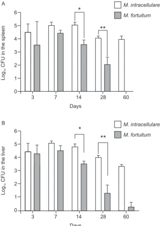
Course of M. fortuitum and M. intracellulare infec-tion in BALB/c mice - The comparative bacterial loads
in the spleens and liver of M. fortuitum and M.
intra-cellulare-infected mice were assessed. We observed a progressive reduction in the colony forming unit (CFU)
count in both the M. fortuitum and M. intracellulare
-infected mice; however, the mice -infected with M.
for-tuitum more efficiently controlled the bacteria than the
mice infected with M. intracellulare. The bacterial load
was 1.47 log lower in the spleen and 1.29 log lower in
the livers of M. fortuitum-infected mice 14 days
post-in-fection than in those of M. intracellulare-infected mice
(p= 0.0079, Mann-Whitney). At 28 days post-infection,
a similar difference was observed: M. fortuitum-infected
mice had a 1.98 log lower bacterial load in the spleen and
a 2.65 log lower level in the liver than M. intracellulare
-infected mice (p= 0.0159, Mann-Whitney) (Fig. 1A, B).
Granulomatous response inM. fortuitum and M. in-tracellulare-infected mice - The numbers of granulomas
in the liver sections of M. fortuitum or M. intracellulare
-infected mice were compared at the same time points that the bacterial load was determined. At day seven of infection, poorly structured aggregates containing a small number of macrophages and sparse inflammatory infiltrates consisting primarily of lymphocytes were ob-served in the liver sections of mice infected with either bacterium (Fig. 2A, B). At day 14, the granulomatous
response in the liver of M. fortuitum-infected mice was
characterized by lymphoid aggregates and macrophag-es, whereas the granulomas consisted primarily of
mac-rophages in M. intracellulare-infected mice (Fig. 2C,
D). In addition, in the M. intracellulare-infected mice,
the portal tract contained a moderate to extensive lym-phocytic infiltrate at day 28 after infection (Fig. 2E, F).
Overall, the numbers of granulomas in M. fortuitum
-infected mice were five-fold, seven-fold and fifteen-fold
lower than the number of granulomas in M.
intracellu-lare-infected mice at days 14, 28 and 60 post-infection,
respectively (p= 0.0159 and p= 0.0159 for day 28 and
60, respectively, Mann-Whitney) (Fig. 2I). We observed a progressive reduction in the granuloma area over time in M. fortuitum-infected mice. In contrast, in M. intra-cellulare-infected mice, the granuloma area enlarged approximately two-fold from day 14-60 after infection. In M. intracellulare-infected mice, the granulomas con-sisted of tightly grouped epithelioid macrophages in a round structure rimmed by lymphocytes (Fig. 2J).
Immunohistochemistry of granulomatous lesions in M. fortuitum or M. intracellulare-infected mice - The cellular composition of the hepatic granulomas was as-sessed by immunohistochemistry using specific mono-clonal antibodies against macrophages, B cells, T cells and plasmocytes. Greater numbers of macrophages,
6
5
4
3
2
1
0
3 7 14 28 60
Log
CFU in the spleen
10
Days
*
**
A
6
5
4
3
2
1
0
3 7 14 28 60
Log
CFU in the liver
10
Days
*
**
B M. intracellulare
M. fortuitum M. intracellulare
M. fortuitum
Fig. 1: bacterial load in the spleen or liver from Mycobacterium fortuitum or Mycobacterium intracellulare-infected BALB/c mice. Animals were infected with 107 bacteria intravenously (retro-orbital plexus). A: the number of colony forming units (CFU) in the spleen of mice infected with M. fortuitum was smaller than the number in mice infected with M. intracellulare at all time points examined (*: p = 0.0079, 14 days; **: p = 0.0159, 28 days; Mann-Whitney); B: the number of CFUs in the liver of mice infected with M. fortuitum
was smaller than the number in mice infected with M. intracellulare
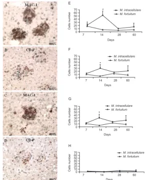
B cells, T cells and plasmocytes were observed in the
M. intracellulare-infected mice than in the M. fortuitum -infected mice and the saline-inoculated mice. In both groups of infected mice, granulomas were predominantly
composed of Mac-1+ cells (Fig. 3E) and CD4+ cells (Fig.
3F), in comparison to the other cell types present in the
granulomas, such as CD8+ (Fig. 3G) and B220+ (Fig. 3H)
cells. By day 14, the granulomas of M. intracellulare
-infected mice contained approximately eight times more
Mac-1+ cells and seven times more CD4+ cells than the
granulomas in M. fortuitum-infected mice. In addition,
at day 14, the numbers of Mac-1+ cells and CD4+ cells
in M. intracellulare-infected mice were approximately three-fold higher than at seven days post-infection. In
contrast, in the M. fortuitum-infected mice, we observed
an equivalent number of Mac-1+ cells and CD4+ cells at
seven, 14 and 60 days infection. On day 28
post-infection, however, the number of CD4+ cells in the liver
granulomas was approximately three-fold higher than
the number of Mac-1+ cells.
Cytokine expression in the liver of M. fortuitum or M. intracellulare-infected mice - qRT-PCR was used to mea-sure the expression of several cytokines that determine
mycobacterial infection outcome (IFN-γ, TNF-α and
IL-10) and of iNOS, known to be expressed by activated macrophages (MacMicking et al. 1997). This technique was chosen instead of others, such as semi-quantitative non-competitive RT-PCR, northern blot analysis and enzyme linked immunosorbent assay, because it offers several advantages. The most notable is a high degree of sensitivity, allowing for the detection of basal cytokine expression even under experimental conditions in which cytokine production is very low (Hein et al. 2001). qRT-PCR was performed using liver samples taken at the time points at which the bacterial load and granuloma
morphology were assessed. In both M. fortuitum and M.
intracellulare-infected mice, the mRNA expression
lev-els of TNF-α, IL-10 and iNOS were very low and similar
to the levels of expression in the control group. At day
seven post-infection, the mRNA expression of IFN-γ in
M. fortuitum-infected mice was three-fold higher than in M. intracellulare-infected mice (p= 0.0115, Kruskal-Wallis, Dunn’s post-test) (Fig. 4). Similarly, a two-fold
increase in IFN-γ mRNA expression was observed at
day 28 post-infection.
20
10
0
3 14 28 60
Number of granuloma
Days *
I
M. intracellulare
M. fortuitum
*
5000
4000
3000
2000
1000
0
7 14 28 60 Days
J
*** *** Granuloma area
M. intracellulare
M. fortuitum
μm
2
Fig. 2: morphometric analysis of the granuloma present in the liver of BALB/c mice infected with Mycobacterium intracellulare or Myco-bacterium fortuitum. Representative images of granulomas (arrow) in liver sections from M. intracellulare and M.fortuitum-infected mice. After seven (A, B), 14 (C, D), 28 (E, F) or 60 days (G, H) of infection, animals were euthanized and the liver was fixed in a 10% formol solution. Liver sections were prepared and stained with hae- hae-matoxylin and eosin in order to characterize the tissue lesion. I: the granulomas were counted and each column represents the mean of three (7 days), four (14 days) or five experiments (28 and 60 days), containing 3-5 animals per group. A total of 10 fields per lamina per animal were counted. The columns represents the mean number of granulomas per experiment (*: p = 0.0159; Mann-Whitney); J: the granuloma area were measured and each column represents the mean of five (7 days), 10 (14 and 28 days) and seven sections (60 days). A total of 2-3 sections per experiment were used and the area of 10 granulomas was measured for each section. The data is presented as the mean ± standard error of granuloma area expressed in mm2 (***: p = 0.0006; t test).
70 60 50 40 30 20 10 0
7 14 28 60
Cells number
M. intracellulare M. fortuitum
F
*
Days
70 60 50 40 30 20 10 0
7 14 28 60 Days
Cells number
M. intracellulare M. fortuitum
G
*
70 60 50 40 30 20 10 0
7 14 28 60
Cells number
M. intracellulare M. fortuitum
H
Days 70
60 50 40 30 20 10 0
7 14 28 60
Cells number
M. intracellulare M. fortuitum
E
*
Days
Fig. 3: phenotypic profile of cells in the inflammatory infiltrate in the liver of BALB/c mice infected with Mycobacterium intracellulare orMycobacterium fortuitum. Representative images of the liver of mice infected with M. intracellulare (A, B) or M.fortuitum (C, D) at 14 days after infection. Note the predominance of Mac-1+ cells (E) and CD4+ T cells (F) in the inflammatory infiltrate in both groups of infected animals. CD8+ cells (G) and B220+ cells (H) are present in smaller numbers. In the group infected with M. intracellulare, the number of Mac-1+ cells starts to decrease at day 14 post-infection (E). On the other hand, in the group infected with M. fortuitum the num-ber of CD4+ T cells begin to increase at 28 days post-infection (F). At 14 days post-infection, the group infected with M. intracellulare
DISCUSSION
In the present study, we observed several quantitative and qualitative differences between the granulomatous
response of M. fortuitum and M. intracellulare-infected
mice. We observed that BALB/c mice more efficiently
controlled the M. fortuitum infection than the M.
intra-cellulare infection. A progressive reduction in the CFU
counts was observed in both groups; however, M.
for-tuitum was almost completely cleared from mice by 60 days post-infection. In contrast, at this time point,
bac-teria could still be detected in the liver and spleen of M.
intracellulare-infected mice, although to a lesser extent than at earlier time points. In accordance with these re-sults, a similar approach employed by other authors de-scribed a model of BALB/c mice that were intravenously
infected with M. fortuitum. The authors observed a rapid
decrease in the bacterial load in the livers and spleens of infected mice (Parti et al. 2005). However, in the
pre-vious study, the authors used a Mycobacterium load in
the inoculum that was five-fold greater than that of the present study but observed a complete disappearance of bacterial load from the infected livers after 10 days. This was much faster than was observed in the present study,
perhaps due to the different Mycobacterium strains used
in the two studies. In agreement with the bacterial load quantification, there were more granulomas in the liver
of the M. intracellulare-infected mice compared to the
M. fortuitum-infected mice (Fig. 2). It is well known that most of the inoculum is trapped in the liver after an in-travenous infection (Khalil et al. 1975).
In addition to macrophages, T cells are important for granuloma formation. T cell-deficient mice do not form granulomas and form loose inflammatory lesions incapable of controlling bacterial growth. The
adop-tive transfer of CD4+ T cells reconstitutes granuloma
formation in RAG-deficient mice infected with
Myco-bacterium (Ladel et al. 1995). The T cell repertoire of a single granuloma is very diverse and similarly sized granulomas in the same organ from the same animal can
have different T cell contents in Mycobacterium
-infect-ed mice (Hogan et al. 2001). In order to determine the
cellular components of the granulomas in the liver of M.
intracellulare or M. fortuitum-infected mice, immuno-histological staining was performed using specific anti-bodies against macrophages, B cells or T cells. In mice
infected with either M. intracellulare or M. fortuitum,
macrophages and CD4+ T cells were the main
compo-nents of the granulomas. The numbers of macrophages,
CD4+ and CD8+ T cells, and B cells in the liver of M.
in-tracellulare-infected mice were higher than the numbers
in the liver of M. fortuitum-infected mice (Fig. 2A-H).
These data can be correlated with the higher number
and larger size of granulomas observed in the liver of M.
intracellulare-infected mice (Fig. 2I, J). On day 28
post-infection, the number of CD4+ cells was approximately
three-fold higher than the number of Mac-1+ cells in the
liver of M. fortuitum-infected mice. It has been reported
that granuloma formation is accelerated by
antigen-spe-cific CD4+ T cells and depends, to a large extent, on the
TNF-α and IFN-γ levels reached within the infected tis -sues (Appelberg et al. 1994, Hänsch et al. 1996, Chan et al. 2010, unpublished observations).
In order to elucidate the mechanisms underlying the
differences in the tissue response induced by M.
intracel-lulare and M. fortuitum in mice, quantitative RT-PCR for
IFN-γ and TNF-α was performed on liver samples taken
at the same time that the bacterial load and granuloma morphometry were assessed (Fig. 3). At day 24
post-in-fection, compared to M. intracellulare-infected mice, M.
fortuitum-infected mice expressed more IFN-γ, which is
known to be essential for the control of mycobacterial infection (MacMicking et al. 1997). These data are con-sistent with the increased number of granulomas and the
higher number of CD4+ T cells in the hepatic
granulo-mas. It is likely that the increased number of CD4+ T
cells was responsible for the increased IFN-γ mRNA
level that was observed. Furthermore, the increased
ex-pression of IFN-γ could explain the accelerated recruit -ment of inflammatory cells and the increased number of granulomas. We speculate that the observed increase
in CD4+ cells and IFN-γ may be responsible for this en
-hanced antibacterial response, which leads to the clear-ance of mycobacteria by 60 days after infection. We
ex-pected to detect TNF-α mRNA in the liver tissue of both groups. It has been demonstrated that TNF-α-deficient
mice have disorganized granulomas following mycobac-terial infection, indicating that this cytokine is important for granuloma formation and maintenance (Algood et al. 2005). Unexpectedly, in the liver of
mycobacterium-infected mice, we detected a low TNF-α mRNA level,
similar to the level of the control uninfected mice. Previ-ously, it has been demonstrated that the conventional use of an entire infected organ to study granuloma-specific gene expression can yield data that may not genuinely reflect intralesional events (Zhu et al. 2003). In this pre-vious work, the authors identified a downregulation of nine genes that are known to regulate the inflammatory 4.5
4.0 3.5 3.0 2.5 2.0 1.5 1.0 0.5 0.0
7 14 28 *
M. intracellulare
M. fortuitum
Days
IFN-
related to control (Log )
2
Fig. 4: IFN-γ mRNA expression in the liver of BALB/c mice infected with Mycobacterium fortuitum or Mycobacterium intracellulare.
The mRNA expression of IFN-γ in liver from M. fortuitum-
response to M. tuberculosis, as determined by qRT-PCR analyses of microdissected granuloma-derived cDNA (up to 27-fold) (Zhu et al. 2003). This result points to the idea that use of total liver RNA most likely resulted in the dilution of the gene of interest by nongranulomatous
liver tissue, preventing the detection of TNF-α and iNOS
mRNA expression in our study.
We did not actually explore the mechanism responsi-ble for the differences in the granulomatous responses of
M. fortuitum and M. intracellulare-infected mice. Gran-uloma formation may depend on the genetic background of the host (Orrell et al. 1992). It has been demonstrated
that the Nramp1/Slc11A1 gene exerts a major influence
on the early response to infections caused by various
atypical mycobacteria such as M. fortuitum and M.
in-tracellulare (Denis et al. 1986) by affecting granuloma
development (Sato et al. 1990). In addition,
Mycobacte-rium properties (van der Sar et al. 2004) related to
dif-ferences in pathogen virulence factors, such as cell wall composition, may be responsible for differences in the immune response (Chan et al. 2010, unpublished obser-vations) and probably in granuloma formation. The cell wall of mycobacteria consists of a highly complex ar-ray of distinctive lipids, glycolipids and proteins, which are believed to play important roles in the physiology of the bacterium and in the modulation of the host response during infection (Russell et al. 2002, Brennan 2003). Li-poarabinomannans (LAMs) are ubiquitous in mycobac-teria and appear to be the most potent non-peptidic cell wall molecules that modulate the host immune response. The LAM structure differs depending on the mycobacte-rial species. In slow-growing mycobacteria, the arabinan domain is capped by mannose residues (ManLAM) and in rapidly growing mycobacteria, the domain is capped by phosphoinositide motifs (PILAMs) (Chatterjee & Khoo 1998, Nigou et al. 2002). It has been observed that purified lipids from different strains induce differ-ent patterns of cytokines (Mohan et al. 2001). PILAMs more potently induce the secretion of proinflammatory
cytokines, such as TNF-α and IL-1, and the production
of microbicidal radicals than ManLAMs. In addition,
ManLAMs inhibit the IFN-γ-induced activation of mac -rophages (Vercellone et al. 1998). These data reinforce the idea that, in our study, the differences observed in
the BALB/c mouse responses to either M. fortuitum or
M. intracellulare infections were related to the
composi-tion of the Mycobacterium cell wall.
In summary, this paper compared granuloma
forma-tion in M. fortuitum and M. intracellulare-infected mice.
Well-structured granulomas were observed in both groups of infected mice at two weeks after infection as along with a progressive reduction in the bacterial load.
Only the group infected with M. fortuitum, however,
completely cleared the infection and 60 days after infec-tion, the granulomas were resolved, suggesting that the cells in the granulomas were efficiently activated to clear the bacteria. We believe that BALB/c mice are more
ca-pable of controlling M. fortuitum than M. intracellulare
infection. Our data reinforce the idea that early pathogen clearance can depend on the mycobacterial species and that some mycobacteria can live within the granuloma in
a state of dormancy with the potential to resurface later. While it is generally accepted that granulomas represent a component of the protective immune response against mycobacteria, the precise mechanism underlying the formation and maintenance of granulomas remains to be determined. Understanding the factors that contribute to this long and complex relationship between the pathogen and host is essential to be able to successfully modulate clinical outcomes.
ACKNOWLEDGEMENTS
To Dr Washington Luis Conrado dos Santos, for valuable advice and help in the statistical analyses used herein, and to Luana Palma, for photographic helpful support.
REFERENCES
Algood HM, Lin PL, Flynn JL 2005. Tumor necrosis factor and chemokine interactions in the formation and maintenance of gran-ulomas in tuberculosis. Clin Infec Dis 41 (Suppl. 3): S189-193.
Alvarez-Uria G 2010. Lung disease caused by nontuberculous myco-bacteria. Curr Opin Pulm Med16: 251-256.
Appelberg R, Castro AG, Pedrosa J, Silva RA, Orme IM, Minóprio P 1994. Role of gamma interferon and tumor necrosis factor alpha during T-cell-independent and dependent phases of Mycobacte-riumavium infection. Infect Immun62: 3962-3971.
ATS - American Thoracic Society 1997. Diagnosis and treatment of disease caused by nontuberculous mycobacteria. Am J Respir
Crit Care Med156: 1-25.
Bouley DM, Ghori N, Mercer KL, Falkow S, Ramakrishnan L 2001. Dynamic nature of host-pathogen interactions in Mycobacterium marinum granulomas.Infect Immun69: 7820-7831.
Brennan PJ 2003. Structure, function and biogenesis of the cell wall of Mycobacterium tuberculosis. Tuberculosis (Edinb) 83: 91-97.
Brown-Elliott BA, Wallace RJ Jr 2005. Infections caused by nontuber-culous mycobacteria. In GL Mandell, JC Bennett, R Dolin (eds.), Mandell, Douglas and Bennett’s: Principles and practice of infec-tious diseases, Vol. 2, 6th ed., Elsevier, Philadelphia, p. 2909-2916.
Chan ED, Bai x, Kartalija M, Orme IM, Ordway DJ 2010. Host im-mune response to rapidly growing mycobacteria, an emerging cause of chronic lung disease. Am J Respir Cell Mol Biol: in press.
Chatterjee D, Khoo KH 1998. Mycobacterial lipoarabinomannan: an extraordinary lipoheteroglycan with profound physiological ef-fects. Glycobiology8: 113-120.
Da Silva TR, De Freitas JR, Silva QC, Figueira CP, Roxo E, Leão SC, De Freitas LA, Veras PS 2002. Virulent Mycobacterium fortuitum restricts NO production by a gamma interferon-ac-tivated J774 cell line and phagosome-lysosome fusion. Infect Immun70: 5628-5634.
Denis M, Forget A, Pelletier M, Turcotte R, Skamene E 1986. Control of Bcg gene of early resistance in mice to infections with BCG sub-strains and atypical mycobacteria. Clin Exp Immunol 63: 517-525.
Flynn JL, Scanga CA, Tanaka KE, Chan J 1998. Effects of aminoguani-dine on latent murine tuberculosis. J Immunol160: 1796-1803.
Griffith DE, Aksamit T, Brown-Elliott BA, Catanzaro A, Daley C, Gordin F, Holland SM, Horsburgh R, Huitt G, Iademarco MF, Iseman M, Olivier K, Ruoss S, von Reyn CF, Wallace RJ Jr, Win-throp K 2007. An official ATS/IDSA statement: diagnosis, treat-ment and prevention of nontuberculous mycobacterial diseases.
Hänsch HC, Smith DA, Mielke ME, Hahn H, Bancroft GJ, Ehlers S 1996. Mechanisms of granuloma formation in murine Myco-bacterium avium infection: the contribution of CD4+ T cells. Int Immunol8: 1299-1310.
Hein J, Schellenberg U, Bein G, Hackstein H 2001. Quantification of murine IFN-gamma mRNA and protein expression: impact of real-time kinetic RT-PCR using SYBR green I dye. Scand J Im-munol 54: 285-291.
Ho MH, Ho CK, Chong LY 2006. Atypical mycobacterial cutaneous infections in Hong Kong: 10-year retrospective study. Hong Kong Med J12: 21-26.
Hogan LH, Macvilay K, Barger B, Co D, Malkovska I, Fennelly G, Sandor M 2001. Mycobacterium bovis strain bacillus Calmette-Guérin-induced liver granulomas contain a diverse TCR reper-toire, but a monoclonal T cell population is sufficient for protec-tive granuloma formation. J Immunol166: 6367-6375.
Jarzembowski JA, Young MB 2008. Nontuberculous mycobacterial infections.Arch Pathol Lab Med132: 1333-1341.
Khalil A, Bourut C, Halle-Pannenko O, Mathé G, Rappaport H 1975. Histologic reactions of the thymus, spleen, liver and lymph nodes to intravenous and subcutaneous BCG injections.
Biomedicine22: 112-121.
Ladel CH, Daugelat S, Kaufmann SH 1995. Immune response to My-cobacterium bovis bacille Calmette Guérin infection in major his-tocompatibility complex class I and II-deficient knock-out mice: contribution of CD4 and CD8 T cells to adquired resistance. Eur J Immunol25: 377-384.
MacMicking JD, North RJ, LaCourse R, Mudgett JS, Shah SK, Nathan CF 1997. Identification of nitric oxide synthase as a protective lo-cus against tuberculosis. Proc Natl Acad SciUSA 94: 5243-5248.
Mohan VP, Scanga CA, Yu K, Scott HM, Tanaka KE, Tsang E, Tsai MM, Flynn JL, Chan J 2001. Effects of tumor necrosis factor al-pha on host immune response in chronic persistent tuberculosis: possible role for limiting pathology. Infect Immun69: 1847-1855.
Nigou J, Gilleron M, Rojas M, García LF, Thurnher M, Puzo G 2002. Mycobacterial lipoarabinomannans: modulators of dendritic cell function and the apoptotic response. Microbes Infect4: 945-953.
Orrell JM, Brett SJ, Ivanyi J, Coghill G, Grant A, Beck JS 1992.
Mor-phometric analysis of Mycobacterium tuberculosis infection in mice suggests a genetic influence on the generation of the granu-lomatous inflammatory response. J Pathol 166: 77-82.
Palwade PK, Dhurat RS, Tendolkar UM, Dethe GR, Jerajani HR 2006. Chronic cutaneous disease caused by the rapid growers Myco-bacterium fortuitum and chelonae. Br J Dermatol 154: 774-775.
Parti RP, Srivastava S, Gachhui R, Srivastava KK, Srivastava R 2005. Murine infection model for Mycobacterium fortuitum. Microbes Infect 7: 349-355.
Runyon EH 1959. Anonymous mycobacteria in pulmonary disease.
Med Clin North Am43:273-290.
Russell DG, Mwandumba HC, Rhoades EE 2002. Mycobacterium
and the coat of many lipids. J Cell Biol158: 421-426.
Sato IY, Kobayashi K, Kasama T, Kaga S, Kasahara K, Kanemitsu H, Nakatani K, Takahashi T, Nakamura RM, Skamene E, Yoshida T 1990. Regulation of Mycobacterium bovis BCG and foreign body granulomas in mice by the Bcg gene. Infect Immun 58: 1210-1216.
Sungkanuparph S, Sathapatayavongs B, Pracharktam R 2003. Rap-idly growing mycobacterial infections: spectrum of diseases, antimicrobial susceptibility, pathology and treatment outcomes. J Med Assoc Thai86: 772-780.
Uslan DZ, Kowalski TJ, Wengenack NL, Virk A, Wilson JW 2006. Skin and soft tissue infections due to rapidly growing mycobacte-ria: comparison of clinical features, treatment and susceptibility.
Arch Dermatol142: 1287-1292.
van der Sar AM, Abdallah AM, Sparrius M, Reinders E, Vanden-broucke-Grauls CM, Bitter W 2004. Mycobacterium marinum
strains can be divided into two distinct types based on genetic diversity and virulence. Infect Immun 72: 6306-6312.
Vercellone A, Nigou J, Puzo G 1998. Relationships between the struc-ture and the roles of lipoarabinomannans and related glycoconju-gates in tuberculosis pathogenesis. Front Biosci3: 149-163.
Weitzul S, Eichhorn PJ, Pandya AG 2000. Nontuberculous mycobac-terial infections of the skin. Dermatol Clin 18: 359-377.