Hypoxia Enhances Proliferation of Human
Adipose-Derived Stem Cells via HIF-1
ɑ
Activation
Natsuko Kakudo1*, Naoki Morimoto1, Takeshi Ogawa1, Shigeru Taketani2, Kenji Kusumoto1
1Department of Plastic and Reconstructive Surgery, Kansai Medical University, Osaka, Japan, 2Department of Biotechnology, Kyoto Institute of Technology, Kyoto, Japan
Abstract
Background
Adipose tissue-derived stem cells (ASCs) have been recently isolated from human subcuta-neous adipose tissue. ASCs may be useful in regenerative medicine as an alternative to bone marrow-derived stem cells. Changes in the oxygen concentration influence physiolog-ical activities, such as stem cell proliferation. However, the effects of the oxygen concentra-tion on ASCs remain unclear. In the present study, the effects of hypoxia on ASC
proliferation were examined.
Methods
Normal human adipose tissue was collected from the lower abdomen, and ASCs were pre-pared with collagenase treatment. The ASCs were cultured in hypoxic (1%) or normoxic (20%) conditions. Cell proliferation was investigated in the presence or absence of inhibitors of various potentially important kinases. Hypoxia inducible factor (HIF)-1αexpression and
MAP kinase phosphorylation in the hypoxic culture were determined with western blotting. In addition, the mRNA expression ofvascular endothelial growth factor(VEGF) and fibro-blast growth factor(FGF)-2in hypoxic or normoxic conditions were determined with real-time RT-PCR. The effects of these growth factors on ASC proliferation were investigated. Chromatin immunoprecipitation (ChIP) of the HIF–1α-binding hypoxia responsive element
inFGF–2was performed. HIF–1αwas knocked down by siRNA, and FGF–2 expression
was investigated.
Results
ASC proliferation was significantly enhanced in the hypoxic culture and was inhibited by ERK and Akt inhibitors. Hypoxia for 5–15 minutes stimulated the phosphorylation of ERK1/ 2 among MAP kinases and induced HIF–1αexpression. The levels of VEGF and FGF–2
mRNA and protein in the ASCs were significantly enhanced in hypoxia, and FGF–2 increased ASC proliferation. The ChIP assay revealed an 8-fold increase in the binding of OPEN ACCESS
Citation:Kakudo N, Morimoto N, Ogawa T, Taketani S, Kusumoto K (2015) Hypoxia Enhances Proliferation of Human Adipose-Derived Stem Cells via HIF-1ɑActivation. PLoS ONE 10(10): e0139890. doi:10.1371/journal.pone.0139890
Editor:Masuko Ushio-Fukai, University of Illinois at Chicago, UNITED STATES
Received:June 20, 2014
Accepted:September 18, 2015
Published:October 14, 2015
Copyright:© 2015 Kakudo et al. This is an open access article distributed under the terms of the Creative Commons Attribution License, which permits unrestricted use, distribution, and reproduction in any medium, provided the original author and source are credited.
Data Availability Statement:All relevant data are within the paper and its Supporting Information files.
Funding:This research was supported in part by the Japanese Ministry of Education, Science, Sports and Culture and a Grant-in-Aid for Young Scientists (B) (26861515, Natsuko Kakudo).
HIF–1αtoFGF–2in hypoxia. HIF–1αknockdown by siRNA partially inhibited the FGF–2
expression of ASCs induced by hypoxia.
Conclusion
ASC proliferation was enhanced by hypoxia. HIF–1αactivation, FGF–2 production, and the
ERK1/2 and Akt pathway were involved in this regulatory mechanism.
Introduction
Adipose tissue was recently shown to be a source of multipotent adult stem cells, providing enriched adipose-derived stem cells (ASCs). ASCs have the potential to differentiate into bone, cartilage, tendons, nerves, and fat when cultured under lineage-specific conditions [1] [2]. Because of the convenience of isolation and extensive proliferative and differentiation capaci-ties in vitro, ASCs are a promising source of human stem cells for regenerative medicine. To date, various cell culture methods have been developed to more efficiently obtain stem cells while minimizing the risks to donors[3] [4].
Recent studies revealed that low oxygen tension or hypoxia affects various types of stem cells, such as embryonic stem cells [5], induced pluripotent stem cells [6], and bone marrow-derived stem cells (BMCs) [7] [8] [9]. A low oxygen environment is physiologically normal not only for most mammalian embryos, but also for adult somatic stem cells [8]. In mammalian cells, the transcriptional response to oxygen deprivation is largely mediated by hypoxia induc-ible factor 1 (HIF–1), which gradually increases as the oxygen concentration decreases. Expres-sion of genes such asvascular endothelial growth factor(VEGF) anderythropoietinis induced to stimulate angiogenesis and hematopoiesis. ASC proliferation is enhanced in hypoxia com-pared with normoxia [10] [8]. Secretion of VEGF and fibroblast growth factor (FGF)-2 pro-teins from ASCs is increased in hypoxia [11]. However, the detailed mechanisms remain unknown. The relationship between the response of ASCs to hypoxia and cell proliferation in this process remains unclear. Proliferation of ASCs is closely related to self-renewal and FGF signaling [12].
We hypothesized that hypoxic conditions are beneficial for ASC proliferation due to self-renewal-mediated autocrine FGF–2 signaling. In the present study, ASC proliferation and the associated signaling pathways in hypoxic conditions were examined. HIF–1αexpression and FGF–2 production in hypoxia were examined. A chromatin immunoprecipitation (ChIP) assay for HIF–1αbinding to the hypoxia responsive element (HRE) inFGF–2was performed. HIF–1αwas knocked down by siRNA in ASCs under hypoxia, and the mRNA expression of HIF–1α, FGF–2, and VEGF was investigated. Finally, FGF–2 and VEGF were added to ASCs, and the proliferation response was examined.
These results provide important insight into how hypoxic culture favors the ex vivo expan-sion of human ASCs, which will be important for maximizing the cell yield for clinical-scale ASC expansion.
Materials and Methods
Materials
Rabbit antibody against Erk1/2 was from Cell Signaling Technology (Beverly, MA). Rabbit antibody anti-phospho-nuclear factor kappa B (NF-ĸB) was from Abcam (Cambridge, UK).
Rabbit antibodies for NF-ĸB and FGF–2 were from GeneTex Inc. (Irvine, CA). Rabbit antibody
against beta-actin was from BioVision (Milpitas, CA). Mice antibody Histone H3 was Cell Sig-naling Technology (Beverly, MA). PD98059, an inhibitor of the MEK pathway, LY294002, an inhibitor of phosphatidylinositol-3-kinase-Akt, and SB203580, an inhibitor of p38, were from Calbiochem Novabiochem (San Diego, CA). Recombinant human VEGF and FGF–2 were pur-chased from PeproTech Ltd. (London, UK). Other reagents were from Sigma-Aldrich
(St. Louis, MO) unless otherwise stated.
Cell culture
The Ethics Review Board of Kansai Medical University has approved all research involving human participants and all patients provided their written consent to participate in this study. Human abdominal subcutaneous fat was collected from excess tissues excised during plastic and reconstructive surgery. ASCs were prepared as described previously [13–16]. Briefly, adi-pose tissue was washed extensively three times with 20 ml phosphate-buffered saline (PBS), cut into small pieces, and the extracellular matrix was digested with 0.1% collagenase solution with shaking at 37°C for 40 minutes. After adding basal medium consisting of Dulbecco’s modified Eagle’s medium (DMEM), 10% fetal bovine serum, and 1% penicillin, the cell pellet was centri-fuged at 1600 rpm for 3 minutes. After removing cellular debris by filtering the cell suspension through a 100-μm nylon mesh, the cells were incubated in control medium in a dish. The adherent ASCs were maintained until passage 3 in control medium, and nearly all cells formed fibroblast-like morphology.
Hypoxic culture experiments were performed in a multigas incubator (ASTEC, Hukuoka, Japan) at 37°C in an atmosphere containing 5% CO2balanced with nitrogen to reach an
oxy-gen concentration of 1%. Normoxic culture was performed in a standard incubator in an atmo-sphere containing 5% CO2and 20% O2.
Cell proliferation assay
Cell proliferation was assessed using the commercial kits Cell Counting Kit–8 (Dojindo Molec-ular Technologies, Inc., Gaithersburg, MD) and DNAIdU Labeling and Detection Kit (Takara Bio, Otsu, Japan).
The rationale for the cell counting assay using the Cell Counting Kit–8 kit is that the color-developing substrate WST–8 contained in the kit is reduced by intracellular dehydrogenase to water-soluble formazan, which can be directly quantitated photometrically. ASCs (1 × 10⁴ cells/well) were plated in 24-well plates and incubated for 1–7 days in 1% or 20% O2. FGF–2
(1–100 ng/mL) and VEGF (50–200 ng/mL) were added to the DMEM. The absorbance was measured at 450 nm (n = 3). The spectrometry was converted into cell number. The DNA IdU Labeling and Detection Kit is a colorimetric immunoassay based on the measurement of 5-iodo–20-deoxyuridine (IdU) incorporation during DNA synthesis. ASCs (2 × 10³cells/well)
were plated in 96-well plates, incubated for 24 hours in 1% or 20% O2, and labeled with IdU.
Cells were fixed, incubated with peroxidase-conjugated anti-IdU antibody, incubated with the peroxidase substrate 3,30,5,50-tetramethylbenzidine, and IdU incorporation was quantitated by
measuring the optical density at 450 nm. Proliferation of ASCs in 1% O2in the presence of an
Western blotting
Total cell protein extracts were obtained using M-PER (Mammalian protein extraction reagent; Thermo Fisher Scientific Inc.) for the detection of phospho-Erk1/2, Erk1/2, phospho-Akt, Akt, phospho-p38, p38, phospho-NF-ĸB, NF-ĸB, and beta-actin. Nuclear protein extracts was
obtained using NE-PER Nuclear and Cytoplasmic Extraction Kit (Thermo Fisher Scientific Inc., Waltham, MA) for detection of HIF–1αand Histone H3. Protein concentrations were measured using a BCA Protein assay kit (Pierce, Rockford, IL). Ten micrograms of protein extracts were separated with SDS-PAGE using a NuPAGE electrophoresis system (Invitrogen, Carlsbad, CA). Proteins were transferred to polyvinylidene difluoride membranes using the iBlot Dry Blotting System (Invitrogen) in accordance with the manufacturer’s protocol. Mem-brane blocking and immunodetection of proteins was performed with the WesternBreeze Chemiluminescent Detection Kit containing a secondary antibody solution of alkaline phos-phatase-conjugated antibody (Invitrogen). Antibodies raised against the following proteins were used: HIF–1α(1:500), phospho-Erk1/2 (1:1000), Erk1/2 (1:1000), phospho-Akt (1:1000), Akt (1:1000), phospho-p38 (1:1000), p38 (1:1000), phospho-NF-ĸB (1:1000), NF-ĸB (1:1000),
beta-actin (1:500) and Histone H3 (1:2000). We performed densitometric analysis for the west-ern blotting result.
RNA isolation and real-time reverse transcription-polymerase chain
reaction (RT-PCR)
ASCs were plated in 60-mm cell culture dishes at 4.25× 10⁵cells/dish. Confluent ASCs were cultured in 1% O2or 20% O2for 24 hours. RNA was extracted using Trizol (Life Technologies,
Carlsbad, CA). Real-Time RT-PCR was performed using the One Step SYBR PrimeScript RT-PCR Kit II (TAKARA BIO INC., Otsu, Japan), according to the manufacturer’s protocol. Briefly, RT-PCR was performed in a total volume of 25μl containing 10–100 ng total RNA, 12.5μl 2× One Step SYBER RT-PCR Master Mix, and 1μl PrimeScript 1-step Enzyme Mix. Each sample was analyzed in duplicate. Thermal cycler conditions were 42°C for 5 minutes and 95°C for 10 seconds, followed by 40 cycles of 95°C for 5 seconds and 60°C for 30 seconds. Amplification of the housekeeping geneß-actinmRNA, which served as a normalization stan-dard, was carried out withß-actinforward (50- TGGCACCCAGCACAATGAA -30) and
reverse (50- CTAAGTCATAGTCCGCCTAGAAGCA -30) primers. Side-stand-specific primers
forVEGFandFGF–2wereVEGFforward (50-TGCTTCTGAGTTGCCCAGGA–30) and reverse (50-TGGTTTCAATGGTGTGAGGACATAG–30), andFGF
–2forward (50- CCATCC TTTCTCCCTCGTTTCTT -30), and reverse (50- GATGTTTCCCTCCAATGTTTCATTC -30).
Quantification of target cDNA (VEGFandFGF–2) and the housekeeping gene (ß-actin) was performed using Real-time PCR Opticon 2 (Bio-Rad Laboratories, Inc., CA, USA). Data collection and analyses were performed using the software included with the system.VEGF
andFGF–2mRNA levels were measured as CT threshold levels and normalized to the individ-ualß-actincontrol CT values.
ELISA for secreted growth factors
ASCs were plated in 60-mm cell culture dishes at 4.25 × 10⁵cells/dish. Confluent ASCs were cultured in 1% O2or 20% O2for 24 hours. The amounts of VEGF and FGF–2 in the
ChIP assay
ChIP assays were performed using the EpiScope ChIP Kit (anti-mouse IgG) (Takara Bio) according to the manufacturer’s protocol. Briefly, ASCs were cultured in 1% or 20% O2for 24
hours. Cells were cross-linked with 1% formaldehyde for 5 minutes, and reactions were stopped by adding Quenching solution. Then, the cells were washed, lysed, and sonicated with Bioruptor UCD–200 (COSMO BIO Co., Ltd., Tokyo, Japan) for 30 seconds 5 times in an ice bath with 60-second cooling periods between sonications to shear chromatin into smaller DNA fragments. Lysates were centrifuged, and an aliquot of supernatant was saved as input DNA. Supernatants were then immunoprecipitated with anti-HIF–1α. Immunoprecipitates were recovered by adding MagnosphereTManti-mouse IgG beads (Takara Bio). After extensive washing, 100μl chelating resin solution was added to the beads and boiled for 15 minutes. Finally, the purified DNA was analyzed with real-time PCR for the presence of the HIF–1α -binding HRE. The PCR primers were designed to cover the HIF–1α-binding site of the human
FGF–2fragment (nucleotides 75142 to 75264): forward, 5'-TTGGGGGAGCTGGTAACTG ATG–3'; reverse, 5'-CAGTAGATGTTTCCCTCCAATG–3'. Ten percent of the lysate was used as the input control for PCR. The ChIP-precipitated DNA and input DNA were subjected to real-time PCR analyses using the One Step SYBR PrimeScript RT-PCR Kit II (TAKARA BIO), and samples from two individual ChIP assays were analyzed in triplicate. The results were nor-malized to the input and expressed as the n-fold increase over those of the normoxic controls.
siRNA transfection
One day before transfection, 25,000 cells of ASCs were plated in 2500μl of growth medium without antibiotics in a 6-well plate. The cell density was 30–50% confluent at the time of trans-fection. For each well to be transfected, RNAi duplex-Lipofectamine™RNAiMAX complexes were prepared as follows: 150 pmol RNAi (10μl, esiRNA human HIF1A, Sigma-Aldrich, St. Louis, MO, USA) was diluted in 1250μl of Opti-MEM1I Reduced Serum Medium without serum (Invitrogen, Carlsbad, CA, USA) gently. Lipofectamine™RNAiMAX (25μl) was diluted in Opti-MEM1I Reduced Serum Medium (1250μl) gently. RNAi duplex with the diluted Lipofectamine™RNAiMAX was combined and incubated for 10–20 minutes at room tempera-ture. RNAi duplex-Lipofectamine™RNAiMAX complexes (500μl) were added to each well containing cells. This gave a final volume of 3,000μl and a final RNA concentration of 10 nM. The cells were incubated for 48 hours at 37°C in a CO2incubator until the time of the assay for
gene knockdown. Knockdown was evaluated by real-time RT-PCR of HIF–1αand FGF–2. The real-time PCR method and primers of FGF–2 and the housekeepingβ-actin gene were the same as those for the above-mentioned RT-PCR described in Materials & Methods. Strand-specific primers were as follows: HIF–1αforward (5’-TTGCTCATCAGTTGCCACTTCC–3’) and reverse (5’-AGCAATTCATCTGTGCTTTCATGTC–3’). The universal negative control (Nippon Gene, Co., Ltd., Tokyo) was used as siRNA control in the experiments.
Statistical analysis
The Mann-Whitney U test was used for comparisons between groups, with p<0.05
consid-ered significant. Data are the means ± SD.
Results
Hypoxia promotes proliferation of ASCs
normoxia, ASCs cultured in 1% O2showed 1.5-fold higher proliferation on day 7. As shown in
Fig 1B, in DNA synthetic quantity of hypoxia ASC was increased significantly compared with normoxia. Cell proliferation in hypoxia was significantly suppressed by PD98059 and LY294002 but not SB203580 (Fig 1C).
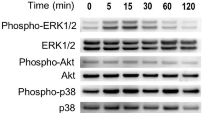
Hypoxia induces Erk and Akt phosphorylation and induces HIF
–
1
α
expression in ASCs
We investigated the activation of Erk1/2, Akt, p38, and NF-ĸB in ASCs in hypoxia.
Phosphory-lation of Erk1/2 and Akt was mostly seen after 5–15 minutes in hypoxia. Phosphorylation of p38 (Fig 2). Phosphorylation of NF-ĸB was not detected in hypoxia (data not shown). In the
densitometric analysis, the intensity of the phospho-ERK1/2 and phospho-Akt protein signal increased significantly with under hypoxia as sown inS1 Fig. Therefore, we concluded that only phospho-ERK 1/2 and Akt were responsible for ASC proliferation. We also evaluated the amount of HIF–1αprotein to demonstrate that the cells were actually exposed to low oxygen.
Fig 1. (A) Effects of hypoxia on the proliferation of human adipose-derived stem cells (ASCs). Proliferation was measured using Cell Counting Kit–8 according to the manufacturer’s instructions. A significant difference in ASC proliferation was observed between the hypoxia and normoxia groups on days 3 and 7. (B) Effects of hypoxia on the proliferation of human ASCs. DNA synthetic quantity was measured using the DNA IdU Labeling and Detection Kit according to the manufacturer’s instructions. A significant difference in DNA synthetic quantity of ASC was observed between the hypoxia and normoxia groups. (C) Effects of PD98059, LY294002, and SB203580 on the proliferation of human ASCs in hypoxia.Cell proliferation during hypoxia was significantly suppressed by PD98059 (10μM) and LY294002 (10μM) but not SB203580. Data are the means±SD.*p<0.05 vs. control.
HIF–1αprotein was increased in hypoxia. In addition, we examined whether Erk and Akt inhibitors suppressed the HIF–1αexpression. The expression of HIF–1αof ASCs in 1% O2
with inhibitor (PD98059: 10μM; LY294002: 20μM) was not detected by western blotting, as shown inFig 3.
Hypoxia induces the mRNA expression of
VEGF
and
FGF
–
2
To determine the induction of expression of hypoxia-associated growth factor genes, we mea-sured mRNA levels ofVEGFandFGF–2with real-time RT-PCR. TheVEGFandFGF–2 expression levels were significantly increased 4.28-fold and 1.21-fold, respectively, in hypoxia compared to normoxia at 24 hours (Fig 4).
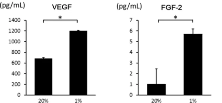
Hypoxia induces the secretion of VEGF and FGF
–
2
Secreted growth factors in the medium were measured with ELISA in hypoxia and normoxia at 24 hours. Protein levels of VEGF and FGF–2 were significantly increased in hypoxia (Fig 5). Therefore, hypoxia promoted the secretion of both VEGF and FGF–2. When culture media under hypoxia was treated with antibody for FGF–2, ASC proliferation almost diminished (Fig 6). These results confirm that FGF–2 promotes ASC proliferation. The results above were added in the text.
FGF
–
2 induces ASC proliferation
VEGF and FGF–2 were added to the medium to examine their effects on proliferation of ASCs. Compared with the control group that was not treated with VEGF and FGF–2, no significant
Fig 2. Effects of hypoxia on the expression of Erk, Akt, p38, and NF-ĸB in ASCs.Hypoxia activated the Erk and Akt pathways. Cell lysates were prepared from ASCs exposed to hypoxia for the indicated times, and the phosphorylation levels of ERK1/2, Akt, p38 and were determined with western blotting.
doi:10.1371/journal.pone.0139890.g002
Fig 3. Effects of hypoxia on the expression of HIF–1αin ASCs.Nuclear protein was prepared from ASCs
exposed to hypoxia for 6 hours, and western blotting was performed. HIF–1αwas increased in hypoxia.
However, the expression of HIF–1αof ASCs in hypoxia with inhibitor (PD98059: 10μM; LY294002: 20μM)
difference in ASC proliferation was observed in groups treated with VEGF. In contrast, FGF–2 significantly promoted cell proliferation in a dose-dependent manner (Fig 7).
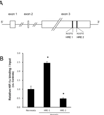
Functional HIF
–
1
α
binding to the HRE in
FGF
–
2
in ASCs
The HIF–1α-binding responsive element (5'-ACGTG–3') called the HRE inFGF–2is shown in Fig 8A. HRE was present at 2 sites (HRE1 and HRE2) in exon 3 of the FGF–2 gene. To clarify the interaction of HIF–1αwith one of its target genes in ASCs, we examined HIF–1αbinding toFGF–2and the promoter. The protein-DNA interaction was examined at the HRE found in
FGF–2in ASCs using the ChIP assay. The ChIP assay of HIF–1αwas performed for each of HRE1 and HRE2, but amplification by PCR was confirmed at only one site (HRE1). In response to hypoxia, the binding for HRE1 was enhanced about 2.5-fold (Fig 8B). In contrast, the ChIP assay for HIF–1αin theFGF–2promoter revealed that HIF–1αdid not bind to the HRE site (data not shown).
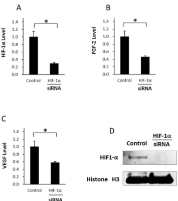
HIF
–
1
α
knockdown significantly decreases the expression of FGF
–
2 on
ASCs
For further determination of the effect of HIF–1αknockdown in hypoxia, we measured the expression levels of HIF–1α, FGF–2, and VEGF (Fig 9A, 9B and 9C). Transfection with HIF–
1αsiRNA in hypoxic culture inhibited protein expression of HIF–1αin western blotting. Transfection with HIF–1αsiRNA in hypoxic culture inhibited HIF–1αmRNA expression by about 70%, and the levels of FGF–2 and VEGF were inhibited by about 54% and 43%,
Fig 4. Effects of hypoxia on mRNA expression ofVEGFandFGF–2in ASCs.After incubation for 24 hours, the mRNA expression of the indicated genes was analyzed using real-time RT-PCR. The fold-change during hypoxia is expressed relative to the level in normoxia.VEGFandFGF–2expression in hypoxia was
significantly higher than in normoxia. Data are the means±SD.*p<0.05.
doi:10.1371/journal.pone.0139890.g004
Fig 5. Effects of hypoxia on the secretion of VEGF and FGF–2 from ASCs.The secretion of VEGF and FGF–2 into the medium in hypoxia was significantly higher than in normoxia. Data are the means±SD. *p<0.05.
respectively, showing that HIF–1αexpression under hypoxia is closely related to FGF–2 and VEGF expressions. Furthermore, we showed the inhibition level of HIF–1αby siRNA using western blotting (Fig 9D).
Discussion
This study firstly demonstrated that hypoxia enhanced the proliferation of ASCs via HIF–1α activation. The hypoxic culture stimulated the phosphorylation of ERK1/2 and Akt-induced HIF–1αexpression. The levels ofVEGFandFGF–2mRNA and their proteins in the ASCs were significantly enhanced in the hypoxic condition. Of these growth factors, FGF–2 affected ASC proliferation. The ChIP assay revealed that the binding of HIF–1αtoFGF–2increased in hypoxia. HIF–1αactivation, FGF–2 production, and the ERK1/2 and Akt pathway were involved in this regulatory mechanism.
ASCs were recently isolated from human subcutaneous adipose tissue [1] [13] [14]. ASCs may be useful in regenerative medicine as an alternative to BMCs. Changes in the oxygen con-centration influence physiological activities, such as stem cell proliferation. For example,
Fig 6. Effects of anti FGF–2 antibodies on proliferation of ASCs.ASCs were cultured in media with 0 (control), 5 and 25μg/mL of the antibodies under 20% O2and 1% O2conditions for 48h. Proliferation was
measured using Cell Counting Kit–8 according to the manufacturer’s instructions and the absorbance was converted into cell number. The proliferation of ASCs under 1% O2was inhibited by the antibodies. Data are
the means±SD.*p<0.01.
doi:10.1371/journal.pone.0139890.g006
Fig 7. Effects of VEGF and FGF–2 on proliferation of ASCs.Proliferation was measured using Cell Counting Kit–8 1 day after addition growth factors according to the manufacturer’s instructions. Compared with the control group that was not treated with VEGF and FGF–2, no significant difference in ASC proliferation was observed in groups treated with VEGF. In contrast, FGF–2 significantly promoted cell proliferation in a dose-dependent manner.
Lennon et al. showed that culturing BMCs from 6- to 12-week-old rats in 5% pO2resulted in
an approximately 40% higher cell number at the first passage compared with culturing these cells in 21% pO2[17]. Grayson et al. also reported that human BMCs show enhanced
prolifera-tion in hypoxia (2% pO2) over seven passages, resulting in a 30-fold increase in cell number
compared with normoxia [7]. However, the effects of the oxygen concentration on ASCs remain unclear. In the present study, we found that the proliferation-inducing effects of hyp-oxia on ASCs were similar to those on BMCs.
Several researchers have reported that ASC proliferation is more markedly influenced under hypoxic versus normoxic conditions in vitro. Lee et al. reported that the proliferation of human ASCs is significantly increased in hypoxia (2%) [11]. Yamamoto et al. also reported that ASCs cultured in 2% O2show a 1.5-fold increase in proliferation over 6 weeks of culture
[8]. In addition, Rasmussen compared the effects of prolonged hypoxic culture on growth of human ASCs cultured in 1, 5, and 21% oxygen. They concluded that culturing ASCs in 5% oxy-gen significantly lowers the ASC doubling time among the groups [18]. Using the IdU incorpo-ration assay and cell number counting assay, we demonstrated that ASC prolifeincorpo-ration in hypoxia (1%) was increased compared with normoxia. An oxygen tension of 1–5% in culture is reported to be sufficient to increase the proliferation of ASCs. Collectively, these results support the idea that hypoxic culture conditions are favorable for ASCs. Hypoxic culture allows the
Fig 8. (A) The HIF–1α-binding responsive element (5'-ACGTG–3') is called the HRE. HRE was present
at 2 sites in the FGF–2 gene, and these were designated as HRE1 and HRE2. (B) Functional HIF–1α
binding to hypoxia responsive element (HRE) found in the proximal region ofFGF–2in hypoxia.The relative association of HIF–1αwith humanFGF–2was analyzed with ChIP in ASCs incubated in normoxia
(20% O2) or hypoxia (1% O2) for 24 hours. Sheared chromatin was immunoprecipitated with anti-HIF–1α. The
enrichment of HIF–1αwas quantified with real-time PCR using HRE-specific primers forFGF–2. In response
to hypoxia, the binding for HRE 1 was significantly enhanced about 2.5-fold. Data were normalized to the total amount of added DNA and are the means±SD of two independent experiments performed in triplicate. *p<0.05 vs. normoxia.
production of many ASCs from a few donor cells, providing a useful culture method for the large-scale production of ASCs that will be required in regenerative medicine.
HIF is induced by insufficient oxygen supply to cells and functions as a transcription factor. HIF-α(HIF–1α, -2α, and -3α) binds to constitutively expressed HIF–1βin cells, forming a het-erodimer. HIF–1αis also produced under normoxic conditions, but does not function because it is degraded by the 26S proteasome, a proteolytic enzyme complex. HIF–1αis unlikely to be degraded in hypoxia, and it migrates into the nucleus to form a heterodimer with HIF–1βor binds to histone acetylation enzymes, such as CBP (CREB1-binding protein) /p300 [19]. The resulting complexes bind to a response element called the HRE (5'-ACGTG–3'). Asp803 of HIF–1αis involved in the transport of the molecular complex CBP/p300 with histone acetyl-transferase activity to HRE on DNA, promoting transcription of various genes. HIF–1α induces the expression of VEGF, platelet-derived growth factor, and basic fibroblast growth factor, enhancing angiogenesis and eventually improving the oxygen environment [20]. How-ever, the roles of HIF–1αin ASCs remain unclear.
The expression of HIF–1αin ASCs during hypoxia has been reported by several researchers [8] [21]. Stubbs et al. reported that HIF–1αbecomes stabilized during hypoxia due to an increase in VEGF-A protein secretion [21]. Yamamoto et al. also reported that HIF–1αprotein is increased in hypoxic conditions due to significantly enhanced VEGF secretion [8]. Lee et al. demonstrated that expression ofVEGFandFGF–2mRNA is enhanced in ASCs in hypoxia,
Fig 9. Influence of HIF–1αknockdown by siRNA in ASCs under hypoxia (A) The relative mRNA
expression levels of HIF–1αin ASCs after transfection for 48 h under hypoxia. HIF–1αlevel was
suppressed significantly compared with that in the control. (B) The relative mRNA expression levels of FGF–2 in ASCs after transfection for 48 h under hypoxia. FGF–2 level was suppressed significantly compared with that in the control. (C) The relative mRNA expression levels of VEGF in ASCs after transfection for 48 h under hypoxia. VEGF level was suppressed significantly compared with that in the control. Data are the means±SD.*p<0.05. (D) Expression of HIF–1αwas inhibited by siRNA
using western blotting.
promoting the secretion of these growth factors [11]. However, the relationship between the mRNA and protein expression of FGF–2 and the expression of HIF–1αin hypoxia was unclear. In the present study, we first demonstrated that HIF–1αbinds to an HRE in FGF–2 gene in ASCs to enhance mRNA and protein expression of FGF–2, thereby promoting proliferation.
Akt and p38 phosphorylation lead to the stabilization of HIF–1αand the survival response in BMCs [22]. HIF–1αstabilization leads to the induction of HIF–1α-responsive genes includ-ing proangiogenic factors such as VEGF and interleukin–6 [23]. In the present study, we dem-onstrated that Akt was phosphorylated in the hypoxic culture of ASCs. Activated Akt may stabilize HIF–1αas a survival response in ASCs, as is seen in BMCs. Kim et al. reported that the expression levels of phosphorylated ERK1/2 and Akt are increased as well as the prolifera-tion of ASCs in hypoxia [24]. Our results also demonstrated that ERK1/2 and Akt were phos-phorylated along with the proliferation of ASCs in hypoxia.
FGF–2 plays a critical autocrine/paracrine role in human ASC self-renewal [12]. The effects of VEGF on ASCs vary in different published reports. Suga et al. reported no significant effect after the addition of VEGF to the medium of ASCs [25]. In contrast, VEGF treatment signifi-cantly increased bromodeoxyuridine incorporation, indicating increased proliferation of ASCs. In this study, VEGF did not affect the cell proliferation of ASCs, although FGF–2 promoted proliferation. Thus, of the growth factors produced by ASCs in hypoxia, FGF–2 was involved in cell proliferation.
The relationship between ASC proliferation and differentiation and reactive oxygen species (ROS) has recently been attracting attention [26], and a role for ROS generation as a key medi-ator of ASC proliferation under hypoxia has been proposed [24] [27] [28]. Further investiga-tion is necessary to clarify the relainvestiga-tionship between ASC proliferainvestiga-tion under hypoxia and ROS.
In conclusion, the mRNA and protein expression of FGF–2 was enhanced as well as the pro-liferation of ASCs in hypoxia. HIF–1αexpression and ERK1/2 and Akt phosphorylation were observed in hypoxia. HRE, a binding site fir HIF–1α, was present inFGF–2in ASCs. These results demonstrate that HIF–1αexpression is strongly involved in cell proliferation in hyp-oxia, revealing a component of the kinetics and cell regulatory mechanisms of ASCs in hypoxia. Hypoxic culture may be a convenient method to enhance the proliferative capacity of stem cells for transplantation. Although the effects of hypoxic culture on ASCs should be further investigated, ASCs cultured on a large scale in hypoxia using this method of controlling prolif-eration may be clinically useful.
Supporting Information
S1 Fig. The quantities of phospho-ERK1/2, ERK1/2, Phospho-Akt, Akt, phospho-p38 and p38 were determined using densitometry.Data are the means ± SD of 4 independent experi-ments. The expression levels of phospho-ERK1/2 were normalized to ERK1/2 levels in the same sample.p<0.05 compared with 0 hours. The expression of Akt and p38 was analyzed
in the same manner. The intensity of the phospho-ERK1/2 and phospho-Akt protein signal increased significantly with under hypoxia.
(TIF)
Acknowledgments
Author Contributions
Conceived and designed the experiments: NK TO NM ST KK. Performed the experiments: NK TO. Analyzed the data: NK TO. Contributed reagents/materials/analysis tools: NK TO. Wrote the paper: NK TO NM KK.
References
1. Zuk PA, Zhu M, Ashjian P, De Ugarte DA, Huang JI, Mizuno H, et al. Human adipose tissue is a source of multipotent stem cells. Molecular biology of the cell, 2002. 13: 4279–4295. PMID:12475952 2. Mizuno H, Tobita M, Uysal AC. Concise review: Adipose-derived stem cells as a novel tool for future
regenerative medicine. Stem Cells, 2012. 30: 804–810. doi:10.1002/stem.1076PMID:22415904 3. Rajala K, Lindroos B, Hussein SM, Lappalainen RS, Pekkanen-Mattila M, Inzunza J, et al. A defined
and xeno-free culture method enabling the establishment of clinical-grade human embryonic, induced pluripotent and adipose stem cells. PloS one, 2010. 5: e10246. doi:10.1371/journal.pone.0010246 PMID:20419109
4. Zeng G, Lai K, Li J, Zou Y, Huang H, Liang J, et al. A rapid and efficient method for primary culture of human adipose-derived stem cells. Organogenesis, 2013. 9: 287–295. doi:10.4161/org.27153PMID: 24280895
5. Ezashi T, Das P, Roberts RM. Low O2tensions and the prevention of differentiation of hES cells.
Pro-ceedings of the National Academy of Sciences of the United States of America, 2005. 102: 4783–
4788. PMID:15772165
6. Yoshida Y, Takahashi K, Okita K, Ichisaka T, Yamanaka S. Hypoxia enhances the generation of induced pluripotent stem cells. Cell Stem Cell, 2009. 5: 237–241. doi:10.1016/j.stem.2009.08.001 PMID:19716359
7. Grayson WL, Zhao F, Bunnell B, Ma T. Hypoxia enhances proliferation and tissue formation of human mesenchymal stem cells. Biochem Biophys Res Commun, 2007. 358: 948–953. PMID:17521616 8. Yamamoto Y, Fujita M, Tanaka Y, Kojima I, Kanatani Y, Ishihara M, et al. Low oxygen tension
enhances proliferation and maintains stemness of adipose tissue-derived stromal cells. Biores Open Access, 2013. 2: 199–205. doi:10.1089/biores.2013.0004PMID:23741631
9. Hung SP, Ho JH, Shih YR, Lo T, Lee OK. Hypoxia promotes proliferation and osteogenic differentiation potentials of human mesenchymal stem cells. J Orthop Res, 2012. 30: 260–266. doi:10.1002/jor. 21517PMID:21809383
10. Dos Santos F, Andrade PZ, Boura JS, Abecasis MM, da Silva CL, Cabral JMS. Ex vivo expansion of human mesenchymal stem cells: a more effective cell proliferation kinetics and metabolism under hyp-oxia. Journal of cellular physiology, 2010. 223: 27–35. doi:10.1002/jcp.21987PMID:20020504 11. Lee EY, Xia Y, Kim WS, Kim MH, Kim TH, Kim KJ, et al. Hypoxia-enhanced wound-healing function of
adipose-derived stem cells: increase in stem cell proliferation and up-regulation of VEGF and bFGF. Wound repair and regeneration: official publication of the Wound Healing Society [and] the European Tissue Repair Society, 2009. 17: 540–547.
12. Zaragosi LE, Ailhaud G, Dani C. Autocrine fibroblast growth factor 2 signaling is critical for self-renewal of human multipotent adipose-derived stem cells. Stem Cells, 2006. 24: 2412–2419. PMID:16840552 13. Kakudo N, Shimotsuma A, Miyake S, Kushida S, Kusumoto K. Bone tissue engineering using human
adipose-derived stem cells and honeycomb collagen scaffold. J Biomed Mater Res A, 2008. 84: 191–
197. PMID:17607760
14. Kakudo N, Shimotsuma A, Kusumoto K. Fibroblast growth factor–2 stimulates adipogenic differentia-tion of human adipose-derived stem cells. Biochem Biophys Res Commun, 2007. 359: 239–244. PMID:17543283
15. Kakudo N, Minakata T, Mitsui T, Kushida S, Notodihardjo FZ, Kusumoto K. Proliferation-promoting effect of platelet-rich plasma on human adipose-derived stem cells and human dermal fibroblasts. Plas-tic and reconstructive surgery, 2008. 122: 1352–1360. doi:10.1097/PRS.0b013e3181882046PMID: 18971718
16. Kakudo N, Kushida S, Suzuki K, Matsumoto N, Kusumoto K. Effect of C3 transferase on human adi-pose-derived stem cells. Hum Cell, 2011. 24: 165–169. doi:10.1007/s13577-011-0033-0PMID: 21984005
18. Rasmussen JG, Frobert O, Pilgaard L, Kastrup J, Simonsen U, Zachar V, et al. Prolonged hypoxic cul-ture and trypsinization increase the pro-angiogenic potential of human adipose tissue-derived stem cells. Cytotherapy, 2011. 13: 318–328. doi:10.3109/14653249.2010.506505PMID:20795759 19. Yamashita K, Discher DJ, Hu J, Bishopric NH, Webster KA. Molecular regulation of the endothelin–1
gene by hypoxia. Contributions of hypoxia-inducible factor–1, activator protein–1, GATA–2, AND p300/ CBP. The Journal of biological chemistry, 2001. 276: 12645–12653. PMID:11278891
20. Rey S, Semenza GL. Hypoxia-inducible factor-1-dependent mechanisms of vascularization and vascu-lar remodelling. Cardiovascuvascu-lar research, 2010. 86: 236–242. doi:10.1093/cvr/cvq045PMID: 20164116
21. Stubbs SL, Hsiao ST, Peshavariya HM, Lim SY, Dusting GJ, Dilley RJ. Hypoxic preconditioning enhances survival of human adipose-derived stem cells and conditions endothelial cells in vitro. Stem cells and development, 2012. 21: 1887–1896. doi:10.1089/scd.2011.0289PMID:22165914
22. Kanichai M, Ferguson D, Prendergast PJ, Campbell VA. Hypoxia promotes chondrogenesis in rat mes-enchymal stem cells: a role for AKT and hypoxia-inducible factor (HIF)-1alpha. Journal of cellular physi-ology, 2008. 216: 708–715. doi:10.1002/jcp.21446PMID:18366089
23. Hu X, Yu SP, Fraser JL, Lu Z, Ogle ME, Wang JA, et al. Transplantation of hypoxia-preconditioned mesenchymal stem cells improves infarcted heart function via enhanced survival of implanted cells and angiogenesis. The Journal of thoracic and cardiovascular surgery, 2008. 135: 799–808. doi:10.1016/j. jtcvs.2007.07.071PMID:18374759
24. Kim JH, Park SH, Park SG, Choi JS, Xia Y, Sung JH. The pivotal role of reactive oxygen species gener-ation in the hypoxia-induced stimulgener-ation of adipose-derived stem cells. Stem cells and development, 2011. 20: 1753–1761. doi:10.1089/scd.2010.0469PMID:21265612
25. Suga H, Eto H, Shigeura T, Inoue K, Aoi N, Kato H, et al. IFATS collection: Fibroblast growth factor-2-induced hepatocyte growth factor secretion by adipose-derived stromal cells inhibits postinjury fibro-genesis through a c-Jun N-terminal kinase-dependent mechanism. Stem Cells, 2009. 27: 238–249. doi:10.1634/stemcells.2008-0261PMID:18772314
26. Kim JH, Kim SH, Song SY, Kim WS, Song SU, Yi TG, et al. Hypoxia induces adipocyte differentiation of adipose-derived stem cells by triggering reactive oxygen species generation. Cell biology international, 2014. 38: 32–40. doi:10.1002/cbin.10170PMID:23956071
27. Kim JH, Song SY, Park SG, Song SU, Xia Y, Sung JH et al. Primary involvement of NADPH oxidase 4 in hypoxia-induced generation of reactive oxygen species in adipose-derived stem cells. Stem cells and development, 2012. 21: 2212–2221. doi:10.1089/scd.2011.0561PMID:22181007