Mechanism of hif-1
a
mediated hypoxia-induced
permeability changes in bladder endothelial cells
C. Liu
1, C.L. Shui
2, Q. Wang
3, H. Luo
1and C.G. Gu
41Department of Urology, The Second People
’s Hospital of Deyang City, Deyang, Sichuan Province, China 2Department of Anesthesiology, Af
filiated Yongchuan Hospital of Chongqing Medical University, Yongchuan, Chongqing, China 3Department of Pathology, Cancer Center of Guangzhou Medical University, Guangzhou, Guangdong Province, China
4Department of Laboratory Medicine, The Fifth People
’s Hospital of Chengdu, Chengdu, Sichuan Province, China
Abstract
This study aimed to investigate the mechanism of hypoxia-inducible factor-1 alpha (HIF-1a) mediated hypoxia-induced permeability changes in bladder endothelial cells. Models ofin vitrohypoxic cell culture of bladder cancer, bladder cancer cells with low HIF-1aexpression and HIF-1aRNA interference (RNAi) expression vector were established. Western blot and reverse transcription polymerase chain reaction (RT-PCR) were used to detect the expression of HIF-1a and vascular endothelial growth factor (VEGF) in each group. Bladder cell permeability was determined. Results showed that protein and mRNA expression of HIF-1aand VEGF at 3 and 12 h of hypoxia were significantly higher than normal control (Po0.05), and peaked
at 12 h. HIF-1a and VEGF expression in the hypoxic group and hypoxic+3-(50-hydroxymethyl-20-furyl)-1-benzyl indazole (YC-1) group were significantly higher than normal control (Po0.05), while expression in the hypoxic+YC-1 group was
significantly lower than the hypoxic group (Po0.05). Bladder cell permeability in the hypoxic and hypoxic+YC-1 group were
significantly increased compared to normal control (Po0.05), while in the hypoxic+YC-1 group was significantly decreased
compared to the hypoxic group (Po0.05). Most of the cells in the stably transfected HIF-1aRNAi expression vector
pcDNA6.2-GW/EmGFP-miR-siHIF-1a expressed green fluorescence protein (GFP) under fluorescence microscope. pcDNA6.2-GW/ EmGFP-miR-siHIF-1acould significantly inhibit HIF-1agene expression (Po0.05). HIF-1aand VEGF expression in the hypoxic
group and siHIF-1a hypoxic group were significantly higher than normal group (Po0.05), while expression in the siHIF-1a
hypoxic group was significantly lower than the hypoxic group (Po0.05). Findings suggest that HIF-1ais an important factor
in the increase of bladder cancer cell permeability.
Key words: Bladder cancer; Hypoxia inducible factor-1a; VEGF; Cell permeability; YC-1
Introduction
Bladder cancer is the most common malignancy of the urinary tract, having a high incidence rate and a recurrence rate above 80%. Patients not only have to bear the burden of high medical expenses, but also the stress of psycho-logical distress caused by relapses. Non-muscle invasive bladder cancer (NMIBC) comprises about 70% of all newly diagnosed bladder cancers. NMIBC tumor stages include papillary lesions that have not invaded sub-mucosal lamina propria (Ta), papillary lesions that have invaded the lamina propria but not the muscle (T1), and carcinoma in situ (CIS). At present, transurethral resection followed by instillation of adjuvant intravesical chemotherapy has been the main treatment of choice for NMIBC. However, NMIBC still has a recurrence rate of 70–80% (1,2).
Tumor cells regulate and adapt to the hypoxic environ-ment by initiating a series of hypoxic adaptive responses. A number of studies reported that hypoxia-inducible
factor-1 (HIF-1) is the central mediator of adaptive responses to hypoxia in tumors. HIF-1 is a heterodimer transcription factor consisting ofaandbsubunits. Among these, HIF-1adetermines the activity of HIF-1 and is the sole oxygen regulator. HIF-1 can adapt to hypoxia by regulating the expression of various target genes and involves in the process of tumor growth, invasion and metastasis (3). HIF-1ahas been found to be highly expressed in a variety of malignant tumors and precancerous lesions, indicating that it is closely related with tumor angiogenesis, invasion and metastasis (4).
The synthetic compound hypoxic+3-(50 -hydroxy-methyl-20-furyl)-1-benzyl indazole (YC-1) has superior anti-angiogenic and anti-tumor activities. YC-1 was first discovered by Ko et al. (5), who reported that its role is achieved by activating platelet soluble guanylate cyclase and upregulation of adenosine monophosphate and cyclic
Correspondence: C.G. Gu:<[email protected]>
guanosine monophosphate. Vascular endothelial growth factor (VEGF), also known as vascular permeability factor, is an efficient and specific growth factor for vascular endothelial cells. It has a strong effect on promoting mitosis and chemotaxis of vascular endothelial cells, stimulating angiogenesis, and increasing vascular permeability. In normal tissue, its expression is very low or absent, but it is highly expressed in malignant solid tumors and positively correlated with the malignancy grade of the tumor (6). This paper describes for thefirst time the role of HIF-1a/VEGF in cancer.
This study aimed to investigate the role of HIF in the recurrence of bladder cancer at the gene level, in order to provide a new therapeutic approach for the disease. We hypothesized that during hypoxia, an increase in the HIF-1a and VEGF protein expression would lead to an increase in bladder cancer cell permeability, and that HIF-1ais an important factor in the increase of bladder cancer cell permeability.
Material and Methods
Experiments were performed at the Central Laboratory of the Affiliated Yongchuan Hospital of Chongqing Medical University and Department of Pathology, Cancer Center of Guangzhou Medical University. Cells collection period were from February 2015 to February 2016.
All procedures were approved by the Ethics Commit-tee of The Second People’s Hospital of Deyang City, Sichuan, China (Reference number [2014]-20) and in accordance with the 1964 Helsinki declaration and its later amendments or comparable ethical standards.
Material
Cell lines. Bladder cancer cells were purchased from Cell Bank of the Chinese Academy of Sciences (Shanghai, China).
Main reagents and instruments. Rabbit HIF-1a poly-clonal antibodies (Upstate Biotechnology, USA); VEGF polyclonal antibodies, albumin-fluorescein isothiocyanate conjugate (FITC-albumin),b-actin monoclonal antibodies (Sigma-Aldrich Corporation, USA); IgG secondary anti-bodies (Beijing Zhongshan Gold Bridge Biotechnology Corporation Ltd, China); newborn calf serum (Chengdu Harry Biotechnology Corporation Ltd, China); primer synthe-sis (Sangon Biotechnology (Shanghai) Corporation Limited, China); Roswell Park Memorial Institute (RPMI) 1640 culture medium (HyClone Laboratories, USA); Bicinchoni-nic acid (BCA) protein assay kit (PierceTM, Thermo Fisher Scientific, USA); Mini Trans-Blotselectro transfer system, fluorescence quantitative PCR instrument, ChemiDoc XRS Gel Analysis System, ‘‘Quantity one’’ software (Bio-Rad Laboratories, USA); MilicellTMelectrical resistance system (ERS) instrument (Millipore, USA); inverted fluorescence microscope model TH4-200 (Olympus Corporation, Japan); ultra-sensitive enhanced chemiluminescence (ECL) kit
(Pierce Biotechnology, USA); HeraeusCO2constant tem-perature incubator (Heraeus, Germany); DUs-640 ultra-violet spectrophotometer (Beckman Inc., USA); modular incubator chamber (Billups-Rothenberg Inc., USA).
Methods
Model ofin vitrohypoxic cell culture of bladder cancer. Bladder cancer cells were cultured in a cultureflask with primary culture medium and kept at 37°C in a 5% CO2 incubator. The culture medium was replaced every 2 days. When the cell culture reached around 90% confluence, it was digested with 0.25% trypsin-0.01% ethylenediamine-tetraacetic acid (EDTA) solution and subcultured. Cells were then seeded at a concentration of 1.5105/cm2in a 0.1% gelatin-coated TranswellTMpolycarbonate membrane. Culture medium was replaced every 2 days after 24 h of inoculation. The transendothelial electrical resistance (TER) of the monolayer endothelial cells was measured with MilicellTM ERS for cell integrity. When the TERs were stable, we proceeded to the next step of experiment. Cells seeded in the 6-well plates were examined and placed in a hypoxic tankfilled with 1% O2of mixed gas (94% N2, 5% CO2, 1% O2) and cultured in a 37°C cell culture incubator. Observation was done at 0, 3, and 12 h post-hypoxia.
Model of hypoxic HIF-1a inhibitor. After the bladder
cancer cells in the 6-well plate reached complete con-fluence or the monolayer bladder cancer cells in the TranswellTMhad been tested for TER stabilization, these were randomly assigned into normal control group (normal culture under normoxia with 5% CO2 and O2 in air), hypoxic group (cultured in 1% O2for 6 h) and hypoxic+ 3-(50-hydroxymethyl-20-furyl)-1-benzyl indazole (YC-1) group (addition of 100mM YC-1, followed by incubation in 1% O2 for 6 h). Time of incubation with YC-1 was 48 h.
Model of HIF-1aRNA interference (RNAi) expression vector. RNAi expression vector was established. Amplifi-cation of recombinant plasmid and DNA extraction, plasmid transfection, screening of SiHIF-1apositive cell clones and identification of pcDNA6.2-GW/EmGFP-miR-siHIF-1astable expression cell lines were performed.
HIF-1a and VEGF mRNA expression (detected with RT-PCR). RNA was extracted with TRIzon, followed by reverse transcription and detection with RT-PCR. Expres-sion ratio and relative content of the HIF-1a, VEGF mRNA and internal reference (b-actin) was calculated. Primers used were as follows: HIF-1a-F: TCAAAGTCGGACAGC CTCA, HIF-1a-R: CCCTGCAGTAGGTTTCTGCT; VEGF-F: GAAGTGGTGAAGTTCATGGATGTC, VEGF-R: CGAT CGTTCTGTATCAGTCTTTCC;b-actin-F: TGAGACCTTC AACACCCCAG,b-actin-R: GCCATCTCTTGCTCGAAGTC. Determination of bladder cancer cell permeability. Culture medium was discarded. FITC-albumin (100mL, 1 mg/mL dissolved in D-Hank solution) was added to the upper part of the double-chamber, while 500 mL D-Hank solution was added to the lower part at 37°C. Then, the culture plate was placed back into the incubator and continued to culture for 1 h. Following this, the solutions from the upper and lower chambers were sucked out.
The excitation and emission wavelength of the fluores-cence spectrophotometer were set at 490 and 525 nm, respectively. Fluorescence intensity of each sample was determined to analyze permeability changes.
Statistical analysis
Data are reported as means±SD. Analysis was performed using SPSS 19.0 statistical software (SPSS Inc., USA). Comparison between groups were performed
with ANOVA test. Po0.05 was considered statistically significant.
Results
Effects of hypoxia on HIF-1aand VEGF expression in
bladder endothelial cells
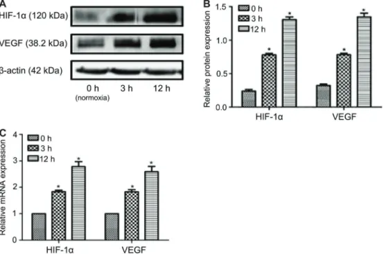
Compared with the normal control group, the protein and mRNA expression of HIF-1a and VEGF were sig-nificantly higher at 3 and 12 h of hypoxia, and peaked at 12 h (Po0.05; Figure 1A–C).
YC-1 reduced the protein and mRNA expression of HIF-1aand VEGF expression
Protein and mRNA expression of HIF-1aand VEGF in the hypoxic group and hypoxic+YC-1 group were signif-icantly higher than the normal control group (Po0.05), while expression in the hypoxic+YC-1 was significantly lower than the hypoxic group (Po0.05; Figure 2A–C). Protein and mRNA expression of HIF-1aand VEGF were significantly reduced by YC-1 (Figure 2B and C).
Bladder cancer cell permeability
Cell permeability in the hypoxic group and hypoxic+ YC-1 group were significantly increased compared to the normal control group (Po0.05), while in the hypoxic+ YC-1 group, it was significantly decreased compared to the hypoxic group (Po0.05; Figure 3).
Effects of HIF-1aRNAi expression vector on VEGF
expression and bladder cancer cell permeability
Most of the cells in the stably transfected HIF-1aRNAi expression vector pcDNA6.2-GW/EmGFP-miR-siHIF-1a expressed green florescence protein (GFP) (Figure 4A).
This vector could significantly inhibit HIF-1agene expres-sion (Po0.05; Figure 4B and C).
Effects of stable transfection on HIF-1aand VEGF
expression
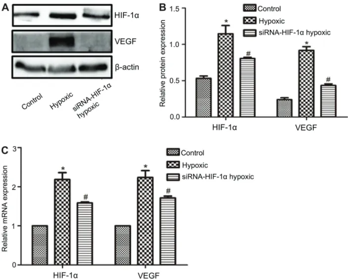
The HIF-1a and VEGF expression in the hypoxic group and siHIF-1ahypoxic group was significantly higher (Po0.05) than the normal control group (Figure 5A). The protein (Figure 5B) and mRNA expression (Figure 5C) in the siHIF-1a hypoxic group were significantly lower than the hypoxic group (Po0.05).
Discussion
Bladder cancer is one of the most common urologic cancer with high incidence rate. During development, many solid tumors loose regulation of cell proliferation and apoptosis. This accelerates its growth, and when growth rate is higher than the process of angiogenesis, local ischemia and hypoxia of the tumor can occur.
HIF-1a wasfirst discovered by Semenza et al. (7) in 1992. A large number of studies showed that HIF-1a, which has long been recognized as one of the important transcription factor that mediate cellular response to hypoxia, plays an important role in the regulation of pathophysio-logical changes in ischemic and hypoxic conditions (8).
Figure 2.A, HIF-1aand VEGF expression.B, Histogram of HIF-1aand VEGF protein expression.C, Histogram of HIF-1aand VEGF mRNA expression. Data are reported as means±SD. *Po0.05 compared with normal control group;#Po0.05 compared with hypoxic
group (ANOVA). Time of incubation with YC-1=48 h.
Figure 3.Effect of YC-1 on increased bladder cancer cell per-meability by hypoxia. Data are reported as means±SD. *Po0.05
compared with normal control group; #P
o0.05 compared with
The expression of HIF-1 is subjected to hypoxia, tumor genes and various regulatory factors, and is an important factor in hypoxia-induced adaptive gene expression.
HIF-1 is composed of two subunits, the HIF-1a and HIF-1bsubunits. The intracellular protein level of the HIF-1b subunits is not affected by the level of oxygenation while the HIF-1a gene is subjected to the regulation of hypoxia signals. Under normoxic conditions, theasubunit undergoes post-translational modifications and O2 -depen-dent prolyl hydroxylation, and is degraded via the ubiquitin-proteasome pathway mediated by pVHL (von Hippel-Lindau tumor suppressor protein). In hypoxic condition, HIF-1a does not undergo self-hydroxylation and degradation, and thus it is stable. It forms a transcriptionally active hetero-dimer complex with the HIF-1b subunit and binds to the
hypoxia-response element of the target genes. It involves the regulation of adaptive response to hypoxia through a variety of target gene expressions (9–11). YC-1 is a specific inhibitor of HIF-1a. It can reduce the accumulation of HIF-1aand inhibit HIF-1 transcriptional activity through a pathway that is independent of soluble guanylate cyclase (12). It can also promote protein degradation of HIF-1a(13).
Vascular endothelium is the inner lining of blood vessels with semi-selective permeable membrane. Endothelial cells regulate the tension of the blood vessel, maintain their normal structure, secretefibrinolytic protein, anticoagulant and other substances, and have anti-inflammatory effects (14). Recent studies have confirmed that there are many factors involved in the whole process of angiogenesis in
Figure 4.Effects of HIF-1aRNAi expression vector on VEGF expression and bladder cancer cell permeability.A, Stably transfected HIF-1aRNAi expression vector in normal control group and hypoxic group.B, HIF-1agene expression after stable transfection of HIF-1a
RNAi expression vector pcDNA6.2-GW/EmGFP-miR-siHIF-1a. C, Histogram of HIF-1agene expression after stable transfection of HIF-1aRNAi expression vector pcDNA6.2-GW/EmGFP-miR-siHIF-1a. Data are reported as means±SD. *Po0.05 compared with
solid tumors. Among these, VEGF is known to be the most active and the most specific angiogenesis inducing factor. The increase of HIF-1aand VEGF were parallel, indicating a correlation between the two. Thus, it is speculated that HIF-1a may stimulate angiogenesis by increasing the expression of VEGF to adapt to the growth environment of hypoxic and low-sugar content, so that the tumor can continue to grow and metastasize distantly (15).
A number of studies has identified high expression of HIF-1ain gastric cancer and various other cancers, and it is found to be closely associated with the biological behavior of tumors (16,17). HIF-1ashows high expression in hypoxic environment. This stimulates the release of VEGF in tumor cells and promotes angiogenesis (18,19). RNAi can silence the expression of target genes. It can specifically knock down or turn off the expression of a specific gene, participate in the protection of genome
against invasion and thus maintain the genome stability. This is a technique of reverse genetics, which plays an important role in functional genomics research (20–25).
In thefirst part of this study, a model ofin vitrohypoxic cell culture of bladder cancer was established. We preli-minarily postulated that increase in HIF-1a and VEGF expression is one of the mechanisms that leads to the increase of cell permeability in hypoxia. In the second part of the study, an HIF-1ainhibitor YC-1 was added and we found that a decrease in the HIF-1a expression resulted in a decrease in VEGF expression and an increase in the permeability of bladder cancer cells during hypoxia. To further clarify whether the activation of HIF-1awas involved in the increase of bladder cancer cells permeability during hypoxia, we used an established bladder cancer cell model with low HIF-1 expression to study the changes of tumor cell permeability in hypoxia.
Figure 5.Effects on HIF-1aand VEGF expression after stable transfection.A, HIF-1aand VEGF expression.B, Histogram of HIF-1a
and VEGF protein expression.C, Histogram of HIF-1aand VEGF mRNA expression. Data are reported as means±SD. *Po0.05
This studyfinally found that hypoxia induced an increase in bladder cancer cell permeability. HIF-1ainhibitor YC-1 and HIF-1a RNAi could significantly inhibit the increase of hypoxia-induced cell permeability. HIF-1ais an important factor in the increase of bladder cancer cell permeability.
Acknowledgements
This study was supported by the Deyang City Science and Technology Bureau Support Plan (Project No. 2017 SZ097-1).
References
1. Anastasiadis A, de Reijke TM. Best practice in the treatment of nonmuscle invasive bladder cancer.Ther Adv Urol2012; 4: 13–32, doi: 10.1177/1756287211431976.
2. Nepple KG, O’Donnell MA. The optimal management of T1 high-grade bladder cancer.Can Urol Assoc J2009; 3: S188–S192, doi: 10.5489/cuaj.1194.
3. Masoud GN, Li W. HIF-1a pathway: role, regulation and intervention for cancer therapy.Acta Pharm Sin B2015; 5: 378–389, doi: 10.1016/j.apsb.2015.05.007.
4. Jubb AM, Pham TQ, Hanby AM, Frantz GD, Peale FV, Wu TD, et al. Expression of vascular endothelial growth factor, hypoxia inducible factor 1alpha, and carbonic anhydrase IX in human tumours.J Clin Pathol2004; 57: 504-512, doi: 10.1136/jcp.2003.012963.
5. Ko FN, Wu CC, Kou SC, Lee FY, Teng CM. YC-1, a novel activator of platelet guanylate cyclase. Blood 1994; 84: 4226–4233.
6. Bussolati B, Mason JC. Dual role of VEGF-induced heme-oxygenase-1 in angiogenesis.Antioxid Redox Signal2006; 8: 1153–1163, doi: 10.1089/ars.2006.8.1153.
7. Semenza GL, Agani F, Booth G, Forsythe J, Iyer N, Jiang BH, et al. Structural and functional analysis of hypoxia-inducible factor 1.Kidney Int1997; 51: 553–555, doi: 10.1038/ ki.1997.77.
8. Semenza G. Signal transduction to hypoxia-inducible factor 1. Biochem Pharmacol2002; 64: 993–998, doi: 10.1016/S0006-2952(02)01168-1.
9. Semenza GL. HIF-1 and mechanisms of hypoxia sensing. Curr Opin Cell Biol2001; 13: 167–171, doi: 10.1016/S0955-0674(00)00194-0.
10. Cummins EP, Taylor CT. Hypoxia-responsive transcription factors.Pflügers Arch2005; 450: 363–371, doi: 10.1007/ s00424-005-1413-7.
11. Semenza GL. Life with oxygen.Science2007; 318: 62–64, doi: 10.1126/science.1147949.
12. Chun YS, Yeo EJ, Choi E, Teng CM, Bae JM, Kim MS, et al. Inhibitory effect of YC-1 on the hypoxic induction of erythropoietin and vascular endothelial growth factor in Hep3B cells.Biochem Pharmacol2001; 61: 947–954, doi: 10.1016/ S0006-2952(01)00564-0.
13. Kim HL, Yeo EJ, Chun YS, Park JW. A domain responsible for HIF-1alpha degradation by YC-1, a novel anticancer agent. Int J Oncol2006; 29: 255–260, doi: 10.3892/ijo.29.1.255. 14. Mullin JM, Agostino N, Rendon-Huerta E, Thornton JJ.
Keynote review: epithelial and endothelial barriers in human
disease.Drug Discov Today2005; 10: 395–408, doi: 10.1016/ S1359-6446(05)03379-9.
15. Maslennikova AV, Orlova AG, Prianikova TI, Kostenikov NA, Vinogradova IuN, Denisenko AN. Clinical significance of and diagnostic methods for tumoral hypoxia.Vopr Onkol2011; 57: 413–420.
16. Huang C, Sun Z, Sun Y, Chen X, Zhu X, Fan C, et al. Association of increased ligand cyclophilin A and receptor CD147 with hypoxia, angiogenesis, metastasis and prog-nosis of tongue squamous cell carcinoma.Histopathology 2012; 60: 793–803, doi: 10.1111/j.1365-2559.2011.04130.x. 17. Horiuchi A, Hayashi T, Kikuchi N, Hayashi A, Fuseya C, Shiozawa T, et al. Hypoxia upregulates ovarian cancer invasiveness via the binding of HIF-1ato a hypoxia-induced, methylation-free hypoxia response element of S100A4 gene. Int J Cancer2012; 131: 1755–1767, doi: 10.1002/ijc.27448. 18. Huang X, He Z, Jiang X, Hou M, Tang Z, Zhen X, et al. Folic
acid represses hypoxia-induced inflammation in THP-1 cells through inhibition of the PI3K/Akt/HIF-1apathway.PLoS One 2016; 11: e0151553, doi: 10.1371/journal.pone.0151553. 19. Zhang HZ, Zhao JH, Zhao XH, Zhu SC, Liu B. Effect of
hypoxia on HIF-1aand VEGF expression and radiosensi-tivity in esophageal cancer cell line Eca109.J Hebei Med Univ2008; 29: 815–818.
20. Fire A, Xu S, Montgomery MK, Kostas SA, Driver SE, Mello CC. Potent and specific genetic interference by double-stranded RNA inCaenorhabditis elegans.Nature1998; 391: 806–811, doi: 10.1038/35888.
21. Meister G, Tuschl T. Mechanisms of gene silencing by double-stranded RNA. Nature 2004; 431: 343–349, doi: 10.1038/ nature02873.
22. Matzke MA, Birchler JA. RNAi-mediated pathways in the nucleus. Nat Rev Genet 2005; 6: 24–35, doi: 10.1038/ nrg1500.
23. Lage H. Therapeutic potential of RNA interference in drug-resistant cancers. Future Oncol 2009; 5: 169–185, doi: 10.2217/14796694.5.2.169.
24. Bobbin ML, Rossi JJ. RNA interference (RNAi)-based therapeutics: delivering on the promise?Annu Rev Pharmacol Toxicol 2016; 56: 103–122, doi: 10.1146/annurev-pharmtox-010715-103633.