The impact of interleukin-13 receptor expressions in cell
migration of astrocytomas
Isabele Fattori MorettiI, Roseli SilvaI, Sueli Mieko Oba-ShinjoI, Priscila Oliveira de CarvalhoI, Lais Cavalca CardosoI, Isac
de CastroII, Suely Kazue Nagahashi MarieI,III
DOI: 10.5935/MedicalExpress.2015.05.05
I Universidade de São Paulo, Faculdade de Medicina, Departamento de Neurologia, Laboratório de Biologia Molecular e Celular (LIM 15), Sao Paulo, SP, Brazil II Universidade de São Paulo, Faculdade de Medicina, Departamento de Clínica Médica, Disciplina de Medicina Molecular, Sao Paulo, SP, Brazil
III Universidade de São Paulo, Núcleo de Estudos de Terapia Celular e Molecular, Sao Paulo, SP, Brazil
INTRODUCTION: Astrocytomas are common brain tumors. Increased expression levels of Interleukin-13 Receptor α2 (IL-13RA2) have been reported in astrocytomas. The Interleukin-13 signaling pathway may be associated with cell migration when binding to Interleukin-13 Receptor α1.
OBJECTIVE: To investigate Interleukin-13 Receptor α1 (IL-13RA1) and IL13RA2 expression levels in human difusely iniltrative astrocytomas and test the involvement of Interleukin-13 levels in cell migration in two glioblastoma cell lines.
METHODS:IL13RA expression levels were accessed by quantitative real time PCR in 128 samples of astrocytomas and 18 samples of non-neoplastic brain tissues from temporal lobe epilepsy surgery. The impact of IL-13 levels (10 and 20 ng/mL) on cell migration was analyzed by the wound assay in U87MG and A172 cells.
RESULTS: Glioblastoma presented higher IL13RA1 and IL13RA2 expression levels compared to lower grades astrocytomas and to non-neoplastic cases. U87MG and A172 cells presented higher expression levels of IL-13RA1 vs.IL-13RA2. A signiicant diference in migration rate was observed in A172 cells treated with 10 ng/mL of IL-13
vs. control: treated cells presented slower migration than non-treated cells. U87MG cells treated with IL-13 20ng/ mL presented slower migration than non-treated cells. This indicates that the IL13Rα1 signaling pathway was not activated, indeed inhibited by the decoy IL-13Rα2, slowing cell migration. This impact occurred with a lesser concentration of IL-13 on the A172 than on the U87MG cell line, because A172 cells have a higher IL-13RA2/A1 ratio. CONCLUSION: The present results suggest IL-13 receptors as possible targets to decrease tumor cell migration.
KEYWORDS: Astrocytoma; Interleukin-13; Interleukin-13 Receptors.
Moretti IF, Silva R, Oba-Shinjo SM, Carvalho PO, Cardoso LC, Castro I, Marie SKN. The impact of interleukin-13 receptor expression in cell migration of astrocytomas. MedicalExpress (São Paulo, online). 2015;2(5):M150505
Received for Publication on July 17, 2015; First review on July 31, 2015; Accepted for publication on August 11, 2015
E-mail: [email protected]
■
INTRODUCTIONAstrocytomas are the most frequent adult brain tumors,1 and are classified into four grades of malignancy based on morphological characteristics, according to the World Health Organization (WHO): grade I or pilocytic astrocytoma (AGI) with low proliferation rate; grade II orlow grade astrocytoma (AGII) with higher mitotic rate and potential progression to more malignant grades; grade III or anaplastic astrocytoma (AGIII) with higher cell density, heterogeneity and vascular proliferation,
represented by contrast enhancement on neuroimaging exams; grade IV or glioblastoma (GBM) with the highest proliferation rate, neovascularization, and presence of necrotic areas, corresponding to rimmed contrast enhanced areas on neuroimaging.2-4
purity was evaluated using the 260/280 absorbance ratio on Nanodrop (ThermoScientific, Wilmington, DE, USA), and samples presenting values ranging from 1.8 to 2.0 were considered satisfactory. The reverse transcription of RNA was performed using DNase I (FPLC-pure, GE Healthcare, Uppsala, Sweden), random oligonucleotides (dT), RNase inhibitors and the reverse transcriptase SuperScript III (Life Technologies, Carlsbad, CA, USA). All samples were treated with RNase H (GE Healthcare) and stored in TE buffer at -20°C until use.
Quantitative Real Time PCR (qRT-PCR)
The expressions of IL13RA1 and IL13RA2 were
determined by qRT-PCR on an ABI Prism 7500 real time PCR system (Life Technologies) using SYBR Green I with a mixture of 12µl (3µl of cDNA, 6µl of 2x Power SYBR Green I Master Mix (Life Tchnologies), and 3µl of forward and reverse primers. The following protocol was used, in duplicate: uracyl N-glycosylase incubation for 2 minutes at 50 °C, polymerase activation for 10 minutes at 95 °C, 40 cycles of 15 seconds at 95 °C and 1 minute at 60 °C. The result was normalized in relation to the geometric mean of three suitable housekeeping genes as previously described.14 The primers were designed to amplify 80-120pb amplicons, and were synthetized by IDT (Integrated DNA Technologies, Coralville, IA, USA). The sequence of each primer
was: IL13RA1 F: CTCTGGAGTAATTGGAGCCAAGA,
I L 1 3 R A 1 R : T G C G A C G A T G A C T G G A A C A A , I L 1 3 R A 2 F : T T G C G T A A G C C A A A C A C C T A , I L 1 3 R A 2 R : T G A A C A T T T G G C C A T G A C T G , H P RT F: T G A G G AT T T G G A A A G G G T G T, H P RT R : A G C A C A C A G A G G G C T A C A A ; G U S B F : G A A A A TA C G T G G T T G G A G A G C T C A T T, G U S B R : C G A G T G A A G A T C C C C T T T T T A ; T B P F : A G G A T A A G A G A G C C A C G A A C C A , T B P R : CTTGCTGCCAGTCTGGACTGT. The expression was calculated according to equation 2-ΔCt where ΔCt = mean Ct of target gene - geometric mean of the Ct from the housekeeping genes.15
Cell culture
Adherent cells of U87MG and A172 cell lines were grown in DMEN (Dulbecco’s Modified Eagle Medium, Life Technologies) plus 10% of fetal bovine serum (FBS) and penicillin and streptomycin, at 37 °C and under 5% of CO2. Viability and concentration of cells were determined by trypan blue staining and automatically analyzed in the CountessTM Automated Cell Counter (Life Technologies).
Migration assay
The migration assay was performed by the scratch wound healing analysis. A total of 2x10⁵ and 1x10⁵ cells of U87MG and A172 cell lines, respectively, were plated to its recurrence.6,7 In this scenario, there is an urge for a
search of new molecular targets to improve therapeutic effectiveness. One candidate molecule is the receptor for interleukine-13 alpha 2 (IL-13Rα2), which has been reported as presenting high expression in astrocytoma according to the grade of malignancy and associated to poor prognosis.8,9 The IL-13Rα2 is a decoy monomeric plasma membrane receptor, presenting fast association and slow dissociation rate with cytokine interleukin-13 (IL-13).10 IL-13 inhibits IL-13Rα2, in proportion to the receptor expression level.11
The IL-13 is also the ligand for IL-13Rα1, and upon this ligation, the heterodimerization with receptor IL-4Rα activates a downstream signaling pathway that interferes with cell migration.10,11 Therefore, the ratio of the expression levels of IL13RA1 and IL13RA2 on the cell membrane and the abundance of IL-13 in the surrounding environment may regulate cell migration.
The present study aims to: 1) analyze the expression levels of the two genes coding for IL-13Rα (IL13RA1 and
IL13RA2) in different grades of human astrocytomas and
correlate their expression ratios to the grade of malignancy and invasiveness; 2) analyze the role of IL-13 in the cell migration in two GBM cell lines with different expression ratios of IL13RA1 and IL13RA2.
■
MATERIALS AND METHODSTissues samples
A series of 128 cases of diffusely infiltrating astrocytomas (26 AGII; 18 AGIII; 84 GBM) and 18 anonymous non-neoplastic (NN) brain tissues from epilepsy surgery were analyzed. Tissues samples were collected by the Neurosurgery Group of the Department of Neurology at Hospital das Clínicas of the School of Medicine of the University of São Paulo, from 2000 to 2007. The samples were classified according to WHO grading system by the Institutional Pathology Division. All samples were macrodissected before being snap-frozen in liquid nitrogen and microdissected before RNA extraction. A 4µm-thick cryosection of each sample was analyzed under light microscopy after hematoxylin-eosin staining to perform the microdissection of cellular debris, necrotic and non-neoplastic areas.12,13
Written informed consent was obtained from all patients included in this analysis, and the study was approved by the ethical committee of School of Medicine of University of São Paulo (case # 0599/10).
RNA Extraction and Reverse Transcription
onto a 24-well plate and cultured to achieve a confluent monolayer. Cells were scraped to create a scratch with p200 pipet tip and washed twice with phosphate buffer saline (PBS). Cells were incubated with DMEN and 0.5% FBS added with 10ng/ml and 20ng/ml of IL-13, in triplicates. Images were acquired on an inverted microscope at 0, 24, 48, 72 hours after the scratch for each sample, and they were analyzed quantitatively by the Image J software, version 1.48 (National Institute of Health, USA).
Statistical Analysis
The normality of the gene expression distribution was analyzed by the Kolmogorov-Smirnov test, and the comparison of the expression levels between different grades of astrocytoma and the non-neoplastic brain tissue was accessed by the non-parametric Kruskal-Wallis and Dunn test (SPSS version 20 - IBM, Armonk, USA, 2011). Differential expression was considered statistically significant when p < 0.05. The variation of specificity and sensibility of the gene expression levels was analyzed using the ROC curve16 comparing the different grades of astrocytoma with the non-neoplastic brain tissue. Sensibility was defined as the probability of the expression of each gene to indicate the diagnosis of astrocytoma, and specificity as the probability of not being astrocytoma. The cut off value of expression level was selected at the maximum sensibility and specificity, and these values wereused to determine the status of expression level of each sample. Cases were divided into two groups, namely (a) with gene hyperexpression (expression levels higher than or equal to the cut off value), and (b)with gene hypoexpression (expression levels lower than the cut off value).
Linear and Non-Linear Regression was used to analyze the behavior of samples over time. Values with r square value ≥ 0.8 were considered to exhibit good performance or linearity. The obtained equations were compared by slope values for linear regression and plateau for non-linear regression (second-order models). Statistical significance was considered with p ≤ 0,05.
■
RESULTSIL13RA1andIL13RA2expression levels in human
astrocytoma samples
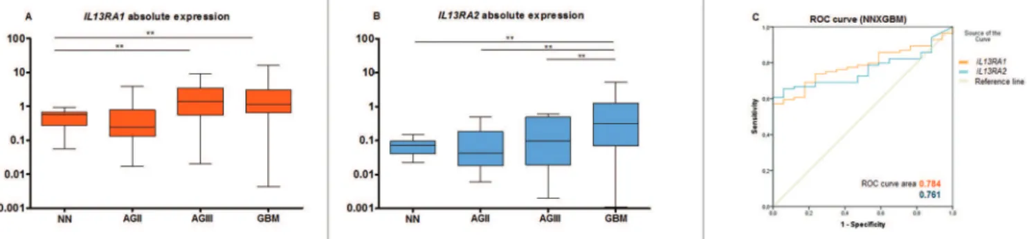
The expression of IL13RA1 by qRT-PCR showed
lower median expression levels in low grade astrocytomas (AGII) as compared to control, non-neoplastic (NN) cases. In contrast, AGIII and GBM cases showed higher expression levels than NN samples, with statistical significance (p < 0.05) as seen in figure 1A. IL13RA1 expression increased according to the level of malignancy. A similar expression
pattern was observed for IL13RA2, with significant
differences in expression levels between GBM vs. NN (p
< 0.005), GBM vs. AGII (p < 0.005), and GBM vs. AGIII (p <
0.05), as shown in figure 1B.
The IL13RA1 and IL13RA2 expression levels
in astrocytomas of different malignant grades are demonstrated as a heatmap in figure 2: the IL13RA2/
IL13RA1 expression ratio was higher in the more malignant
grades of astrocytomas (AGIII and IV). IL13RA2 expression levels showed better sensitivity and specificity presenting an area under ROC curve of 0.784 (p = 0.001), and IL13RA1
expression levels an area of 0.761 (p < 0.001). This is illustrated in figure 1C. No positive correlation between
IL13RA1 and I-13RA2 expressions was detected through
Spearman’s rho test.
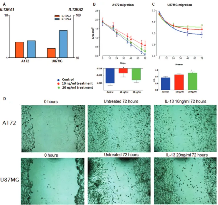
IL13RA1 andIL13RA2expression levels on U87MG and
A172 cells
Both GBM cell lines, U87MG and A172, presented higher expression of IL13RA2 than IL13RA1, and U87MG
cells presented higher expression for IL13RA2 than A172
cells, as shown in figure 3A.
IL-13 alters U87MG and A172 cell migration
Changes in the U87MG and A172 cell migration rates after the treatment with IL-13 were observed. A172 cell migration decreased significantly when 10ng/ml of IL-13 was added to the culture media compared to non-treated cells. However, when IL-13 concentration was increased to 20ng/ml, no impact on cell migration rate was detected, as shown in figure 3B, D. In contrast, U87MG showed a significant decrease of cell migration rate only when treated with 20ng/ml of IL-13, as shown in figure 3C, D.
■
DISCUSSIONIncrement of IL13RA2 expression according to the
increase of malignancy in astrocytomas was confirmed in the present study corroborating the results of previous reports with respect to GBM13,17-19 and other types of cancer.20-23 Moreover, IL13RA1 overexpression was also observed in more malignant astrocytomas, and IL13RA2/
IL13RA1 ratio was higher in more malignant astrocytomas,
particularly in GBM.
T h e ro l e o f I L 1 3 R A 2 in tumorigenesis is
controversial, as high levels of IL13RA2 have been
reported to inhibit tumor progression in breast and pancreatic cancers,24 in contrast to reports of IL13RA2 overexpression leading to increase of cell invasion and metastasis in ovarian21 and pancreatic25 cancer cells. In ovarian cancer, the treatment of overexpressing IL13RA2
Figure 1 - Expression levels of IL13RA1 and IL13RA2 in non-neoplastic cases and difuse iniltrative astrocytomas. Results of qRT-PCR for IL13RA1 (A) and IL13RA2 (B) in 18 non--neoplastic (NN) cases; 26 astrocytomas grade II (AGII); 18 astrocytomas grade III (AGIII) and 84 astrocytomas grade IV (GBM) are presented as boxplot with the horizontal bar representing the median expression value in each group. ** p < 0.05 (Dunn test). (C) ROC curve showing the sensitivity and the speciicity for IL13RA1 (in orange) and IL13RA2 (in blue) expressions to diferentiate GBM from non-neoplastic (NN) brain tissues.
Figure 2 - Heatmap of the IL13RA1 and IL13RA2 expression levels in non-neoplastic (NN) brain tissues, astrocytomas grade II (AGII), astrocytomas grade III (AGIII) and glioblastomas (GBM). The cut ofs to determine hypoexpression (green) or hyperexpression (red) were calculated based on the ROC curves at the maximum sensitivity and speciicity. The gene expression values below 0.01 were spotted in grey (2 NN, 11 AGII, 7 AGIII and 5 GBM for IL13RA2).
metalloproteinases (MMPs) expression levels;21 it also leads to the binding to cell specific nuclear receptor (PNR), participants of signaling pathways related to cell migration and metastasis.26
Invasiveness is one of the malignant characteristics of diffusively infiltrating astrocytomas (AGII to GBM). To analyze whether IL13RA1 and IL13RA2 overexpressions
were related to cell migration, in vitro migration assays
with two GBM cell lines, U87MG and A172, were performed. These assays showed that A172 cells, with lower expression levels of IL13RA2 than U87MGcells presented significant
decrease of cell migration when treated with IL-13 in low concentration (10ng/mL). Differently, U87MG cells required higher concentration of IL-13 to decrease cell migration (20ng/mL).
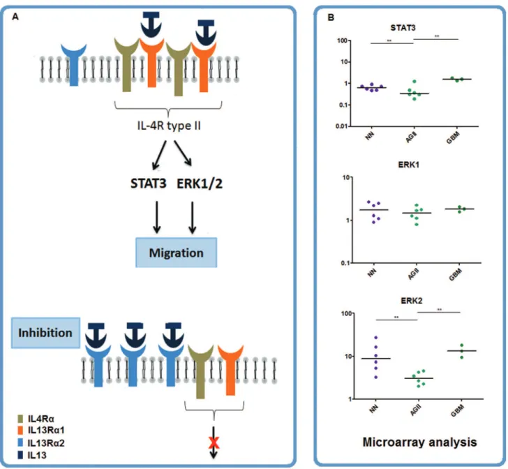
The IL13RA2 signaling pathway is still not
well determined. There is no intracellular binding site in this receptor, but its short cytoplasmic tail may inhibit IL-4Rα through molecular interaction.27 Extracellularly, IL-13Rα2 presents a large number of peripheral interactions, which increase its affinity28 and internalizes IL-13.11 IL13RA2 overexpression in addition to low amount of IL-13 has been described to decrease the signaling pathway through STAT6 and ERK.29 Additionally, colorectal metastatic cancer cells knocked out for IL13RA2 lack invasive properties.23
On the other hand, IL-13 may lead to IL-13Rα1 dimerization with IL-4Rα to form an IL-4R type II receptor, with a proline-rich cytoplasmic tail that associates with Janus kinase-1 (JAK1), Janus kinase-2 (JAK2) and Tyrosine
kinase-2 (TYK2).30 These three kinases recruit signal transducers.31,32 JAK2 may activate ERK 1/2,29,33 as well as STAT3, which is involved in the Rho-GTPases signaling34 in cell migration pathway. ERK modulates cell migration by membrane protrusion as a result of myosin light-chain kinase (MLCK) phosphorylation,33 and also by interacting with calpain, and focal adhesion kinase (FAK), both proteins involved in cellular adhesion.
Our previous oligonucleotide microarray data of GBM13 showed that this cascade including ERK2 and STAT3 was upregulated in GBM cases (figure 4), but STAT6 and ERK1 were not. In this context and considering the present results of migration assay with GBM cell lines, we hypothesize that the IL-13 binding to IL-13Rα2 may prevent its ligation to IL-13Rα1/IL-4Rα, depending on the ratio
of IL13RA2/IL13RA2 expression levels and the available
amount of IL-13, and consequently halt the downstream activation of migration signaling cascade (figure 4).
Previous reports have also demonstrated the role of IL-13Rα2 in apoptosis,34,35 and explored this aspect in therapeutic trial as targeting 13Rα2 by cytotoxin IL-13 fused with Pseudomonas exotoxin,11,18,36,37 or through construction of specific TCD8+ lymphocyte genetically modified to IL13RA2,8 or yet silencing IL13RA2 by siRNA and
allowing IL-13 interaction with IL13RA1 leading to apoptosis
by the expression of 15-LOX-1, an apoptosis regulator.35 In addition to these observations of IL13RA2 in the apoptotic process, the present results may suggest an additional role of IL13RA2 in cell migration process,
Figure 3 - (A) IL13RA1 (orange) and IL13RA2 (blue) expressions in A172 and U87MG cells by qRT-PCR. Variation of cell migration of A172 (B, D) and U87MG (C, D) cells treated with 10ng/ml and 20ng/ml of IL-13 based on the calculation of the remaining area at 0, 24, 48 and 72hs after the scratch. * p < 0.05
spectrum of targetable mechanisms to better control the tumor progression in GBM.
■
ACKNOWLEDGMENTSThis study was supported by grants from São Paulo Research Foundation (FAPESP 2001/12898-4, 2004/12133-6, 2013/02162-8, 2013/06315-3), and from Conselho Nacional de Desenvolvimento Cientifico e Tecnologico (CNPq 83467/2011-1). We sincerely thank Emerson Bernardes for the gift of IL-13.
■
AUTHOR PARTICIPATIONFigure 4 - IL-13 network. (A) Schematic representation of IL-13 signaling pathway emphasizing of the downstream cascade including IL13Rs, extracellular-signal-regulated kinases-1 and 2 (ERK1/2), and signal transducer activator of transcription-3 (STAT3). These genes expression levels are presented based on oligonucleotide microarray data(13) (B).
■
CONFLICT OF INTERESTNo potential conflict of interest relevant to this manuscript is reported.
O IMPACTO DA EXPRESSÃO DOS RECEPTORES DE INTERLEUCINA-13 NA MIGRAÇÃO CELULAR EM ASTROCITOMAS
INTRODUÇÃO: Astrocitomas são os tumores cerebrais mais frequentes. Nestes tumores foi observada maior expressão do receptor de Interleucina-13 α2
(IL13RA2). A cascata de sinalização da Interleucina-13 pode
estar associada com a migração celular, após sua ligação com o receptor de Interleucina-13 α1 (IL13Rα1).
OBJETIVO: Investigar os níveis de expressão dos receptores de Interleucina-13 (IL13RA1 e IL13RA2)
em astrocitomas difusamente infiltrativos e avaliar o envolvimento da Interleucina-13 na migração celular de duas linhagens de glioblastoma.
MÉTODOS: A expressão dos receptores IL13RA
RESULTADOS: As amostras de Glioblastoma apresentaram maior expressão de IL13RA1 and IL13RA2
comparados com astrocitomas de baixo grau e os casos não-neoplásicos. Nas células U87MG e A172 foi observado maior nível de expressão deIL-13RA1 do que IL-13RA2.
Uma diferença significativa na taxa de migração foi obtida em células A172 tratadas com 10 ng/mL comparadas com o controle: as células tratadas apresentaram menor migração que as células não tratadas. As células U87MG tratadas com 20ng/mL de IL-13 apresentaram menor migração celular que as células não tratadas. A diferença na migração celular indica que o caminho de sinalização de IL13Rα1 não foi ativado e foi inibido pelo IL-13Rα2, diminuindo a migração celular. Esse impacto ocorreu com uma concentração menor de IL-13 nas células A172 ao contrário da U87MG, porque as células A172 possuem uma razão IL-13RA2/A1 maior.
CONCLUSÃO: os resultados sugerem que os receptores de IL-13 podem ser utilizados como possíveis alvos para a diminuição da migração celular tumoral.
PALAVRAS-CHAVE: Astrocitomas, Interleucina-13, Receptores de Interleucina-13
■
REFERENCES1. Huang PH, Cavenee WK, Furnari FB, White FM. Uncovering Therapeutic Targets for Glioblastoma: a System Biology Aproach. Cell Cycle. 2007;6(22):2750-4.
2. Caffo M, Caruso G, Passalacqua M, Angileri FF, Tomasello F. Antisense Oligonucleotides Therapy in the Treatment of Cerebral Gliomas: A Review. Genetic Syndromes & Gene Therapy. 2013;4(10):1-10. 3. Dunn GP, Rinne ML, Wykosky J, Genovese G, Quayle SN, Dunn IF,
et al. Emerging Insights into the Molecular and Cellular Basis of Glioblastoma. Genes Dev. 2012;26(8):756-84.
4. Louis DN, Ohgaki H, Wiestler OD, Cavenee WK, Burger PC, Jouvet A, et al. The 2007 WHO Classification of Tumours of the Central Nervous System. Acta Neuropathol. 2007;114(2):97-109.
5. Ewelt C, Goeppert M, Rapp M, Steiger H-J, Stummer W, Sabel M. Glioblastoma multiforme of the elderly: the prognostic effect of resection on survival. J Neurooncol. 2011;103(3):611-8.
6. Xie ZG. Brain Tumor Stem Cells. Neurochem Res. 2009;34(12):2055-66. 7. Berger F, Gay E, Pelletier L, Tropel P, Wion D. Development of gliomas: potential role of asymmetrical cell division of neural stem cells. Lancet Oncology. 2004;5(8):511-4.
8. Brown CE, Starr R, Aguilar B, Shami AF, Martinez C, D’Apuzzo M, et al. Stem-like Tumor-Initiating Cells Isolated from IL13R alpha 2 Expressing Gliomas Are Targeted and Killed by IL13-Zetakine-Redirected T Cells. Clin Cancer Res. 2012;18(8):2199-209.
9. Debinski W, Slagle B, Gibo DM, Powers SK, Gillespie GY. Expression of a restrictive receptor for interleukin 13 is associated with glial transformation. J Neuro-Oncol. 2000;48(2):103-11.
10. Andrews AL, Holloway JW, Puddicombe SM, Holgate ST, Davies DE. Kinetic analysis of the interleukin-13 receptor complex. J Biol Chem. 2002;277(48):46073-8.
11. Kawakami K, Taguchi J, Murata T, Puri RK. The interleukin-13 receptor alpha 2 chain: an essential component for binding and internalization but not for interleukin-13-induced signal transduction through the STAT6 pathway. Blood. 2001;97(9):2673-9.
12. Oba-Shinjo SM, Bengtson MH, Winnischofer SMB, Colin C, Vedoy CG, de Mendonca Z, et al. Identification of novel differentially expressed genes in human astrocytomas by cDNA representational difference analysis. Mol Brain Res. 2005;140(1-2):25-33.
13. Marie SKN, Okamoto OK, Uno M, Hasegawa APG, Oba-Shinjo SM, Cohen T, et al. Maternal embryonic leucine zipper kinase transcript abundance correlates with malignancy grade in human astrocytomas. Int J Cancer. 2008;122(4):807-15.
14. Valente V, Teixeira SA, Neder L, Okamoto OK, Oba-Shinjo SM, Marie SKN, et al. Selection of suitable housekeeping genes for expression analysis in glioblastoma using quantitative RT-PCR. Bmc Mol Biol. 2009;10:17. 15. Livak KJ, Schmittgen TD. Analysis of relative gene expression data
using real-time quantitative PCR and the 2(T)(-Delta Delta C) method. Methods. 2001;25(4):402-8.
16. Pencina MJ, D’Agostino RB Sr, D’Agostino RB Jr, Vasan RS. Evaluating the added predictive ability of a new marker: From area under the ROC curve to reclassification and beyond. Stat Med. 2008;27(2):157-72.
17. Debinski W, Gibo DM, Hulet SW, Connor JR, Gillespie GY. Receptor for interleukin 13 is a marker and therapeutic target for human high-grade gliomas. Clin Cancer Res. 1999;5(5):985-90.
18. Kawakami M, Kawakami K, Puri RK. Intratumor administration of interleukin 13 receptor-targeted cytotoxin induces apoptotic glioma tumor xenografts. Mol Cancer Ther. 2002;1(12):999-1007.
19. Joshi BH, Plautz GE, Puri RK. Interleukin-13 receptor alpha chain: A novel tumor-associated transmembrane protein in primary explants of human malignant gliomas. Cancer Res. 2000;60(5):1168-72. 20. Minn AJ, Gupta GP, Siegel PM, Bos PD, Shu WP, Giri DD, et al.
Genes that mediate breast cancer metastasis to lung. Nature. 2005;436(7050):518-24.
21. Fujisawa T, Joshi BH, Puri RK. IL-13 regulates cancer invasion and metastasis through IL-13R alpha 2 via ERK/AP-1 pathway in mouse model of human ovarian cancer. Int J Cancer. 2012;131(2):344-56. 22. Fujisawa T, Nakashima H, Nakajima A, Joshi BH, Puri RK. Targeting
IL-13R alpha 2 in human pancreatic ductal adenocarcinoma with combination therapy of IL-13-PE and gemcitabine. Int J Cancer. 2011;128(5):1221-31.
23. Barderas R, Bartolomé RA, Fernandez-Aceñero MJ, Torres S, Casal JI. High expression of IL-13 receptor alpha2 in colorectal cancer is associated with invasion, liver metastasis, and poor prognosis. Cancer Res. 2012;72(11):2780-90.
24. Kawakami K, Kawakami M, Snoy PJ, Husain SR, Puri RK. In vivo overexpression of IL-13 receptor alpha 2 chain inhibits tumorigenicity of human breast and pancreatic tumors in immunodeficient mice. J Exp Med. 2001;194(12):1743-54.
25. Fujisawa T, Joshi B, Nakajima A, Puri RK. A Novel Role of Interleukin-13 Receptor alpha 2 in Pancreatic Cancer Invasion and Metastasis. Cancer Res. 2009;69(22):8678-85.
26. Zhao Z, Wang L, Xu W. IL-13Ra2 mediates PNR-induced migration and metastasis in ERa-negative breast cancer. Oncogene. 2015;34(12):1596-607..
27. Rahaman SO, Sharma P, Harbor PC, Aman MJ, Vogelbaum MA, Haque SJ. IL-13R(alpha)2, a decoy receptor for IL-13 acts as an inhibitor of IL-4-dependent signal transduction in glioblastoma cells. Cancer Res. 2002;62(4):1103-9.
28. Lupardus PJ, Birnbaum ME, Garcia KC. Molecular Basis for Shared Cytokine Recognition Revealed in the Structure of an Unusually High Affinity Complex between IL-13 and IL-13Ralpha2. Structure. 2010;18(3):332-42.
29. Mandal D, Levine AD. Elevated IL-13Ralpha2 in intestinal epithelial cells from ulcerative colitis or colorectal cancer initiates MAPK pathway. Inflamm Bowel Dis. 2010;16(5):753-64.
30. Hershey GKK. IL-13 receptors and signaling pathways: An evolving web. J Allergy Clin Immunol. 2003 Apr;111(4):677-90.
31. Kaptein A, Paillard V, Saunders M. Dominant negative stat3 mutant inhibits interleukin-6-induced Jak-STAT signal transduction. J Biol Chem. 1996;271(11):5961-4.
32. Scott MJ, Godshall CJ, Cheadle WG. Jaks, STATs, Cytokines, and Sepsis. Clin Diagn Lab Immunol. 2002;9(6):1153-9.
34. Hanna S, El-Sibai M. Signaling networks of Rho GTPases in cell motility. Cellular Signal. 2013;25(10):1955-61.
35. Hsi LC, Kundu S, Palomo J, Xu B, Ficco R, Vogelbaum MA, et al. Silencing IL-13Ra2 Promotes Glioblastoma Cell Death via Endogenous Signaling. Mol Cancer Ther. 2011;10(7):1149-60.
36. Van N, Conyers JM, Zhu D, Gibo DM, Dorsey JF, Debinski W, et al. IL-13Ra2-Targeted Therapy Escapees: Biologic and Therapeutic Implications. Transl Oncol. 2011;4(6):390-400.