Caserta NMG et al. / Cesarean scar ectopic pregnancy
Radiol Bras. 2017 Mai/Jun;50(3):197–198 197
0100-3984 © Colégio Brasileiro de Radiologia e Diagnóstico por Imagem
Case Report
Cesarean scar ectopic pregnancy: invasion of the bladder wall
detected by magnetic resonance imaging
Gravidez ectópica em cicatriz de cesariana: invasão da parede vesical detectada pela ressonância magnética
Caserta NMG, Bacha AM, Grassiotto OR. Cesarean scar ectopic pregnancy: invasion of the bladder wall detected by magnetic resonance imaging. Radiol Bras. 2017 Mai/Jun;50(3):197–198.
Abstract
R e s u m o
Although cesarean scar ectopic pregnancy continues to be the rarest form of ectopic pregnancy, its incidence is increasing because of the worldwide increase in the number of cesarean deliveries. If the diagnosis is delayed, there is a high risk of severe hemorrhage and death, whereas early diagnosis can minimize the complications associated with the condition. Here, we report a case in which invasion of the bladder wall was identified by magnetic resonance imaging.
Keywords: Pregnancy, ectopic; Cesarean section; Urinary bladder; Hematuria; Magnetic resonance imaging.
Gestação em cicatriz de cesariana é a forma mais rara de gravidez ectópica, mas com aumento devido ao maior número de cesarianas. O diagnóstico tardio pode provocar hemorragia grave, com risco de morte, mas se precoce pode reduzir as complicações. Relatamos um caso com invasão da parede da bexiga, provocando hematúria, e que foi demonstrada pela ressonância magnética.
Unitermos: Gravidez ectópica; Cesariana; Bexiga urinária; Hematúria; Ressonância magnética.
Study conducted at the Hospital de Clínicas and at the Centro de Atenção Inte-gral à Saúde da Mulher (CAISM) of the Faculty of Medical Sciences, FCM-Unicamp, Campinas, SP, Brazil.
1. PhD, Tenured Associate Professor, Department of Radiology, Faculty of Medi-cal Sciences, FCM-Unicamp, Campinas, SP, Brazil.
2. PhD, Professor, Department of Gynecology and Obstetrics, Faculty of Medical Sciences, FCM-Unicamp, Campinas, SP, Brazil.
Mailing address: Dr. Nelson M. G. Caserta. Departamento de Radiologia – FCM-Unicamp. Rua Vital Brasil, 251, Cidade Universitária Zeferino Vaz. Campinas, SP, Brazil, 13083-888. E-mail: [email protected].
Received July 4, 2014. Accepted after revision September 3, 2014.
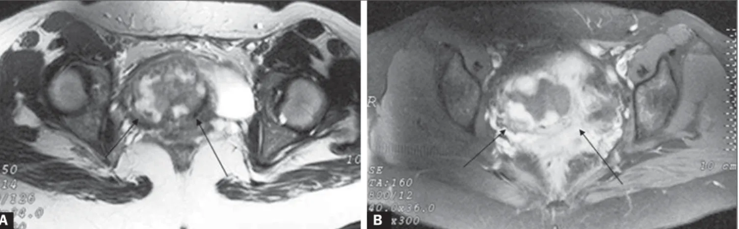
for a normal pregnancy at 10 weeks is 25,700–288,000 mIU/ mL). MRI revealed a heterogeneous hyperintense mass of the myometrium in the lower anterior uterine segment (Fig-ure 1). At one point, the mass had invaded the bladder wall and opened through an orifice in the bladder mucosa, as identified on the MRI scan (Figure 2). Cystoscopy confirmed the opening of the fistula, and a biopsy of this site revealed chronic cystitis. The patient was submitted to hysterectomy with resection of the bladder wall lesion. Anatomopathological examination confirmed the diagnosis of ectopic pregnancy in a cesarean scar with invasion of the bladder wall. She de-veloped no complications during the postoperative period.
DISCUSSION
Implantation of a pregnancy within the scar of a previ-ous cesarean section is a potentially life-threatening condi-tion and is considered the rarest form of ectopic pregnancy(4).
It is known that cesarean section represents one of the risk factors for ectopic pregnancy and placental abnormalities in subsequent pregnancies(5). Although many hypotheses have
been proposed for this rare condition, the most reasonable explanation would be that the trophoblast penetrates the myometrium along a microscopic tract(6).
Early diagnosis with ultrasound can offer treatment options that could prevent uterine rupture and hemorrhage and thus preserve the uterus(4). Curettage seems contraindi-cated because the trophoblastic tissue is outside the uterine cavity(4). Nonsurgical treatment options include
administra-tion of systemic and local methotrexate, as well as potassium chloride and hyperosmolar glucose, which have reportedly met with some success (4,5,7). However, primary surgical
treat-Nelson Marcio Gomes Caserta1, Angela Maria Bacha2, Oswaldo R. Grassiotto2
http://dx.doi.org/10.1590/0100-3984.2014.1855
INTRODUCTION
Cesarean scar ectopic pregnancy is a rare form of ec-topic pregnancy that is considered a potentially life-threat-ening condition(1,2). Invasion of the myometrium may lead
to massive uterine bleeding(3). We report a case of cesarean
scar ectopic pregnancy that invaded the bladder wall, which was confirmed by magnetic resonance imaging (MRI).
CASE REPORT
Caserta NMG et al. / Cesarean scar ectopic pregnancy
Radiol Bras. 2017 Mai/Jun;50(3):197–198 198
ment by laparotomy and hysterotomy, as soon as the diag-nosis is confirmed, would be the best treatment option(4).
Clinical history and endovaginal ultrasound are quite useful for differentiating cesarean scar ectopic pregnancy from incomplete abortion or cervico-isthmic gestation. Our pa-tient presented with macroscopic hematuria, which is not expected as a symptom of cesarean scar ectopic pregnancy. Approximately 40% of patients with cesarean scar ectopic pregnancy experience only painless vaginal bleeding(1).
Some authors have used MRI as an additional diagnos-tic modality. A recent report indicated that contrast-enhanced MRI can be used as the initial imaging modality to diag-nose cesarean scar ectopic pregnancy, in selected cases, al-lowing a more accurate diagnosis before the specific treat-ment is instituted(8). Because MRI has excellent tissue
reso-lution, it can be used in order to locate the implantation in the cesarean section scar, determine the thickness of the anterior uterine wall, and provide an accurate view of the vesicouterine space. Although invasion of the bladder wall is a known possibility in cesarean scar ectopic pregnancy, we know of no other reports of this complication diagnosed by MRI. In the case presented here, MRI clearly
demon-strated that the hematuria was caused by the penetration of the ectopic pregnancy into the bladder wall.
REFERENCES
1. Rotas MA, Haberman S, Levgur M. Cesarean scar ectopic pregnan-cies: etiology, diagnosis, and management. Obstet Gynecol. 2006; 107:1373–81.
2. Kung FT, Huang TL, Chen CW, et al. Image in reproductive medi-cine. Cesarean scar ectopic pregnancy. Fertil Steril. 2006;85:1508–9. 3. Reyftmann L, Vernhet H, Boulot P. Management of massive uterine bleeding in a cesarean scar pregnancy. Int J Gynaecol Obstet. 2005; 89:154–5.
4. Fylstra DL. Ectopic pregnancy within a cesarean scar: a review. Obstet Gynecol Surv. 2002;57:537–43.
5. Maymon R, Halperin R, Mendlovic S, et al. Ectopic pregnancies in a Caesarean scar: review of the medical approach to an iatrogenic com-plication. Hum Reprod Update. 2004;10:515–23.
6. Lee CL, Wang CJ, Chao A, et al. Laparoscopic management of an ectopic pregnancy in a previous Caesarean section scar. Hum Reprod. 1999;14:1234–6.
7. Shufaro Y, Nadjari M. Implantation of a gestational sac in a cesarean section scar. Fertil Steril. 2001;75:1217.
8. Huang Q, Zhang M, Zhai RY. The use of contrast-enhanced mag-netic resonance imaging to diagnose cesarean scar pregnancies. Int J Gynaecol Obstet. 2014;127:144–6.
Figure 2.A: Coronal T2-weighted MRI showing that the myometrium (arrows) was ruptured by the gestational mass. B:
Sagittal T2-weighted MRI scan along the midline, showing the empty endometrial cavity and the opening (arrow) caused by the cesarean scar ectopic pregnancy in-vading the bladder.
A B
Figure 1.A: Axial T2-weighted MRI showing a heterogeneous mass on the right side of the uterine isthmus (arrows). B: After administration of gadolinium, there was pronounced, heterogeneous impregnation of this mass (arrows).