285
Radiol Bras. 2017 Set/Out;50(5):285–290
Spectrum of indings on magnetic resonance imaging
of the brain in patients with neurological manifestations
of dengue fever
O espectro de achados de ressonância magnética do encéfalo em pacientes com manifestações neurológicas de dengue
Tejeshwar Singh Jugpal1, Rashmi Dixit1, Anju Garg1, Swati Gupta1, Virendra Jain1, Ronak Patel1, Shobhit Agarwal2
Jugpal TS, Dixit R, Garg A, Gupta S, Jain V, Patel R, Agarwal S. Spectrum of indings on magnetic resonance imaging of the brain in patients with neurological manifestations of dengue fever. Radiol Bras. 2017 Set/Out;50(5):285–290.
Abstract
Resumo
Objective: To describe the spectrum of magnetic resonance imaging (MRI) indings in patients with neurological manifestations of dengue.
Materials and Methods: We included nine patients with dengue fever (three females and six males; age range, 9–30 years), all of whom presented with neurological manifestations. The MRI examinations, performed in 1.5 T or 3 T scanners, included T1-weighted, T2-T1-weighted, and luid-attenuated inversion recovery (FLAIR) sequences. Diffusion-weighted imaging with apparent diffu-sion coeficient mapping was also employed. Fast low-angle shot and susceptibility-weighted gradient-recalled echo sequences, as well as contrast-enhanced T1-weighted scans, were also obtained in order to assess parenchymal enhancement. MRI scans were analyzed for lesion distribution and imaging features.
Results: All patients showed areas of altered signal intensity that appeared as hyperintensity on T2-weighted and FLAIR sequences. The most commonly affected site was the basal ganglia-thalamus complex. Other affected sites were the cerebellum, cerebral cortex, white matter, and brainstem. In all cases, we observed patchy areas of restricted diffusion and focal areas of hemorrhage. Conclusion: Dengue encephalitis commonly affects the basal ganglia, thalamus, cerebellum, cerebral cortex, and white matter. Therefore, MRI should be an indispensable part of the evaluation of patients with neurological complications of dengue fever. Keywords: Dengue; Encephalitis; Magnetic resonance imaging; Leukoencephalitis, acute hemorrhagic; Cerebellar diseases/diag-nosis.
Objetivo: Descrever o espectro dos achados de ressonância magnética (RM) em pacientes com manifestações neurológicas de dengue.
Materiais e Métodos: Foram incluídos nove pacientes com dengue (três do sexo feminino e seis do sexo masculino; faixa etária: 9–30 anos), todos com manifestações neurológicas. Os exames de RM, realizados em aparelhos de 1,5 T ou 3 T, incluíram sequên-cias ponderadas em T1 e em T2, assim como luid-attenuated inversion recovery (FLAIR). Também foi empregada a imagem ponde-rada em difusão com mapeamento de coeicientes de difusão aparente. Além disso, foram obtidas sequências gradiente-eco pon-deradas por suscetibilidade e em fast low-angle shot, bem como imagens ponderadas em T1 pós-contraste, para avaliar o realce parenquimatoso. As imagens de RM foram analisadas quanto à distribuição de lesões e características de imagens.
Resultados: Todos os pacientes apresentaram áreas de intensidade de sinal alteradas que apareceram como hiperintensidade em sequências ponderadas em T2 e sequências FLAIR. O local mais comumente afetado foi o complexo gânglios basais-tálamo. Outros locais afetados foram o cerebelo, o córtex cerebral, a substância branca e o tronco encefálico. Em todos os casos observamos áreas irregulares de difusão restrita e áreas focais de hemorragia.
Conclusão: A encefalite por dengue geralmente afeta os gânglios basais, o tálamo, o cerebelo, o córtex cerebral e a substância branca. Portanto, a RM deve ser uma parte indispensável da avaliação de pacientes com complicações neurológicas da dengue.
Unitermos: Dengue; Encefalite; Ressonância magnética; Leucoencefalite hemorrágica aguda; Doenças cerebelares/diagnóstico.
Study conducted in the Department of Diagnostic Radiology of the Maulana Azad Medical College, New Delhi, India.
1. MD, Department of Diagnostic Radiology, Maulana Azad Medical College, New Delhi, India.
2. MD, Department of General Medicine, Maulana Azad Medical College, New Delhi, India.
Mailing address: Tejeshwar Singh Jugpal, MD. Maulana Azad Medical College – Department of Diagnostic Radiology. 2-Bahadur Shah Zafar Marg, New Delhi- 110002, India. E-mail: [email protected].
Received March 20, 2016. Accepted after revision August 22, 2016.
INTRODUCTION
(DEN1 through DEN4), and is transmitted to humans
by the Aedes aegypti mosquito(1–3). Although the dengue
virus is generally thought to be non-neurotrophic, the in-cidence of early neurological manifestations of dengue
has increased in recent years(4). The reported incidence
of neurological complications of dengue has been found
to vary between 0.5% and 6.2%(3). The neurological
com-plications of dengue are broadly classiied into the
follow-ing categories(5,6): encephalopathy secondary to systemic
involvement such as coagulopathy, hepatic failure, and systemic hypotension; direct neuronal invasion by the vi-rus causing encephalitis; and immune complex-mediated vasculitis and demyelinating process such as acute dis-seminated encephalomyelitis (ADEM).
There have been only a few studies describing mag-netic resonance imaging (MRI) indings in patients with
dengue fever(7–9). Here, we describe the spectrum of MRI
features in patients with serologically conirmed dengue and neurological manifestations.
MATERIALS AND METHODS
Using MRI, we evaluated nine patients (9–30 years of age) with dengue fever and neurological manifestations including seizures, altered sensorium, and ataxia. The sample comprised three females and six males. The cases were diagnosed based on clinical features, laboratory test results, and serology, all serum samples testing positive for immunoglobulin M (IgM) antibodies or for nonstruc-tural protein 1 antigen. All of the patients gave written informed consent.
The MRI examinations were performed in dedicated 1.5 T or 3 T MRI scanners. All of the patients underwent imaging during the acute phase of the illness (between day 3 and day 6). The MRI scans included T1-weighted fast spin-echo, T2-weighted, and luid attenuated inversion re-covery (FLAIR) sequences. Diffusion weighted (DWI) im-ages were acquired using single-shot fast spin-echo echo-planar sequences with sensitizing gradients applied in all
three orthogonal planes with b factors of 500 s/mm2 and
1000 s/mm2. We also generated apparent diffusion
coef-icient maps using the software supplied by the vendor. To identify hemorrhagic foci, we also acquired
gradient-recalled echo (GRE) fast low-angle shot sequences (in six patients) and susceptibility-weighted imaging (SWI) sequences (in three patients). Contrast-enhanced T1-weighted scans were also obtained in all cases.
RESULTS
All nine patients presented with high-grade fever, headache, and altered sensorium. Seizures were observed in ive patients. Ataxia was seen in three patients, one of whom also had severe vertigo, tremors, and nystagmus. All the patients had normal liver and kidney function. At the time of imaging, we also determined the platelet count. The lowest platelet count noted during the study was 21,000 cells/mL. The clinical indings are summa-rized in Table 1.
The anatomical distribution of the lesions and their MRI characteristics are summarized in Table 2. All the patients showed abnormal signal intensities on MRI. The basal ganglia-thalamus complex was involved in seven cases: isolated involvement of the basal ganglia in one; involvement of the basal ganglia and thalamus in two; and isolated involvement of the thalamus in four. Cerebral and cerebellar involvement was also seen in four patients. The lesions appeared hyperintense on T2-weighted and FLAIR sequences. All cases showed patchy areas of re-stricted diffusion. The GRE/SWI sequences showed focal areas of blooming suggesting hemorrhage in all cases. On contrast-enhanced images, subtle enhancement was seen in six patients.
DISCUSSION
Dengue fever usually presents as febrile myalgia, ar-thralgia, frontal/retro-orbital headache, nausea, vomit-ing, and rash. Dengue fever is typically accompanied by thrombocytopenia. In its severe form, dengue can mani-fest as dengue hemorrhagic fever or dengue shock syn-drome(10).
The dengue virus was initially considered to be non-neurotropic virus. However, there has been an increase in the incidence of neurological manifestations in patients with dengue fever. Recent studies in which the dengue IgM antibody was isolated in cerebrospinal luid suggest
Table 1—Summary of clinical indings and relevant lab investigation.
Patient No. 1 2 3 4 5 6 7 8 9 Age (years) 9 30 17 22 20 21 25 16 22 Sex Female Male Male Male Male Male Male Female Female Neurological symptoms
Fever, headache, seizure Fever, headache, seizure, altered sensorium
Fever, headache, altered sensorium, Fever, headache, seizure, ataxia Fever, headache, seizure, altered sensorium Fever, headache, altered sensorium, seizure Fever, headache, ataxia, nystagmus, vertigo, tremors
Fever, headache, altered sensorium Fever, headache, ataxia, altered sensorium
Duration of illness (days) 5 4 5 5 4 4 6 3 5 Platelet count
(cells per mL)
25,000 20,000 40,000 36,000 30,000 39,000 29,000 68,000 21,000
that the dengue virus is capable of direct neuroinvasion(11). It has also been observed that the DEN2 and DEN3 sero-types have the greatest proclivity for producing
neurologi-cal complications(12). The neurological complications of
dengue fever can be broadly grouped into three catego-ries: direct viral neurotropism (encephalitis, meningitis, and myelitis); systemic complications (encephalopathy and ischemic or hemorrhagic stroke; and postinfectious
complications (ADEM and myelitis). Dengue encephali-tis is now recognized as a clinical entity which is a leading
cause of encephalitis in endemic regions(13–15).
MRI plays an important role in identifying the exact anatomical area of involvement and substantiating a diag-nosis of dengue encephalitis in patients with neurological manifestations. There have been few studies describing the neuroimaging indings of dengue encephalitis on MRI scans. There have been isolated case reports suggesting that the commonly affected regions of brain include the basal ganglia, thalamus, temporal lobes, hippocampus,
cerebellum, and cerebral white matter(7,8,16–19). This is
similar to what we encountered in our case series, in which the most common site of involvement was the basal ganglia-thalamus complex (in seven patients), followed by the cerebrum (in ive) and cerebellum (in four).
In our case series, all of the lesions appeared hypoin-tense on T1-weighted sequences, hyperinhypoin-tense on T2-weighted sequences with evidence of patchy areas of re-stricted diffusion. We also encountered foci of blooming on GRE/SWI sequences, which was indicative of hemor-rhage within the lesions, in all patients (Figures 1 and 2). In the literature, focal lesions caused by dengue appear
Table 2—MRI indings in nine patients with dengue encephalitis.
Patient
No.
1 2 3 4 5 6 7 8 9
Cerebrum
– + (RD, E) + (RD, Hg, E)
– + (RD, Hg, E)
+ (RD, Hg) – – +(RD)
Ganglio-thalamic
complex
+ (RD, Hg, E) + (RD, Hg, E) + (RD, Hg, E) + (RD, Hg, E) + (RD, Hg, E)
– – + (RD, Hg, E)
+ (RD, Hg)
Brain stem
– – + (RD, Hg, E)
– – – – – + (RD)
Cerebellum
– – – + (RD, Hg, E)
-+ (RD, Hg) + (RD, Hg)
– + (RD, Hg) –, Absent; +, present; RD, restricted diffusion on DWI; Hg, hemorrhagic foci; E, enhancement on contrast-enhanced MRI.
Figure 1. 16-year-old female with dengue encephalitis. Sagittal T1-weighted image (a) showing hypointensity involving the right basal ganglia-thalamus complex (arrow), which appears hyperintense on an axial T2-weighted image (b) and a coronal FLAIR image (c), with an associated mass effect, as evidenced by effacement
hyperintense on T2-weighted images and hypointense on T1-weighted images, similar to what we encountered in
our studies(7–9). The DWI characteristics of the lesions
have been reported in only a few previous studies. In those studies, the lesions showed hyperintense signals on DWI, the diffusion being restricted in some cases and facilitated
in others(7,8). In our case series, all of the focal lesions
showed restricted diffusion. Hemorrhagic foci within the lesions have also rarely been reported in dengue
encepha-litis(7,9). On contrast-enhanced T1-weighted sequences,
six of our nine patients showed minimal patchy heteroge-neous enhancement within the lesions. This could be at-tributed to the fact that the permeability of the blood-brain
barrier is reportedly increased in dengue infection(15).
The common differential diagnoses of the MRI neu-roimaging indings in patients with dengue encephalitis include Japanese encephalitis, herpes simplex encephali-tis, and ADEM. The typical anatomical sites include the basal ganglia-thalamus complex (bilaterally) in Japanese encephalitis and the temporal/basifrontal lobes (also
bi-laterally) in herpes simplex encephalitis(20,21).
Hemor-rhagic foci are characteristic of herpes encephalitis but are usually not encountered in Japanese encephalitis. However, hemorrhagic foci within the basal ganglia-thal-amus complex have been rarely reported in cases of
Japa-nese encephalitis(22,23). Therefore it might be dificult to
differentiate among these different types of viral encepha-litis on the basis of MRI indings alone. Analysis of the ce-rebrospinal luid and clinical proile might provide clues to the speciic clinical entity. In a region where dengue is endemic, the possibility of dengue encephalitis should be borne in mind as a potential differential diagnosis, espe-cially during dengue outbreaks.
ADEM is an immune complex-mediated injury to the brain usually seen during the convalescent phase of viral
infection or after vaccination(24). Classically, there is
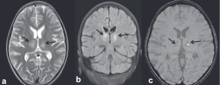
in-Figure 2. 9-year-old female with dengue encephalitis. Axial T2-weighted image (a) and coronal FLAIR image (b) showing a hyperintense signal in bilateral thalami (arrows). SWI image (c) showing hemorrhagic foci in the same region (arrows).
volvement of the cerebral white matter and deep gray matter nuclei in ADEM; although hemorrhagic foci have been reported, they are relatively uncommon. Therefore the MRI indings of ADEM and demyelinating dengue-induced hemorrhagic encephalitis might be similar. How-ever, the temporal relationship between the occurrence of lesions and the acute febrile illness, together with positive serology for dengue, makes the diagnosis of post-dengue
ADEM more plausible(25,26). During the acute phase of
the illness, our patients presented with involvement of the cerebral white matter with hemorrhagic foci scattered throughout the lesion.
Our study included a patient with a high-grade fever and a rash who presented with acute ataxia, dysmetria, and nystagmus. In that patient, the MRI scans showed signal alteration localized to the cerebellar cortex, white matter, and vermis, all of which showed areas of restrict-ed diffusion and hemorrhagic foci on DWI and SWI se-quences, respectively (Figure 3). We identiied only iso-lated case reports of patients with dengue fever
present-ing with symptoms of cerebellitis(7). Cerebellar
involve-ment in dengue patients has been thought to be part of
a postinfectious immune-mediated process(27). However,
there have been reports of cerebellar infection in patients with dengue, in whom pathological studies have revealed
the presence of viral antigen in the cerebellar cells(28).
Therefore, early cerebellar involvement (during the acute phase of the disease) could indicate primary cerebellar infection. Other causes of acute viral cerebellitis include infection with Epstein-Barr virus, Coxsackie virus, or varicella-zoster virus, as well as measles, mumps, herpes,
and, rarely, Japanese encephalitis(29) In our study, the
CONCLUSION
MRI is more sensitive than computed tomography in the evaluation of dengue fever patients with early neu-rological manifestations. MRI can help conirm dengue encephalitis and determine the sites of involvement with high accuracy. The commonly affected areas in dengue encephalitis are the basal ganglia, thalamus, cerebellum, cerebral cortex, and white matter. Most of the lesions en-countered show restricted diffusion on DWI, hemorrhag-ic foci on SWI, and minimal heterogenous enhancement on contrast-enhanced images. In an appropriate clinical setting, MRI can help corroborate the diagnosis of den-gue encephalitis. Although there is no speciic treatment for dengue infection, early identiication of neurological complications by MRI can facilitate the timely institution of supportive management in affected patients.
There-Figure 3. 25-year-old male with dengue fever and acute cerebellitis. Sagittal T1-weighted image (a) showing subtle areas of hypointensity involving the cerebellum (arrow), which is hyperintense on an axial T2-weighted image (b) and a coronal FLAIR image (c). SWI image (d) showing subtle punctate hemorrhagic foci (arrows). DWI image at a b factor of 1000 s/mm2 (e) showing high signal intensity in the same region, which appears dark on an apparent diffusion coeficient map (f), suggesting restricted diffusion.
fore, MRI should be an integral part of the evaluation of cases of dengue fever in which there are neurological complications.
REFERENCES
1. Bhatt S, Gething PW, Brady OJ, et al. The global distribution and burden of dengue. Nature. 2013;496:504–7.
2. Cam BV, Fonsmark L, Hue NB, et al. Prospective case-control study of encephalopathy in children with dengue hemorrhagic fever. Am J Trop Med Hyg. 2001;65:848–51.
3. Hendarto SK, Hadinegoro SR. Dengue encephalopathy. Acta Paedi-atr Jpn. 1992;34:350–7.
4. Arora SK, Aggarwal A, Mittal H. Dengue encephalitis in children. J Neurosci Rural Pract. 2012;3:228–9.
5. Carod-Artal FJ, Wichmann O, Farrar J, et al. Neurological compli-cations of dengue virus infection. Lancet Neurol. 2013;12:906–19. 6. Verma R, Sharma P, Garg RK, et al. Neurological complications of
7. Hegde V, Aziz Z, Kumar S, et al. Dengue encephalitis with predomi-nant cerebellar involvement: report of eight cases with MR and CT imaging features. Eur Radiol. 2015;25:719–25.
8. Bhoi SK, Naik S, Kumar S, et al. Cranial imaging indings in dengue
virus infection. J Neurol Sci. 2014;342:36–41.
9. Nadarajah J, Madhusudhan KS, Yadav AK, et al. Acute hemorrhagic encephalitis: an unusual presentation of dengue viral infection. In-dian J Radiol Imaging. 2015;25:52–5.
10. Rodriguez-Roche R, Gould EA. Understanding the dengue viruses and progress towards their control. Biomed Res Int. 2013;2013: 690835.
11. Puccioni-Sohler M, Soares CN, Papaiz-Alvarenga R, et al.
Neuro-logic dengue manifestations associated with intrathecal speciic im
-mune response. Neurology. 2009;73:1413–7.
12. Malavige GN, Fernando S, Fernando DJ, et al. Dengue viral infec-tions. Postgrad Med J. 2004;80:588–601.
13. Tan le V, Thai le H, Phu NH, et al. Viral aetiology of central ner-vous system infections in adults admitted to a tertiary referral hospital in southern Vietnam over 12 years. PLoS Negl Trop Dis. 2014;8:e3127.
14. Tarantola A, Goutard F, Newton P, et al. Estimating the burden of Japanese encephalitis virus and other encephalitides in countries of the Mekong region. PLoS Negl Trop Dis. 2014;8:e2533.
15. Solomon T, Dung NM, Vaughn DW, et al. Neurological manifesta-tions of dengue infection. Lancet. 2000;355:1053–9.
16. Yeo PS, Pinheiro L, Tong P, et al. Hippocampal involvement in den-gue fever. Singapore Med J. 2005;46:647–50.
17. Kamble R, Peruvamba JN, Kovoor J, et al. Bilateral thalamic in-volvement in dengue infection. Neurol India. 2007;55:418–9. 18. Wasay M, Channa R, Jumani M, et al. Encephalitis and myelitis
associated with dengue viral infection. Clinical and neuroimaging features. Clin Neurol Neurosurg. 2008;110:635–40.
19. Weeratunga PN, Caldera HP, Gooneratne IK, et al. Spontaneously resolving cerebellar syndrome as a sequelae of dengue viral infec-tion: a case series from Sri Lanka. Pract Neurol. 2014;14:176–8. 20. Prakash M, Kumar S, Gupta RK. Diffusion-weighted MR imaging
in Japanese encephalitis. J Comput Assist Tomogr. 2004;28:756–61. 21. Sureka J, Jakkani RK. Clinico-radiological spectrum of bilateral
temporal lobe hyperintensity: a retrospective review. Br J Radiol. 2012;85: e782–92.
22. Mahale R, Mehta A, Rangasetty S. Bilateral isolated basal ganglia bleed: an atypical presentation of Japanese encephalitis. Neurol In-dia. 2015;63:456–7.
23. Sarkar N, Roy BK, Das SK, et al. Bilateral intracerebral haemor-rhages: an atypical presentation of Japanese encephalitis. J Assoc Physicians India. 2005;53:144–6.
24. Murthy JM. Acute disseminated encephalomyelitis. Neurol India. 2002;50:238–43.
25. Yamamoto Y, Takasaki T, Yamada K, et al. Acute disseminated encephalomyelitis following dengue fever. J Infect Chemother. 2002;8:175–7.
26. Gera C, George U. Acute disseminating encephalomyelitis with hemorrhage following dengue. Neurol India. 2010;58:595–6. 27. Karunarathne S, Udayakumara Y, Fernando H. Epstein-Barr virus
co-infection in a patient with dengue fever presenting with post-in-fectious cerebellitis: a case report. J Med Case Reports. 2012;6:43. 28. Ramos C, Sánchez G, Pando RH, et al. Dengue virus in the brain of
a fatal case of hemorrhagic dengue fever. J Neurovirol. 1998;4:465– 8.