Bone attrition: a cause of knee pain in osteoarthritis
*
Fadiga óssea: causa de dor em joelhos na osteoartriteWilson Campos Tavares Júnior1, Fernando Meira de Faria1, Reginaldo Figueiredo2, João Paulo
Kawaoka Matushita3, Luciana Costa Silva4, Adriana Maria Kakehasi5
Knee pain is the most frequent symptom in osteoarthritis, a condition that is the leading cause of chronic disability in the elderly and one of the main sources of morbidity attributable to osteoarthritis in general. The causes of knee pain in individuals with osteoarthritis cannot be easily understood, and the knowledge of such causes is critical for determining future specific interventions. Bone attrition represents remodelling of the subchondral bone envelope in osteoarthritis, leading to a consequential change in bone shape and/or bone loss. However, bone attrition is not a feature that can be easily read, since it is hardly detected in the absence of clear defects of cortical bone integrity and because of overlap of bone structures at radiography. Bone attrition is associated not only with knee pain, but also with stiffness and disability. If attrition occurs prior to advanced osteoarthritis, this would suggest that changes in subchondral bone occur concurrently with cartilage loss and that treatments targeting cartilage loss alone are unlikely to be effective. Association with edema-like bone marrow lesions may be observed and constitute predictive factors for subchondral bone attrition. The present study was aimed at reviewing the literature, demonstrating the relevance of bone attrition and explaining how to diagnose this entity on imaging studies.
Keywords: Magnetic resonance imaging; Osteoarthritis; Knee pain; Bone attrition; Subchondral bone; Diagnosis.
A dor no joelho é o sintoma mais comum na osteoartrite, sendo a principal causa de incapacidade crônica em idosos e uma das principais fontes de morbidade atribuível à osteoartrite em geral. As causas de dor no joelho em pessoas com osteoartrite não são facilmente entendidas e o conhecimento sobre as causas da dor é fundamental para que futuramente sejam realizadas intervenções específicas. A fadiga óssea representa o remodelamento do osso subcon-dral na osteoartrite, levando a uma consequente alteração na forma do osso e/ou perda óssea. No entanto, a fadiga óssea não é algo facilmente interpretado, pois é de difícil detecção na ausência de defeitos claros da cortical e pela sobreposição de estruturas ósseas nas radiografias convencionais. A fadiga óssea está associada não apenas a dor no joelho, mas também a rigidez e incapacidade. Se a fadiga ocorre antes da osteoartrite avançada, isso sugere que alterações no osso subcondral podem ocorrer simultaneamente a alterações da cartilagem e que tratamentos visando sua preservação podem não ser eficazes. Lesões com padrão de edema ósseo estão associadas e são fatores predi-tivos para fadiga óssea. Este trabalho tem por objetivo rever a literatura mostrando a importância da fadiga óssea e de como diagnosticar esta alteração nos exames de imagem.
Unitermos: Ressonância magnética; Osteoartrite; Dor nos joelhos; Fadiga óssea; Osso subcondral; Diagnóstico.
Abstract
Resumo
* Study developed at Hospital das Clínicas da Universidade Federal de Minas Gerais (UFMG), Belo Horizonte, MG, Brazil.
1. MDs, Radiologists, Hospital das Clínicas da Universidade Federal de Minas Gerais (UFMG), Belo Horizonte, MG, Brazil.
2. PhD, Associate Professor, Department of Complementary Diagnostic Work-up, Universidade Federal de Minas Gerais (UFMG), Belo Horizonte, MG, Brazil.
3. PhD, Associate Professor, Department of Complementary Diagnostic Work-up, Universidade Federal de Minas Gerais (UFMG), Belo Horizonte, MG, Brazil.
4. Master, Assistant Professor, Department of Complementary Diagnostic Work-up, Universidade Federal de Minas Gerais (UFMG), Belo Horizonte, MG, Brazil.
5. PhD, Associate Professor, Department of Locomotor Sys-tem, Universidade Federal de Minas Gerais (UFMG), Belo Hori-zonte, MG, Brazil.
Mailing Address: Dr. Wilson Campos Tavares Júnior: Rua Gon-çalves Dias, 750/1803, Funcionários. Belo Horizonte, MG, Brazil, 30140-091. E-mail: [email protected]
Received April 25, 2012. Accepted after revision August 20, 2012.
Tavares Júnior WC, Faria FM, Figueiredo R, Matushita JPK, Silva LC, Kakehasi AM. Bone attrition: a cause of knee pain in osteoarthri-tis. Radiol Bras. 2012 Set/Out;45(5):273–278.
most frequent symptom of osteoarthritis, a condition that is the main cause of chronic disability in the elderly and a big source of deficiency attributable to this condition. The severity of this pain is widely variable, implying from absence of symptoms to immobilization and/or disability of the patient(4).
There is little chance that the severity of knee pain in individuals with osteoarthri-tis constitutes an easily explainable phe-nomenon. The anatomic fundamentals and the molecular pathogenesis of this condi-tion has been centered on the concept that the onset and progression of the disease are related to the thinning and softening of the
INTRODUCTION
Osteoarthritis is a common cause of medical consultations(1). A more
compre-hensive understanding of osteoarthritis, not only regarding symptoms but also regard-ing potentially modifiable risks, could lead to the adoption of new strategies in the management of this disease(2).
Several factors, including biomechan-ics, genetics and inflammation determine the heterogeneous nature of osteoarthri-tis(3). A combination of these factors
joint cartilage, with progressive deteriora-tion of the joint(5). For a long time, the
in-vestigations on the structural cause of pain have been focused on cartilaginous defects, even knowing that the cartilage does not contain pain-sensitive fibers(6). Several
studies have reported variable associations between knee pain and loss of hyaline car-tilage of the joint(7,8).
Although osteoarthritis has been tradi-tionally considered as a primary disease of the articular cartilage, emphasis has nowa-days been put on the concept that this is a very simplistic conclusion(9), since it is a
process which involves articular tissues and structures such as subchondral bone, fibrocartilages, ligaments, muscles, joint capsule and synovial membrane(10).
As one can observe in several publica-tions, the sources of pain in osteoarthritis still remain obscure, and the pain may come from any of the different innervated tissues. It is important to highlight that as-sociation with other abnormalities such as joint effusion, synovitis and meniscal le-sions may be observed. Additionally, sub-chondral bone may also play a significant role in the generation of pain(11).
Currently, emphasis has been given to research involving the causes of knee pain on the grounds of objective findings in the field of imaging diagnosis, particularly, magnetic resonance imaging (MRI)(12,13).
In the investigation of knee pain, it is equally important to correlate patients’ clinical data and age with possible diseases which are more prevalent in that subgroup. Spontaneous knee osteonecrosis, for ex-ample, develops with sudden onset of lo-calized pain, with prevalence in female individuals, notably from the seventh de-cade of life on, with no association with systemic disorders, alcohol abuse, previous corticosteroid therapy, meniscal surgery or local trauma(14). In younger women, the
occurrence of osteonecrosis corroborates the suspicion of any rheumathological dis-ease, particularly systemic lupus erythema-tosus. In such patients, osteonecrosis causes pain prior to articular destruction(15).
Kornaat et al.(8) have evaluated the
as-sociation of clinical findings and structural abnormalities observed at MRI in patients with osteoarthritis of the knee. The pres-ence of a moderate to large amount of
ar-ticular fluid could be found in association with knee pain and rigidity. Concomitantly, the presence of osteophytes in the patellofemoral compartment and/or more than four osteophytes in the whole knee were also associated with pain. On the other hand, focal or diffuse cartilaginous abnormalities, subchondral cysts, bone marrow edema, subluxation and/or me-niscal lesions or Baker cysts were not as-sociated with pain and rigidity.
In summary, the identification of struc-tures actually associated with pain in os-teoarthritis of the knee shall contribute to a deeper understanding of this disease and, in the long term, shall allow the adoption of rational therapeutic approaches to this condition.
OSTEOARTHRITIS GENESIS
In almost of all cases, the joint cartilage is the tissue which presents the major alter-ations found in cases of osteoarthritis which can be observed both in the primary and secondary presentations of the disease. Among its morphological alterations, one observes that the cartilage loses its homo-geneous nature, becoming torn, frag-mented, with fibrillation, clefts and ulcer-ations. Many times, with the progression of the pathological process, the cartilage can-not be seen and, as a consequence, exposed areas of subchondral bone are observed(16).
One of the most frequent etiologies in osteoarthritis is the increase in rigidity of the subchondral bone plate, a fact that could trigger cartilage damage involving particularly fibrillation and clefts. The in-tegrity of this tissue is necessary for the normal functioning of the joint(17).
In spite of the radiographic subchondral sclerosis being considered as secondary to cartilage damage, a series of reports have been published, suggesting that there may be a fundamental change in such a concept and, therefore, bone sclerosis would really lead to cartilage damage, and not vice-versa(17).
Another very interesting information is related to the spongy bone which may be detected adjacent to synovial joints with the important function of cushioning the forces transmitted to the joints. Such forces may be critical in the production of damages to
the cartilage in the setting of osteoarthritis, particularly in case of excessive load on the bone(18).
In experimental models of osteoarthri-tis, subchondral bone changes occur preco-ciously after disease induction, revealing noticeable bone remodeling caused by the disease. Such changes occur about two weeks after surgical procedures involving crossed ligaments and in cases of menis-cectomy, even before the onset of chondral lesions. In the early stages of the disease, focal damages are observed in the cartilage surface of the medial compartment, and it is import to note that, in this phase, the cartilage thinning process is detected only by histomorphometry(19).
Radin et al.(20,21) have described
in-crease in subchondral bone development and decrease in porosity in an animal model of osteoarthritis, in association with a rela-tive rigidity of the bone. After a precocious bone change, the occurrence of deep fibril-lation of the superjacent joint cartilage.
WHAT IS BONE ATTRITION?
Everything indicates that bone attrition might represent subchondral bone remod-eling in cases of osteoarthritis and, as a result, promote changes in the shape and contours of the bone, or even cause bone tissue loss(22). Bone attrition is the least
studied alteration of the subchondral bone, and is evaluated at conventional radiogra-phy as loss of bone density(23) or, at MRI,
as flattening/depression of the articular cortex(24).
The presence of bone attrition was strongly associated with cartilage loss along time, occurring in the bone tissue located in the same region of cartilage loss in the knee(25). Such findings were
consis-tent in both compartments of the knee, in spite of suggesting that the most noticeable effects were observed in specific areas which supported higher load.
such attrition may serve as a marker for an area submitted to compressive forces where cartilage loss becomes inevitable(18,25).
In recent studies approaching the os-teoarthritis etiopathogenesis, Lories(26) and
Brandt et al.(27) have concluded that,
cur-rently, it is commonly accepted that, after an initial aggression, complex interactions between several mechanisms of bone repair and regeneration may explain many of the late joint alterations observed in the disease.
CAUSES OF BONE ATTRITION
Knee misalignment may be associated with an increase in prevalence and incidence of subchondral bone attrition in a specific compartmental presentation(28). For
ex-ample, in cases of varus deformity of knee, the mechanical stress is transmitted by the medial compartment. Such deformity is related with increased risk for progression of the structural damages visualized in knees with pre-existing osteoarthritis and with the loss of cartilage in the involved compartment. Additionally, a multicentric study about osteoarthritis (MOST) has shown that knee misalignment was associ-ated with bone attrition in the compartment submitted to greater stress(18,25,27,29–32).
Attrition may represent alterations in bone remodeling as a response to mechani-cal forces acting on the bone(33). The
con-tribution of bone marrow lesions to the pain severity depends on the presence of subarticular bone attrition(34). Such lesions
are considered as a reflection of the mis-alignment in the specific joint
compart-ment(35,36). Joint stress leads to subchondral
bone remodeling, with consequential attri-tion in these areas undergoing greater stress(37).
CLINICAL MANIFESTATIONS OF BONE ATTRITION
In a cross sectional study, Neogi et al.(25)
have suggested that bone attrition is asso-ciated with knee pain, and such an associa-tion was most noticeable in knees where osteoarthritis could not be visualized at conventional radiography, suggesting that bone attrition participates in the genesis of pain at early or intermediate stages of the disease.
Dieppe et al.(23) have reported evidences
of a possible relation between radiographi-cally observed bone attrition and night pain. In another study utilizing MRI and involving 143 individuals with painful os-teoarthritis of knee, Dieppe(38), reported
that the pain intensity was associated with bone attrition, bone marrow lesion, me-niscal lesions and synovitis/effusion.
In cases where bone attrition is detected at conventional radiography, the probabil-ity of the patient presenting pain, rigidprobabil-ity and physical disability increases in two-three times. As radiographic bone images are interpreted, attention should be paid not only to the reduction of joint space and osteophytes, but also to the subchondral bone(39).
Change in the bone contours and sur-face could result from subchondral bone remodeling, including fibrosis, necrosis
and focal collapse of the bone, being ulti-mately responsible for the joint pain(40–42).
DIAGNOSIS AND CLASSIFICATION
Bone attrition is not easily recognizable. It may be hardly detected in the absence of clear defects of the cortical bone integrity and because of overlap of bone structures at plain radiography.
In 1968, Ahlbäck(43) introduced a
clas-sification system including an evaluation of bone attrition by means of conventional radiography. Considering the subtleness of alterations at conventional radiography and, in the absence of cortical bone abnor-malities, the appearance of such alterations is almost the same either at MRI or com-puted tomography (CT). However, the lat-ter two methods present the advantage of allowing multiplanar visualization.
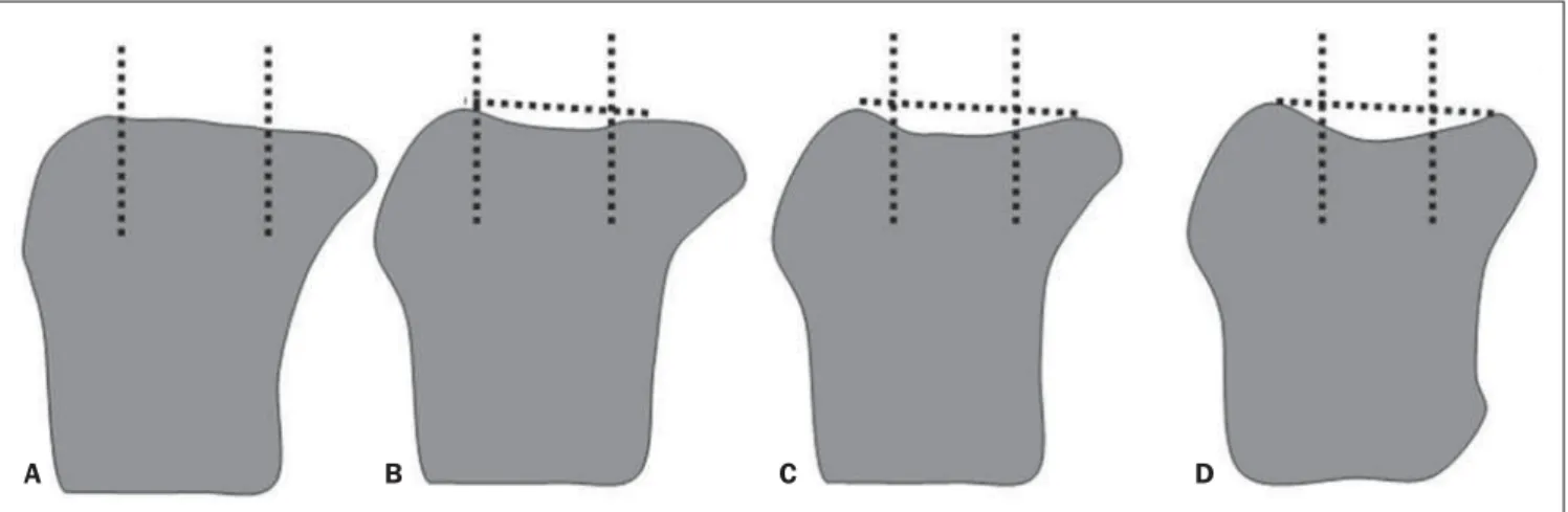
At conventional radiography, MRI and CT, bone attrition is interpreted as flatten-ing or depression of articular surfaces, and classified with basis on a subjective degree of deviation from the normal contour on different images, as follows: grade 0 = normal; grade 1 = moderate; grade 3 = se-vere (Figure 1)(24).
For example, the articular surfaces of the femoral condyles and of the medial aspect of the patella are generally slightly convex. Thus, the normal convexity is shown on Figure 2, while flattening of such structures corresponds to grade 1 (Figure 3), mild concavity corresponds to grade 2 (Figure 4) and severe concavity, to grade 3 (Figure 5).
Figure 1. Grade 0 (normal) (A), grade 1 (mild) (B), grade 2 (moderate) (C), grade 3 (severe) (D). Classification of subarticular bone attrition based on the degree of articular surface flattening or depression as compared with normality. Modified from Peterfy et al.(24).
Figure 2. Grade 0 (normal). Sagittal MRI of knee, T2-weighted image with fat saturation, with no sign of structural alterations.
Figure 3. Grade 1 (mild). Sagittal MRI of knee, T2-weighted image with fat saturation. Signs of subtle infradeleveling of the tibial plateau, suggesting bone attrition observed on the medial tibial plateau.
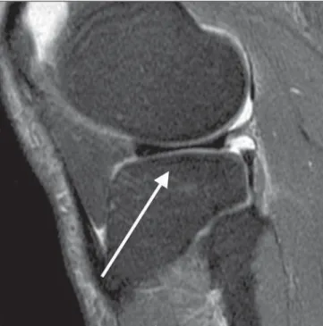
Figure 4. Grade 2 (moderate). Sagittal MRI of the knee, T1-(A) and T2-weighted (B) im-ages with fat suppression. Moderate infradeleveling of the tibial plateau, also with signs of subtle subchondral sclerosis characterized by hyposignal in-tensity on the T1-weighted image, suggesting the pres-ence of bone attrition on the medial tibial plateau.
A B
MRI has shown to be superior to plain radiography because it can demonstrate the knee joint in conjunction with its internal structures(42). CT has been less utilized, since
it lacks clearness in demonstrating some
knee internal structures such as the menis-cus and cartilage. On the other hand, MRI can clearly demonstrate all the internal structures of the knee as well as bone remod-eling and eventual signal alterations(44).
even in cases where the disease is not ad-vanced yet. MRI studies suggest that many knees with radiographically moderate os-teoarthritis, without narrowing of the ar-ticular space, present some evidence of bone attrition(45).
DISCUSSION
Articular pain may become a limiting factor and even a cause of disability in the patient’s life, and may affect any joint in the human body(46,47). The literature presents
discordant information on the actual causes of pain in osteoarthritis patients, but recent multicentric studies, such as MOST(28,30),
have demonstrated the association between bone attrition and pain.
Findings related to bone attrition have ever been present at conventional radiog-raphy, but, maybe because of the difficulty in defining such findings by means of this method, it was not very diffused in the rou-tine clinical assessment(48).
Currently, with the availability of high resolution sectional imaging methods and high-field MRI apparatuses, such alter-ations can be clearly defined, allowing the adoption of an approach to pain in patients with bone attrition.
It is important to highlight that, up to the present moment, the literature does not re-port a specific treatment for pain related to
bone attrition. With the technological progress, a new research field is emerging for treatment and follow-up of these pa-tients.
CONCLUSIONS
Further studies could offer a deeper un-derstanding of the true bone attrition thresholds, preferentially encompassing an independent reading of bone attrition and other characteristics of osteoarthritis.
Cartilage loss is seen as a remarkable characteristic in the development of os-teoarthritis and, up to the present moment, the bone response becomes noticeable only at late stages of the process. Thus, the evaluation of the chondral bone may be-come important in future researches related to the mechanisms of pain generation and also for development of new therapeutic strategies. The involvement of the sub-chondral bone at early stages of the disease may help to explain the failure of chondro-protective agents just like that occurring with disease-modifying drugs. Studies with sodium MRI and with a field higher than the current 1.5T standard could be utilized to investigate relevant data involving the bone attrition biochemistry and physiology. Finally, the prevalence of bone attrition in cases of non-advanced osteoarthritis is sub-stantial, and physicians should be aware of
the relevance and of the ways of diagnos-ing and describdiagnos-ing such finddiagnos-ing on images of these patients of this condition.
REFERENCES
1. Cisternas MG, Yelin E, Katz JN, et al. Ambula-tory visit utilization in a national, population-based sample of adults with osteoarthritis. Arthri-tis Rheum. 2009;61:1694–703.
2. Jüni P, Reichenbach S, Dieppe P. Osteoarthritis: rational approach to treating the individual. Best Pract Res Clin Rheumatol. 2006;20:721–40. 3. Peterfy CG, Gold G, Eckstein F, et al. MRI
proto-cols for whole-organ assessment of the knee in os-teoarthritis. Osteoarthritis Cartilage. 2006;14 Suppl A:A95–111.
4. Clauw DJ, Witter J. Pain and rheumatology: thinking outside the joint. Arthritis Rheum. 2009; 60:321–4.
5. Hudelmaier M, Glaser C, Hohe J, et al. Age-re-lated changes in the morphology and deforma-tional behavior of knee joint cartilage. Arthritis Rheum. 2001;44:2556–61.
6. Felson DT. The sources of pain in knee osteoar-thritis. Curr Opin Rheumatol. 2005;17:624–8. 7. Wluka AE, Wolfe R, Stuckey S, et al. How does
tibial cartilage volume relate to symptoms in sub-jects with knee osteoarthritis? Ann Rheum Dis. 2004;63:264–8.
8. Kornaat PR, Bloem JL, Ceulemans RY, et al. Os-teoarthritis of the knee: association between clini-cal features and MR imaging findings. Radiology. 2006;239:811–7.
9. Brandt KD, Radin EL, Dieppe PA, et al. Yet more evidence that osteoarthritis is not a cartilage dis-ease. Ann Rheum Dis. 2006;65:1261–4. 10. Hunter DJ, Felson DT. Osteoarthritis. BMJ. 2006;
332:639–42.
11. Hill CL, Gale DG, Chaisson CE, et al. Knee effu-sions, popliteal cysts, and synovial thickening:
as-Figure 5. Grade 3 (severe). Sag-ittal MRI of the knee, T1- (A) and T2-weighted (B) images with fat suppression. Signs of marked infradeleveling of the tibial pla-teau and moderate loss of con-cavity with rectification of the femoral condyle, in association with signs of subchondral sclero-sis in the tibial plateau, suggest-ing bone attrition observed on the medial tibial plateau and on the femoral condyle. Signs of involve-ment of the hyaline cartilage rep-resented by changes in signal intensity and thinning.
sociation with knee pain in osteoarthritis. J Rheumatol. 2001;28:1330–7.
12. Torres L, Dunlop DD, Peterfy C, et al. The rela-tionship between tissue lesion and pain severity in persons with knee osteoarthritis. Osteoarthri-tis Cartilage. 2006;14:1033–40.
13. Hunter DJ, Lo GH, Gale D, et al. The reliability of a new scoring system for knee osteoarthritis MRI and the validity of bone marrow lesion as-sessment: BLOKS (Boston Leeds Osteoarthritis Knee Score). Ann Rheum Dis. 2008;67:206–11. 14. Cunha DL, Carvalho ACP, Ribeiro EJS, et al. Res-sonância magnética da osteonecrose do joelho: estudo de 19 casos. Radiol Bras. 2010;43:77–80. 15. Ribeiro DS, Araújo Neto C, D’Almeida F, et al. Achados de imagem das alterações musculoes-queléticas associadas ao lúpus eritematoso sistê-mico. Radiol Bras. 2011;44:52–8.
16. Rezende MU, Hernandez AJ, Camanho GL, et al. Cartilagem articular e osteoartrose. Acta Ortop Bras. 2000;8:100–4.
17. Imhof H, Breitenseher M, Kainberger F, et al. Degenerative joint disease: cartilage or vascular disease? Skeletal Radiol. 1997;26:398–403. 18. Radin EL, Paul IL, Rose RM. Role of
mechani-cal factors in pathogenesis of primary osteoarthri-tis. Lancet. 1972;1:519–22.
19. Hayami T, Pickarski M, Zhuo Y, et al. Character-ization of articular cartilage and subchondral bone changes in the rat anterior cruciate ligament transection and meniscectomized models of os-teoarthritis. Bone. 2006;38:234–43.
20. Radin EL, Martin RB, Burr DB, et al. Effects of mechanical loading on the tissues of the rabbit knee. J Orthop Res. 1984;2:221–34.
21. Radin EL, Rose RM. Role of subchondral bone in the initiation and progression of cartilage dam-age. Clin Orthop Relat Res. 1986;(213):34–40. 22. Burr DB. The importance of subchondral bone in
the progression of osteoarthritis. J Rheumatol Suppl. 2004;70:77–80.
23. Dieppe PA, Reichenbach S, Williams S, et al. Assessing bone loss on radiographs of the knee in osteoarthritis: a cross-sectional study. Arthri-tis Rheum. 2005;52:3536–41.
24. Peterfy CG, Guermazi A, Zaim S, et al. Whole-Organ Magnetic Resonance Imaging Score (WORMS) of the knee in osteoarthritis. Osteoar-thritis Cartilage. 2004;12:177–90.
25. Neogi T, Felson D, Niu J, et al. Cartilage loss oc-curs in the same subregions as subchondral bone attrition: a within-knee subregion-matched ap-proach from the Multicenter Osteoarthritis Study. Arthritis Rheum. 2009;61:1539–44.
26. Lories RJ. Joint homeostasis, restoration, and re-modeling in osteoarthritis. Best Pract Res Clin Rheumatol. 2008;22:209–20.
27. Brandt KD, Dieppe P, Radin EL. Etiopathogen-esis of osteoarthritis. Rheum Dis Clin North Am. 2008;34:531–59.
28. Neogi T, Nevitt M, Niu J, et al. Subchondral bone attrition may be a reflection of compartment-spe-cific mechanical load: the MOST Study. Ann Rheum Dis. 2010;69:841–4.
29. Dieppe P, Cushnaghan J, Young P, et al. Predic-tion of the progression of joint space narrowing in osteoarthritis of the knee by bone scintigraphy. Ann Rheum Dis. 1993;52:557–63.
30. Roemer FW, Neogi T, Nevitt MC, et al. Subchon-dral bone marrow lesions are highly associated with, and predict subchondral bone attrition lon-gitudinally: the MOST study. Osteoarthritis Car-tilage. 2010;18:47–53.
31. Sharma L, Song J, Felson DT, et al. The role of knee alignment in disease progression and func-tional decline in knee osteoarthritis. JAMA. 2001;286:188–95.
32. Hunter DJ, Zhang Y, Niu J, et al. Structural fac-tors associated with malalignment in knee os-teoarthritis: the Boston osteoarthritis knee study. J Rheumatol. 2005;32:2192–9.
33. Hernández-Molina G, Neogi T, Hunter DJ, et al. The association of bone attrition with knee pain and other MRI features of osteoarthritis. Ann Rheum Dis. 2008;67:43–7.
34. Torres L, Dunlop DD, Peterfy C, et al. The rela-tionship between specific tissue lesions and pain severity in persons with knee osteoarthritis. Os-teoarthritis Cartilage. 2006;14:1033–40. 35. Felson DT, McLaughlin S, Goggins J, et al. Bone
marrow edema and its relation to progression of knee osteoarthritis. Ann Intern Med. 2003;139(5 Pt 1):330–6.
36. Hunter DJ, Zhang Y, Niu J, et al. Increase in bone marrow lesions associated with cartilage loss: a longitudinal magnetic resonance imaging study of knee osteoarthritis. Arthritis Rheum. 2006;54: 1529–35.
37. Dequeker J. The relationship between osteoporo-sis and osteoarthritis. Clin Rheum Dis. 1985;11: 271–96.
38. Dieppe P. Subchondral bone should be the main target for the treatment of pain and disease pro-gression in osteoarthritis. Osteoarthritis Cartilage. 1999;7:325–6.
39. Reichenbach S, Dieppe PA, Nüesch E, et al. As-sociation of bone attrition with knee pain, stiff-ness and disability: a cross-sectional study. Ann Rheum Dis. 2011;70:293–8.
40. Zanetti M, Bruder E, Romero J, et al. Bone mar-row edema pattern in osteoarthritic knees: corre-lation between MR imaging and histologic find-ings. Radiology. 2000;215:835–40.
41. Martig S, Boisclair J, Konar M, et al. MRI char-acteristics and histology of bone marrow lesions in dogs with experimentally induced osteoarthri-tis. Vet Radiol Ultrasound. 2007;48:105–12. 42. Bergman AG, Willén HK, Lindstrand AL, et al.
Osteoarthritis of the knee: correlation of subchon-dral MR signal abnormalities with histopatho-logic and radiographic features. Skeletal Radiol. 1994;23:445–8.
43. Ahlbäck S. Osteoarthrosis of the knee. A radio-graphic investigation. Acta Radiol Diagn (Stockh). 1968;Suppl 277:7–72.
44. Yusuf E, Kortekaas MC, Watt I, et al. Do knee abnormalities visualised on MRI explain knee pain in knee osteoarthritis? A systematic review. Ann Rheum Dis. 2011;70:60–7.
45. Reichenbach S, Guermazi A, Niu J, et al. Preva-lence of bone attrition on knee radiographs and MRI in a community-based cohort. Osteoarthri-tis Cartilage. 2008;16:1005–10.
46. Melo Junior CF, Saito OC, Guimarães Filho HA. Avaliação ultrassonográfica dos distúrbios intra-capsulares temporomandibulares. Radiol Bras. 2011;44:355–9.
47. Lima CMAO, Ribeiro EB, Coutinho EPD, et al. Síndrome do impacto do tornozelo na ressonân-cia magnética: ensaio iconográfico. Radiol Bras. 2010;43:53–7.