371 Subungual glomus tumors: imaging findings
Radiol Bras. 2009 Nov/Dez;42(6):371–374 Original Article • Artigo Original
Subungual glomus tumors: imaging findings*
Tumores glômicos subungueais: achados de imagem
Cristiano Montandon1, Juliana da Cunha Costa2, Lorena Andrade Dias2, Fernando Henrique Abrão Alves da Costa2, Ana Carolina Mendes da Costa2, Renato Tavares Daher2, Marcelo Eustáquio Montandon Jr.1
OBJECTIVE: To evaluate main imaging findings of subungual glomus tumors. MATERIALS AND METHODS: Retrospective study of eight cases of subungual glomus tumors from the archives of two private clinics in Goiânia, GO, Brazil. Plain radiographs were obtained in five cases, Doppler ultrasonography in five, and magnetic resonance imaging in five cases. RESULTS: Mean age of the patients included in the present study was 39 years, with female predominance (7:1). Tumors didn’t present prevalence in any specific finger and in most of cases the tumor was located in the medial subungual region. Plain radiography was positive in five cases, demonstrating pressure erosion. Doppler ultrasonography was positive in five cases, demonstrating a hypoechoic and hypervascularized solid nodule. Magnetic resonance imaging was positive in all of the cases, showing a solid nodule, hypointense on T1-, hyperintense on T2-weighted image, with homogeneous contrast uptake. All the patients underwent surgical excision of the lesion with histopathological confirmation. CONCLUSION: In most of cases, glomus tumors are subungual. The diagnosis is clinical and generally is lately achieved. Imaging methods are useful tools for the early diagnosis besides aiding in the surgical planning, considering that the treatment of choice is surgical excision.
Keywords: Subungual glomus tumors; Magnetic resonance imaging; Ultrasonography.
OBJETIVO: Analisar os principais aspectos de imagem dos tumores glômicos subungueais. MATERIAIS E MÉTODOS: Realizado estudo retrospectivo de oito casos de tumores glômicos subungueais, pertencentes a arquivos de duas clínicas particulares de Goiânia, GO, Brasil. Foram obtidas radiografias em cinco casos, ultrassonografia com Doppler em seis casos e ressonância magnética em cinco casos. RESULTADOS: A idade média de acometimento no presente estudo foi de 39 anos, com predomínio do sexo feminino, na proporção de 7:1. Os tumores não apresentaram predileção por nenhum dedo e a maioria localizava-se na região su-bungueal mediana. A radiografia foi positiva em três casos, demonstrando erosões de pressão. A ultrasso-nografia com Doppler foi positiva em cinco casos, evidenciando nódulo sólido, hipoecoico e hipervasculari-zado. A ressonância magnética, em todos os casos, demonstrou nódulo sólido com hipossinal em T1, hiper-sinal em T2 e captação homogênea do meio de contraste. Em todos os pacientes foi realizada excisão cirúr-gica com confirmação anatomopatolócirúr-gica. CONCLUSÃO: A maioria dos tumores glômicos tem localização subungueal. O diagnóstico é clínico, porém geralmente tardio. Os métodos de imagem auxiliam no diagnós-tico precoce, além auxiliar no planejamento terapêudiagnós-tico, cujo tratamento de escolha é a excisão cirúrgica.
Unitermos: Tumor glômico subungueal; Imagem por ressonância magnética; Ultrassonografia.
Abstract
Resumo
* Study developed at Clínica da Imagem and Clínica Multima-gem Diagnósticos, Goiânia, GO, Brazil.
1. MDs, Radiologists, Clínica da Imagem and Clínica Multi-magem Diagnósticos, Titular Members of Colégio Brasileiro de Radiologia e Diagnóstico por Imagem (CBR) and Sociedade Goiana de Radiologia, Goiânia, GO, Brazil.
2. MDs, Residents at Department of Radiology and Imaging Diagnosis, Universidade Federal de Goiás (UFG), Goiânia, GO, Brazil.
Mailing address: Dr. Cristiano Montandon. Rua C-131, nº 670, ap. 401, Jardim América. Goiânia, GO, Brazil, 74255-240. E-mail: [email protected]
Received May 30, 2009. Accepted after revision September 16, 2009.
10 millimeters in diameter(2). Their typical
location is the subungual region of the dis-tal phalanges, but they may be found throughout the body(1,7). Multiple lesions
are extremely rare (2.3%), being most fre-quently found in children(1).
Clinically, these lesions are character-ized by intense, pulsatile, debilitating pain and sensitivity to pressure and tempera-ture(8,9). Ungual alterations are frequently
observed in cases of larger lesions and these tumors are rarely palpable. However, the clinical signs are not always obvious and, considering the small dimensions of these lesions, imaging studies are required Montandon C, Costa JC, Dias LA, Costa FHAA, Costa ACM, Daher RT, Montandon Jr ME. Subungual glomus tumors: imaging findings. Radiol Bras. 2009;42(6):371–374.
blood flow. These bodies can be found in the reticular layer of the dermis throughout the body although they are most numerous in digits, hand palms and feet soles(1).
Glomus tumors are rare benign le-sions(2,3), first described by Wood in
1812(3,4), characterized by hamartomatous
proliferation originated in the neuromyo-arterial glomus bodies(2). This type of
tu-mor, also denominated glomangioma, rep-resents approximately 2% of all the primary soft tissue tumors(5,6) and 1% to 4.5% of
hand neoplasms(3,5).
Usually, these tumors present like a small, reddish blue nodule measuring 3 to
0100-3984 © Colégio Brasileiro de Radiologia e Diagnóstico por Imagem
INTRODUCTION
372
Montandon C et al.
Radiol Bras. 2009 Nov/Dez;42(6):371–374
to elucidate the diagnosis and, above all, to aid in the therapeutic/surgical plan-ning(10,11).
The present study is aimed at analyzing the main imaging findings of subungual glomus tumors.
MATERIALS AND METHODS
Eight cases of subungual glomus tumor from the archives of Clínica Multimagem Diagnósticos and Clínica da Imagem, Goiânia, GO, Brazil, were retrospectively evaluated (Table 1).
Anteroposterior and lateral radiographic images were obtained in five cases. Dop-pler ultrasonography (US) was performed in six patients, and non-contrast- and con-trast-enhanced magnetic resonance imag-ing (MRI) in five cases.
The diagnostic criterion adopted for subungual glomus tumor was the direct visualization of the tumor by Doppler US or MRI. Echogenicity and vascularization were evaluated at color Doppler US. Sig-nal intensity on T1- and T2-weighted im-ages, besides contrast (gadolinium) en-hancement pattern were analyzed at MRI. Additionally, at radiography, the authors utilized an indirect criterion characterized by bone remodeling in the dorsal aspect of the distal phalanx as a positive sign of the presence of a glomus tumor.
The subungual tumors were also clas-sified according to their location, the af-fected digit as well as the tumor situation on the axial plane, either midline or later-ally in the nail bed.
All the patients had not received any treatment, except for the patient number 7 who had already been submitted to surgery five years ago, for resection of a glomus tumor in the middle finger, presenting with tumor recurrence at the time of the present study. All the cases were histopathologi-cally confirmed after the surgical resection. Prognosis evaluation and/or patients’ fol-low-up were not approached by the authors of the present study.
RESULTS
Mean age of the patients with glomus tumors evaluated in the present study was 39 years (26 to 51 years), with a female
Table 1 Subungual glomus tumors.
RX, conventional radiography; US+D, Doppler ultrasonography; MRI, magnetic resonance imaging; Gd, gado-linium; M, male; F, female; N, non-performed study; –, study performed with negative results for glomus tumor; +, study performed with positive results for glomus tumor.
Case 1 2 3 4 5 6 7 8 Sex M F F F F F F F Age (years) 26 42 35 36 43 32 51 47 RX N N + N – + – +
US + D
N + + N + – + + RM + + N + N + + N T1 Low Low Low Low Low T2 High High High High High Gd + + + + + Location
Index finger / middle
Index finger / middle
Little finger / middle
Thumb / lateral
Index finger / lateral
Middle finger / middle
Middle finger / middle
Middle finger / middle
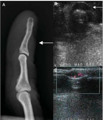
predominance (7:1). Location of the tu-mors was the following: three (37%) in the index finger; two (25%) in the middle fin-ger; two (25%) in the little finfin-ger; and one (12.5%) in the thumb. As regards axial plane location, most of the tumors were in the midline (six cases – 75%) and only two (25%) were laterally located in the nail bed. Plain radiography was positive in three among the five cases evaluated by this method (60%), demonstrating bone remod-eling (pressure erosion) on the dorsal as-pect of the distal phalanx (Figure 1). Dop-pler US was positive in five among the six patients evaluated (83%), all of them with a well-defined, solid, hypoechoic and
hypervascularized subungual nodule (Fig-ures 1 and 2). MRI was positive in the five cases evaluated, demonstrating well-de-fined, solid nodules with hypointense sig-nal on T1-weighted images and sigsig-nal hyperintensity on T2-weighted images with homogeneous intravenous contrast-en-hancement (Figures 3 and 4).
DISCUSSION
Glomus tumors are benign lesions, gen-erally located in the hands, especially in the distal fingertips and particularly in the sub-ungual area since glomus bodies are most numerous in these regions(8). These are rare
373 Subungual glomus tumors: imaging findings
Radiol Bras. 2009 Nov/Dez;42(6):371–374
but debilitating lesions and represent 1% to 4.5% of all hand tumors(5). Like other
re-ports in the literature(6,8,10), the present
study has demonstrated a higher female prevalence (7:1) and mean age of 39 years at the moment of the diagnosis.
Usually, the diagnosis of glomus tumor is clinical, being confirmed in 50% to 78% of cases(12), but many times it is lately
achieved, on average four to seven years after symptoms onset(8,13,14). The evaluation
of patients’ clinical data was not considered by the authors of the present study.
Plain radiography, US and MRI are the imaging methods most frequently utilized in the suspicion of glomus tumor. Such methods contribute for the early diagnosis, reducing the time span between the symp-toms onset and the treatment. Additionally, these methods are useful for the therapeu-tic planning, partherapeu-ticularly in the definition of the surgical approach, reducing the rates
of the main postoperative complication, i.e. nail dystrophia(2).
Signs of bone remodeling in the dorsal aspect of the distal phalanx (pressure ero-sion) were observed at conventional radi-ography. However, this finding is present in only 14% to 60% of cases(5,15–18) (60%
in the present study). Increased distance be-tween the dorsal aspect of the distal pha-lanx and underside of the nail is also de-scribed as a sign of the presence of this type o tumor. However, this finding is rarely observed (25%)(2) and was seen in only one
case of the present study. The low sensitiv-ity of this method is probably related to the small size of the lesions in most of cases. Images magnification and comparison with the contralateral side are useful in the evaluation of these lesions(2).
At Doppler US, a solid, well-delimited, hypoechoic nodule is observed with promi-nent vascularization. The limitations of this Figure 4.Case 7. Female, 51-year-old patient.
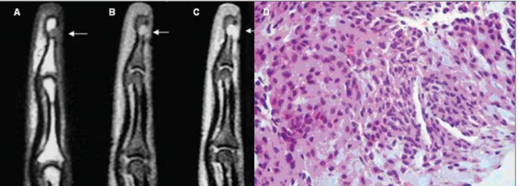
Contrast-enhanced, coronal MRI, T1-weighted se-quence (A) and Sagittal T2-weighted (B): small nod-ule on the dorsal aspect of distal phalanx soft tis-sues of the third finger, with homogeneous contrast uptake and high signal intensity on T2-weighted sequence indicating recurrent/residual tumor.
Figure 3.Case 6. Female, 32-year-old patient. Sagittal MRI, T1-weighted sequence (A) and T1-weighted with fat-saturation, pre- (B) and post-contrast injec-tion (C): small nodule with hypointense signal on T1-weighted sequence, hyperintense signal on T1-weighted sequence with fat-saturation and intense con-trast uptake, determining bone remodeling in the posterior aspect of the distal phalanx. Histological specimen (D): typical aspect of solid glomus tumor. Figure 2.Case 2. Female, 42-year-old patient. US (A): small nodular, hypoechoic, well delimited lesion. Doppler US (B): lesion with increased flow.
374
Montandon C et al.
Radiol Bras. 2009 Nov/Dez;42(6):371–374
method are related to operator dependence and difficulty in the sonographic analysis of the subungual region(6). Thus, a negative
study should not rule out the presence of a small-sized tumor, and investigation should proceed with surgical exploration in the setting of a well-established clinical suspicion(1). In the present study, US was
positive in 83.3% of cases.
At MRI, a solid, well defined nodular lesion is observed with low signal intensity in relation to the dermis of the nail bed on T1-weighted images, and high signal inten-sity on T2-weighted images, with homoge-neous contrast-enhancement(2,7,19). Such
findings were observed in all the patients of the present study evaluated with this imaging method. Additionally, MRI also depicts hyperintense lesions on T1-weighted images, probably as a result of a hemorrhagic component, besides lesions with a hypointense halo and capsular as-pect which have not been observed in the present study(2). MRI has also the
advan-tage of being 3.5 times more sensitive than plain radiography in the detection of bone erosion(2). Presently, it is the most sensitive
imaging method available for detecting glomus tumors, and no false negative case was observed in the present series with this method. Additionally, this method provides satisfactory information on the tumor loca-tion, aiding in the surgical planning(1).
Complete surgical resection is the treat-ment of choice for these lesions(1). The
post-operative incidence of symptoms recurrence ranges between 12–24% of cases(2). In cases
where symptoms recurrence is observed within less than one year postoperatively, many authors attribute the recurrence to in-complete resection or to the presence of a second lesion that had not been previously diagnosed and surgically resected. On the other hand, in cases where the recurrence is observed within more than one year post-operatively, the fact is usually attributed to the development of a new lesion(2).
Most of times, in cases of symptoms re-currence, MRI allows the differentiation between residual/recurrent tumors from postoperative fibrocicatricial alterations. Contrary to postoperative alterations that are characterized by ill-defined margins and low signal intensity on all the se-quences, residual/recurrent glomus tumors correspond to well defined lesions with characteristics similar to the ones of non-treated tumors, likewise the case number 7 of the present study(1).
As regards differential diagnoses, one should mention mucoid and epidermoid inclusion cysts which are avascular cystic lesions, i.e., presenting poor contrast up-take, with no flow at Doppler US and gen-erally with no adjacent bone remodeling. Other rarer entities in this location, such as neuromas and angiomas, represent cases with difficult differential diagnosis through imaging methods. Inflammatory and depo-sition arthropathies, particularly gout, pre-dominantly present alterations in the distal interphalangeal joint(6).
CONCLUSION
Considering that the treatment of glo-mus tumors consists of complete surgical resection of the lesion(1,2), Imaging
meth-ods, especially US and MRI, have demon-strated a significant role in the diagnostic confirmation of this entity. Additionally, such methods allow an accurate determina-tion of the tumor site, facilitating the sur-gical planning as well as ruling out other differential diagnoses. However, it is im-portant to highlight the nonspecific nature of the imaging findings of these lesions and therefore, the diagnosis must be based on correlation with clinical findings(6).
REFERENCES
1. Theumann NH, Goettmann S, Le Viet D, et al. Recurrent glomus tumors of fingertips: MR im-aging evaluation. Radiology. 2002;223:143–51. 2. Drapé JL, Idy-Peretti I, Goettmann S, et al.
Sub-ungual glomus tumors: evaluation with MR im-aging. Radiology. 1995;195:507–15.
3. Vanti AA, Cucé LC, Di Chiacchio N. Tumor glô-mico subungueal: estudo epidemiológico e retros-pectivo, no período de 1991 a 2003. An Bras Der-matol. 2007;82:425–31.
4. Wood W. On painful subcutaneous tubercle. Edinburgh Med J Surg. 1812;8:283–91.
5. Carroll RE, Berman AT. Glomus tumors of the hand: review of the literature and report on twenty-eight cases. J Bone Joint Surg Am. 1972; 54:691–703.
6. Fornage BD. Glomus tumors in the fingers: diag-nosis with US. Radiology. 1988;167:183–5.
7. Al-Qattan MM, Al-Namla A, Al-Thunayan A, et al. Magnetic resonance imaging in the diagnosis of glomus tumours of the hand. J Hand Surg Br. 2005;30:535–40.
8. Van Geertruyden J, Lorea P, Goldschmidt D, et al. Glomus tumours of the hand. A retrospective study of 51 cases. J Hand Surg Br. 1996;21:257– 60.
9. Matsunaga A, Ochiai T, Abe I, et al. Subungual glomus tumour: evaluation of ultrasound imag-ing in preoperative assessment. Eur J Dermatol. 2007;17:67–9.
10. Kale SS, Rao VK, Bentz ML. Glomus tumor of the index finger. J Craniofac Surg. 2006;17:801–4. 11. Chen SH, Chen YL, Cheng MH, et al. The use of ultrasonography in preoperative localization of digital glomus tumors. Plast Reconstr Surg. 2003;112:115–9.
12. Drapé JL, Idy-Peretti I, Goettmann S, et al. Stan-dard and high resolution magnetic resonance im-aging of glomus tumors of toes and fingertips. J Am Acad Dermatol. 1996;35:550–5. 13. Hsu CJ, Wang DY. Glomus tumors. Mid Taiwan
J Med. 2002;7:222–7.
14. Drapé JL, Feydy A, Guerini H, et al. Vascular le-sions of the hand. Eur J Radiol. 2005;56:331–43. 15. Holzberg M. Glomus tumor of the nail. A ‘red herring’ clarified by magnetic resonance imaging. Arch Dermatol. 1992;128:160–2.
16. Matloub HS, Muoneke VN, Prevel CD, et al. Glo-mus tumor imaging: use of MRI for localization of occult lesions. J Hand Surg Am. 1992;17:472– 5.
17. Camirand P, Giroux JM. Subungual glomus tu-mor: radiological manifestations. Arch Dermatol. 1970;102:677–9.
18. Takemura N, Fujii N, Tanaka T. Subungual glo-mus tumor diagnosis based on imaging. J Dermatol. 2006;33:389–93.