ABSTRACT
Micro-shear bond strength and surface
micromorphology of a feldspathic ceramic treated
acid etching
!""#$%&'()#%*1+,-"02$)*##&2, Fabiana Mantovani Gomes FRANÇA2, Flávia Lucisano 6"(7(*(92+*!$:;6(#$&'<2
1- Imperatriz School of Dentistry, Imperatriz, MA, Brazil.
2- School of Dentistry, São Leopoldo Mandic Dental Research Institute, Campinas, SP, Brazil.
222 Profa. Dra. Roberta Tarkany Basting - Faculdade de Odontologia e Instituto de Pesquisas São Leopoldo Mandic - Departamento de Odontologia Restauradora - Dentística - Rua José Rocha Junqueira, 13 - Bairro Swift, Campinas-SP - 13045-755 - Brasil - Phone/Fax: 55-19-3211-3600 - e-mail: [email protected]
#!=7;+7>,(2+(,,#=!+
O
bjective: The aim of this study was to evaluate the effect of feldspathic ceramic surface cleaning on micro-shear bond strength and ceramic surface morphology. Material and ! "# $%&' ( treatment, S: water spray + air drying for 1 minute, US: immersion in ultrasonic bath for 5 minutes, F: etching with 37% phosphoric acid for 1 minute, followed by 1-minute rinse, F+US: etching with 37% phosphoric acid for 1 minute, 1-minute rinse and ultrasonic bath for 5 minutes. Composite cylinders were bonded to the discs following application of silane and hydrophobic adhesive for micro-shear bond strength testing in a universal testing machine at 0.5 mm/min crosshead speed until failure. Stereomicroscopy was used to classify failure type. Surface micromorphology of each treatment type was evaluated by scanning *2* "4 6 89;6<9 " ! $%=>?' failure types were cohesive resin cohesion followed by adhesive failure. Micro-shear bond ! # by the different surface cleaning techniques. Absence of or less residue was observed after @BD@B((! E ! 2 whereas, when cleaning was associated with ultrasound, less residue was observed.Keywords:( GB
INTRODUCTION
The longevity of a ceramic restoration appears to be linked to its physical and mechanical properties, as well as reliable bonding between the restoration and the tooth25.
In order to obtain effective bonding between feldspathic ceramics and the tooth substrate, surface treatment is necessary for both the internal aspect of the ceramic and the tooth structure. The use of methods to pretreat the ceramic surface,
such as mechanical (burs and aluminum oxide !'2 $*K etch and silane) or a combination of both (silicon oxide sandblasting) optimize adhesion between the ceramic and the resin cement6,17.
The combination of hydrofluoric acid and silane on the internal surface of the ceramic has been the most recommended method to increase bond strength between the ceramic and the tooth substrate when using resin cements9,18. However, it
acid removal from the ceramic surface, considerable amounts of deposits are formed, which can affect bond strength13.
Some studies have proposed cleaning techniques for ceramic surface aiming at removing the residues ! 2 of ultrasonic bath3,6,15,17,24, 37% phosphoric acid
smear19, rinsing under running water12,14,23 or a
combination of 37% phosphoric acid smear and ultrasonic bath13.
Considering the lack of an established protocol # ! etching of the ceramic surface, the aims of this study were to evaluate: a) the micro-shear bond strength and failure mode between the feldspathic ceramic and the adhesive system following pretreatment of the ceramic surface with different cleaning methods to remove particles generated by W!' surface micromorphology after using different cleaning methods to remove hydrofluoric acid etching residues by scanning electron microscopy (SEM).
MATERIAL AND METHODS
Forty six feldspathic ceramic discs (Super Porcelain EX-3, Noritake Kizai Co., Ltd, Nishi-Ku, Nagoya, Japan, batch 011906, shade A3,5B) were made, 6 of which were used to assess micromorphology and 40 were used for micro-shear bond strength testing.
The ceramic discs were obtained from 12.5 mm diameter by 3.6 mm thick wax patterns using lost wax casting, which gave room to the feldspathic ceramic. The ceramic powder/liquid ratio was used as instructed by the manufacturer and the mixture was placed into the plaster mold using " ! # ` investment plaster and ceramic set were placed in " by the manufacturer. As the ceramic shrinks by { 2 " dimensions were close to 10 mm in diameter and 3 mm thick.
Micro-shear bond strength test
Forty ceramic discs were included in PVC cylinders using polyester resin (Hobby Automotivo, 3M do Brasil, Sumaré, SP, Brazil). The surfaces of the ceramic discs were sequentially polished using silicium carbide sandpaper (Lixa d’água, 3M, Sumaré, SP, Brazil) of grades 400, 600 and 12005,
mounted on a running water-cooled electric polisher (Aropol 2V, Arotec S/A, São Paulo, SP, Brazil). The discs were randomly divided into 5 experimental &$%&'
The surface of all ceramic discs was treated with
$< # 2 }# 2 B Catarina, Brazil) for 2 minutes, followed by water and water spray rinse for 30 seconds. Then, the discs were treated according to the experimental groups as follows:
Group C: no additional surface treatment; Group S: water spray + air for 1 minute, using the 3-in-1 syringe;
Group US: ultrasonic bath (USC1400, Unique Ind. e Com de Prod Elet. Ltda, São Paulo, SP, Brazil) with distilled water for 5 minutes;
Group F: 37% phosphoric acid smear (Acid Gel, Villevie, Joinville, SC, Brazil), using a disposable brush (Disposable Micro-applicators, Microbrush, Rio de Janeiro, RJ, Brazil) for 1 minute, followed by rinsing under running water for 1 minute;
Group F+US: 37% phosphoric acid smear (Acid Gel, Villevie, Joinville, SC, Brazil), using a disposable brush (Disposable Micro-applicators, Microbrush, Rio de Janeiro, RJ, Brazil) for 1 minute, followed by rinsing under running water for 1 minute, combined with ultrasonic bath (USC1400, Unique Ind e Com de Prod Elet. Ltda, São Paulo, SP, Brazil) with distilled water for 5 minutes.
Five 0.8 mm diameter x 4 mm high composite resin (Filtek Z100, 3M ESPE, St. Paul, MN, USA) cylinders were made for each ceramic disc. Therefore, a layer of silane (Dentsply, Petrópolis, RJ, Brazil) was previously applied to bond the resin onto the ceramic. A layer of adhesive (Adper Scotchbond – Adhesive, 3M ESPE, St. Paul, MN, USA) was applied and light-cured using halogen light (Demetron, LC Kerr Corporation, Orange, CA, USA) for 10 seconds. Then, silicone tubes 4 mm high x 0.8 mm diameter were placed on the ceramic ! " 9 " the tubes, the resin cylinders were light-cured for 40 seconds (Demetron, LC Kerr Corporation, Orange, CA, USA) with the light source placed at the upper end of the cylinders, thus light-curing all of them simultaneously. The light-curing equipment was set at 499 mW/cm2 (from 491 mW/cm2 to 507 mW/cm2)
with the aid of a radiometer (Radiômetro Digital,
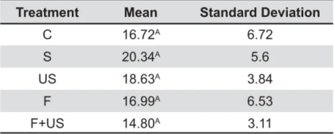
Treatment Mean #PQ
C 16.72A 6.72
S 20.34A 5.6
US 18.63A 3.84
F 16.99A 6.53
F+US 14.80A 3.11
difference
NewDent, Ribeirão Preto, SP, Brazil) and reviewed # "# 8 after light-curing, the silicone tubes were cut using a scalpel blade 12 and disposed of in order to obtain 4 mm high x 0.8 mm diameter composite resin cylinders bonded to the ceramic surface, with a bonding area of 0.5 mm2. The specimens
were stored in a bacteriological incubator (ECB 1.3 digital, Odontobrás Ind. e Com. Equip. Med. Odont. Ltda, Ribeirão Preto, SP, Brazil) at 37°C for 24 hours until the bond strength tests were performed.
"{ #
to the universal testing machine (DL2000, EMIC Ind e Com Ltda, São José dos Pinhais, PR, Brazil). The composite resin cylinders were aligned to a loading cell of 20 KgF. A 0.3 mm diameter stainless steel wire looped around both the extremity of the loading cell and the composite resin cylinder, simultaneously. The wire maintained contact with the inferior semicircle of the cylinders, as close as possible to the area bonded to the ceramic. The test was carried out at a speed of 0.5 mm/min, until bonding failure, for each cylinder sequentially, that is, one at a time.
Y"= C S US F F+US
n % n % n % n % n %
Resin Cohesion 25 62.5 24 60 24 60 28 70 21 52.5
Adhesive 8 20 8 20 8 20 2 5 12 30
Mixed 2 5 4 10 2 5 1 2.5 1 2.5
Ceramic cohesion 1 2.5 3 7.5 0 0 3 7.5 2 5
Failure before testing 4 10 1 2.5 6 15 6 15 4 10
Total 40 100 40 100 40 100 40 100 40 100
Table 2- Failure mode for the different ceramic surface treatments
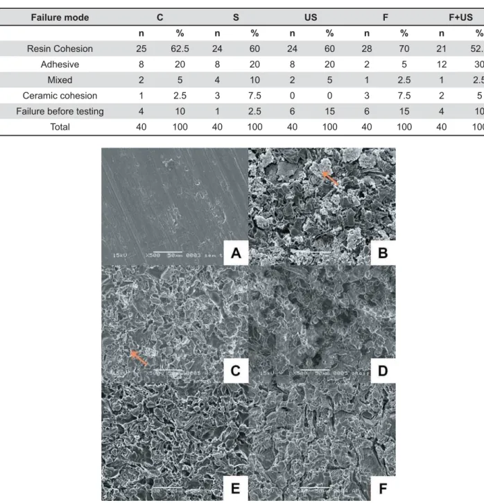
Y Photomicrographs of the ceramic surfaces as seen using a scanning electron microscope (SEM) at 500X
The failure mode was assessed using a stereoscopic lens (EK3ST Model, Eikonal, São Paulo, B2'" " # 2 or resin cohesion failure, or mixed. Cohesion failure " ! or the resin, adhesive failure when there was a fault at the junction between the ceramic and the resin, and mixed when there was both adhesive and cohesion failure, simultaneously.
Micromorphological analysis
One of the six ceramic discs used to evaluate surface micromorphology did not receive any 2 "#
The specimens were then stored in an incubator at 50°C for 3 hours to remove any water particles, 24 hours prior to SEM assessment. The samples were then placed in airtight glass jars containing silica gel and sealed to ensure moisture elimination.
The specimens received a layer of gold/ palladium. The discs were positioned in the SEM
(JSM 5600LV, JEOL, Tokyo, Japan) and images were ! *2*"
Statistical analysis
For each ceramic disc, the mean micro-shear bond strength value for the resin cylinders was considered. After exploratory analysis, the data were tested using one-way analysis of variance (p<0.05) on SAS statistical software (Release 9.2, 2008, Institute Inc., Cary, NC, USA). Descriptive analysis was used for surface micromorphology and failure mode.
RESULTS
Table 1 describes the mean values of micro-shear bond strength and shows no significant difference between the surface treatment methods $%=>?' ! E failure modes following micro-shear bond strength testing. The most frequent failure modes were resin cohesion followed by adhesive failure.
When inspecting the SEM images, the ceramic surface that received no treatment appeared
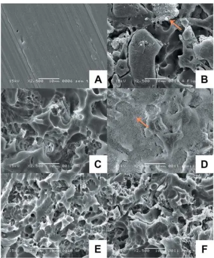
Y Photomicrographs of the ceramic surfaces as seen using a scanning electron microscope (SEM) at 2,500X
smooth without porosity, as a result of the polish obtained when the ceramic disc was prepared (Figure 1A). Regarding the control group (C) and $B'2 rinsed with water and water-air spray, respectively, irregular micrometric structures were observed, which probably consisted of products from the acid etch step (Figures 1B and 1C). Such structures were not encountered on the surfaces of the other groups. Regarding groups US, F and F+US, no " ! # surface micromorphology (Figures 1D, 1E and 1F).
9 "$2*'2@B F+US (Figures 2E and 2F) showed no debris or acid etch residue, whereas, in the two groups in which no ultrasound was used, some debris were still present. This suggests that ultrasonic cleaning was " 9! # when the phosphoric acid smear was used. At lower "2 ` ! # 2 which could have been remains of the phosphoric acid itself, which was not completely removed under 9 "2 obvious that this surface is different from the other groups. It shows a granular or sandy aspect. It is likely that some form of over-etching may have occurred on the ceramic surface, when phosphoric acid was applied. As no ultrasonic treatment was used in this group, the products from over-etching " #
DISCUSSION
Resin cements and internal ceramic surface treatment are used to promote bonding between ceramic surface and tooth7,10. Such treatments are
necessary to modify the surface of these structures, because they are usually smooth and with low surface energy, which prevents the penetration of the bonding agents that create satisfactory chemical or micromechanical retention20.
Dental ceramics have a crystalline microstructure containing large amounts of loose crystals or granules, which form their microtopography2.
Feldspathic ceramic is composed of feldspath (65%), quartz (±25%) and metal oxides (±10%). Sodium and alkaline earth oxides are added as !# " # properties. Potassium oxide (K2O) is one of the main components of feldspathic ceramics, which # " of thermal dilation as similar as possible to the metal alloys used in metal-ceramic restorations4.
The large quantity of glass matrix optimizes the materials vulnerability to acid etching, which allows the formation of microporosities that improve adhesion12. Aluminium oxide sandblasting and
hydrofluoric acid etching promote an increase
of ceramic surface area, creating an irregular topography and increasing surface energy for micromechanical retention of the resin cement13,24.
Hydrofluoric acid etching is widely used to pretreat feldspathic ceramic surfaces, because it acts selectively on the silica glass matrix, forming { 2 to facilitate micromechanical interlocking with the resin cement5,12,23,24. For feldspathic ceramics,
at a concentration of 9% to 10%1,3,8,11,21, and the
etching time ranged between one3,9,15,23-25 and two
minutes15,23. However, studies comparing etching
times reported no difference among the times used, as long as the minimum etching time was one minute15,23.
Following etching of silica-based ceramics, it ! ! # to the formation of insoluble byproducts, such as 2 ceramic surface9. These particles may interfere
with the bond strength between the ceramic and the substrate onto which it is cemented9,16,22. In
the present study, the control groups SEM images $ acid), revealed large quantities of residues when compared to the untreated ceramic and to the groups that received some form of surface cleaning. However, Phark, et al.17 (2009)reported that shear
bond strength is not affected by contamination or cleaning procedures, as shown in this study.
In order to remove byproducts following hydrofluoric acid etching, some authors used ultrasonic washing3,6,15,17,24, 37% phosphoric
acid smear19, rinsing with running water12,14,23,
or a combination of 37% phosphoric acid smear and ultrasonic washing13. The present study
" ! strength using different methods of ceramic surface cleaning, despite the presence of byproducts observed by SEM following surface cleaning with water spray, unlike in groups US and F+US, in which ! 92*"2 ultrasonic washing improved the removal of debris, when compared with the group in which phosphoric acid was used, where residue was observed after rinsing under running water. However, the long-term effect of this interference has not yet been evaluated.
In this study, the main failure mode was resin cohesion followed by adhesive failure, thus showing ! strength. Phark, et al.17 (2009)also observed no
! left by the etching agent on the ceramic surface. In addition, Fabianelli, et al.9 (2010) demonstrated that
have shown increase in bond strength following silane application, regardless of the surface etching technique3,17,23,24. Therefore, the residue does not
seem to interfere with bond strength, as shown in this study.
Regardless of the cleaning technique used on the ceramic surface, the presence or absence of ! ! 8 method may simply consist of spraying with water for 90 seconds.
CONCLUSION
The micro-shear bond strength between the feldspathic ceramic and adhesive system was not influenced by different ceramic cleaning E 2 Micromorphological evaluation of the ceramic surface revealed little to no residue following 2 ! ultrasonic washing.
ACKNOWLEDGEMENTS
To Piracicaba Dental School (UNICAMP) for the use of the SEM to capture images.
REFERENCES
8992 2 and cleaning methods on resin bonding to zirconia ceramic. Dent Mater. 2011;27:207-13.
2- Blatz MB, Sadan A, Kern M. Resin-ceramic bonding: a review of the literature. J Prosthet Dent. 2003;89:268-74.
3- Brentel AS, Özcan M, Valandro LF, Alarça LG, Amaral R, Bottino MA. Microtensile bond strength of a resin cement to feldspathic ceramic after different etching and silanization regimens in dry and aged conditions. Dent Mater. 2007;23:1323-31.
4- Cesar PF, Yoshimura HN, Miranda Júnior WG, Okada CY. Correlation between fracture toughness and leucite content in dental porcelains. J Dent. 2005;33:721-9.
5- Dalkiz M, Sipahi C, Beydemir B. Effects of six surface treatment methods on the surface roughness of a low-fusing and an ultra-low-fusing feldspathic ceramic material. J Prosthodont. 2009;18:217-22.
822` 2 `2 2< LF. Comparison of two bond strength testing methodologies for bilayered all-ceramics. Dental Mater. 2007;23:630-6.
?8 992 B! G2 9! 92 9} " microtensile versus microshear bond testing for evaluation of bond strength of dental adhesive systems to enamel. Dent Mater. 2010;26:848-54.
8- Ersu B, Yuzugullu B, Yazici AR, Canay S. Surface roughness ! 8" 8 using various surface treatments. J Dent. 2009;37:848-56. 9- Fabianelli A, Pollington S, Papacchini F, Goracci C, Cantoro A, Ferrari M, et al. The effect of different surface treatments on bond strength between leucite reinforced feldspathic ceramic and composite resin. J Dent. 2010;38:39-43.
10- Frankenberger R, Reinelt C, Petschelt A, Krämer N. Operator # ! Dent Mater. 2009;25:960-8.
11- Hooshmand T, Parvizi S, Keshvad A. Effect of surface acid !{ { 8 ceramics. J Prosthodont. 2008;17:415-9.
12- Kukiattrakoon B, Thammasitboon K. Optimal acidulated feldspathic porcelain: on shear bond strength to resin composite. Eur J Dent. 2012;6:63-9.
=8 2 ( 8 and connecting porcelain on the microtensile bond strength of composite resin to feldspathic porcelain. J Prosthet Dent. 2006;96:354-61.
14- Melo RM, Valandro L, Bottino MA. Microtensile bond strength of a repair composite to leucite-reinforced feldspathic ceramic. Braz Dent J. 2007;18:314-9.
15- Menezes FC, Borges GA, Valentino TA, Oliveira MA, Turssi CP, Correr-Sobrinho L. Effect of surface treatment and storage on the bond strength of different ceramic systems. Braz J Oral Sci. 2009;8:119-23.
16- Monticelli F, Toledano M, Osorio R, Ferrari M. Effect of temperature on the silane coupling agents when bonding core E"! W8& ?8`}G2 B}2G22B9 ! " Dent Mater. 2009;25:1541-50.
18- Pollington S, Fabianelli A, Noort RV. Microtensile bond strength # 8 different surface treatments. Dent Mater. 2010;26:864-72. 19- Quaas AC, Yang B, Kern M. Panavia F 2.0 bonding to contaminated zirconia ceramic after different cleaning procedures. Dent Mater. 2007;23:506-12.
20- Queiroz JR, Benetti P, Özcan M, Oliveira LF, Della Bona A, Takahashi FE, et al. Surface characterization of feldspathic ceramic using ATR FT-IR and ellipsometry after various silanization protocols. Dent Mater. 2012;28:189-96.
21- Saraç S, Elekdaq-Turk S, Saraç D, Turk T. Surface conditioning methods and polishing techniques effect on surface roughness of a feldspar ceramic. Angle Orthod. 2007;77:723-8.
22- Shimada Y, Yamaguchi S, Tagami J. Micro-shear bond strength of dual-cured resin cement to glass ceramics. Dent Mater. 2002;18:380-8.
=8`2`6229 silanes and acid concentrations on bond strength of brackets to porcelain surfaces. Eur J Orthod. 2009;31:402-6.