ABSTRACT
http://dx.doi.org/10.1590/1678-7757201302136
Effect of the application time of phosphoric acid
and self-etch adhesive systems to sclerotic dentin
Alexandra Patricia MENA-SERRANO1, Eugenio Jose GARCIA2 3, Gislaine Cristine MARTINS4, 5, Alessandro Dourado LOGUERCIO6, Alessandra REIS7
1- DDS, MSc, PhD student, School of Dentistry, Department of Restorative Dentistry, State University of Ponta Grossa, Ponta Grossa, PR, Brazil. 2- Visiting Professor, School of Dentistry, University of São Paulo, São Paulo, SP, Brazil.
3- DDS, MSc, PhD student, School of Dentistry, Department of Restorative Dentistry, State University of Ponta Grossa, Ponta Grossa, PR, Brazil. Adjunctive Professor, University of Valparaíso, Valparaíso, Chile.
4- DDS, MSc, PhD, Visiting Professor of ABOPG, Ponta Grossa, PR, Brazil.
5- DDS, MSc, PhD, Associate Professor, School of Dentistry, Department of Dental Materials, University of São Paulo, São Paulo, SP, Brazil.
6- DDS, MSc, PhD, Adjunctive Professor, School of Dentistry, Department of Restorative Dentistry, State University of Ponta Grossa, Ponta Grossa, PR, Brazil. 7- DDS, MSc, Adjunctive Professor, School of Dentistry, Department of Restorative Dentistry, State University of Ponta Grossa, Ponta Grossa, PR, Brazil.
Corresponding address: Profa. Dra. Alessandra Reis - Universidade Estadual de Ponta Grossa - Mestrado em Odontologia - Rua Carlos Cavalcanti, 4748 - Bloco M - Sala 64A - Uvaranas - 84030-900. Ponta Grossa - PR - Brasil - e-mail: [email protected]
!"#$%"$&'(!)$%"*&+(!;$%"*
O
bjectives: To evaluate the effect of application time on the resin-dentin bond strength (μTBS) and etching pattern of adhesive systems applied on sclerotic dentine. Material and Methods: A total of forty-two bovine incisors had their roots removed. The 1-step self-etch GO (SDI), the 2-step self-etch Adper SE Bond (3MESPE) and the 35% phosphoric acid (3MESPE) from the 2-step etch-and-rinse Adper Single Bond 2 (3MESPE) were applied on the bovine incisal surfaces according to the manufacturer’s instructions or duplicating the recommended conditioning time. After adhesive application, thirty teeth were restored with composite resin, stored for 24 h in distilled water at 37°C, and sectioned into resin-dentin bonded sticks (0.8 mm2) and tested according to the μTBS at 0.5 mm/min. Thescanning electron microscopy. Each tooth was divided into a buccal-to-lingual direction into three thirds, and each third randomly assigned to the groups: control (no treatment), according to the manufacturers’ instructions and duplicating the recommended application time. The μTBS and the relative percentage of the tubule area opening were evaluated by two-way repeated measures ANOVA and Tukey’s tests (α !
of the conditioning time favored only the GO adhesive (p<0.05). Both application methods "# $ & Conclusions: ! "# # '* self-etch adhesive system tested.
Key words: Dentin. Acid etching, Dental. Dentin-bonding agents.
INTRODUCTION
The primary goal of dentin bonding systems is to provide retention of restorative materials to the dental structure as well as to seal the dentin substrate. Even though the immediate bonding effectiveness of most current adhesive systems
is favorable6+ " $
ability to bond sound dentin. Although sound dentin may be a common substrate in the daily practice, a variety of pathological dentin substrates
are also encountered in clinical scenarios, which includes carious-affected and sclerotic dentin7,26.
Irrespective of the bonding strategy used, bonding to pathologically altered substrates such as sclerotic dentin led to compromised bonding17,26.
This has been due to partial or complete obliteration of the dentinal tubules with mineral crystals and due to the presence of an acid-resistant hyper-mineralized layer that acts as an acid resistant substrate17,26.
[etch-and-rinse (ER) and self-etch (SE) adhesives] rely primarily on micromechanical retention, the existence of such obstacles may compromise " dental tissues. Thus, previous studies suggested that bonding to human sclerotic dentin could be improved by changing the adhesive protocol that is typically employed for sound dentin. For ER systems, the duplication of the phosphoric acid conditioning time was suggested; however, the effectiveness of this approach is not unanimous4,18,19. As for the
SE adhesives, phosphoric acid pre-treatment8,16
or surface roughening of the sclerotic dentin with diamond burs8,27, has also been suggested. To the
extent of the author’s knowledge, no study has so far evaluated the effectiveness of duplication of the conditioning time of SE in sclerotic dentin.
Although phosphoric acid conditioning or surface roughening of sclerotic dentin has shown promising results, they increase the number of clinical steps or may produce thicker smear layers when diamond burs are employed. Thicker smear layers were shown to restrict the penetration of some SE adhesives into the dentin16,25. Therefore,
other simpler strategies should be investigated. Thus, the aim of this study was to evaluate the micro-tensile resin-dentin bond strength (μTBS) and etching pattern of ER and SE adhesives to sclerotic bovine dentin applied as recommended by the manufacturers or after duplicating the conditioning time. The null hypothesis tested was that bonding to sclerotic dentin will not be affected by the application time of the ER and SE adhesives.
MATERIAL AND METHODS
The Ethics Committee from the State University of Ponta Grossa (Paraná, Brazil) reviewed and approved this study under protocol number 06289/09. Forty-two bovine incisors, from animals older than 3 years old2 were obtained from a local
slaughterhouse. These teeth exhibit natural dentin exposure in the incisal edges, and therefore, no bur preparation was required to expose the dentin substrate for bonding.
The roots were sectioned with a water-cooled low-speed diamond saw (Isomet 1000, Buehler, Lake Bluff, IL, USA). The coronal pulp was removed $ = " ! smear-layer free incisal surfaces were cleaned with an anionic detergent rubbed with a disposable sponge for 30 s and rinsed in running water for 30 s.
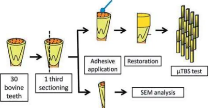
For the μTBS testing, thirty teeth were randomly selected and divided into six groups according to the combination of the main factors Adhesive (3 levels) and Application time (2 levels) so that 5 teeth were employed in each group.
Before the adhesive application, one third of each crown was longitudinally sectioned in a buccal-to-lingual direction using a water-cooled low-speed diamond saw (Isomet 1000) in order to ensure that the bonding substrate was, in fact, sclerotic (Figure 1). In these thirds, no treatment was performed, and the specimens were mounted on aluminum stubs and desiccated in colloidal silica for 24 h. After this period, they were gold-sputtered (Sputter Coater IC 50, Shimadzu, Tokyo, Japan) and examined under the scanning electron microscope (SEM). The SEM was operated in the secondary electrons mode (SSX-500, Shimadzu, Tokyo, Japan) with an accelerating voltage of 12 kV. In case the sclerotic characteristic was not "+
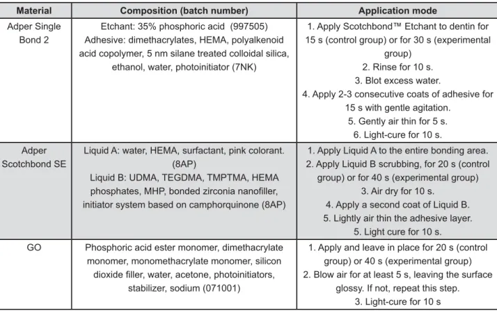
In the other two thirds (not sectioned), the 1-step SE GO (GO, SDI, Bayswater, Victoria, AU), the 2-step SE Adper SE Bond (ASE, 3MESPE, St. Paul, MN, USA), and the 2-step ER Adper Single Bond 2 (SB2, 3MESPE, St. Paul, MN, USA) were applied according to the manufacturers’ instructions or duplicating the conditioning time (Figure 2). A total of 5 tooth specimens were employed for each group. The application mode, composition and batch number of the adhesive systems are shown in Figure 2.
The adhesives were applied by a single and calibrated operator (Figure 2). Composite resin build-ups (Opallis, FGM, Joinville, SC, Brazil) were constructed in three increments of 1 mm, and each one was cured for 40 s (Figure 1). The light-curing unit was set at 500 mW/cm2 (VIP, Bisco,
Schaumburg, IL, USA) and used throughout the restorative procedure.
After 24 h of storage in distilled water at 37°C, the specimens were longitudinally sectioned in both “x” and “y” directions by means of a water-cooled low-speed diamond saw (Isomet 1000, Buehler) in order to obtain approximately 10-14 resin-dentin bonded sticks per tooth, with a cross-sectional area of approximately 0.8 mm2 (Figure
1). All the resin-dentin bonded sticks were tested in a universal testing machine at a crosshead speed
of 0.5 mm/min (Kratos, São Paulo, SP, Brazil). The #> ? " " ' @ H cohesive within composite resin and (3) adhesive or adhesive/mixed (failure at the resin/dentin interface or mixed with cohesive failure of the neighboring substrates). The number of specimens with premature failures during the specimen preparation was also recorded.
For the etching pattern analysis, the remaining teeth per group) according to the material to be used (Figure 2). The crowns of these twelve bovine teeth were longitudinally sectioned in a buccal-to-lingual direction with a water-cooled low-speed diamond saw (Isomet 1000), in order to obtain three crown thirds. One third was used for the evaluation of the sclerotic dentin degree, where no treatment was performed; the second third was treated with one of the adhesives applied according to the manufacturers’ instructions and the last third was treated with the same material but duplicating the conditioning time (Figure 3). The allocation of each third to the subgroup was randomly determined.
In the SE groups, the adhesives were applied as described earlier for the μTBS testing, but they were not light-cured. Then, the resin monomers of the self-etch primer were removed by immediately immersing the specimens in acetone for 5 min followed by immersion in deionized water for 5
min. After this, the specimens were immersed in 96% ethanol for 5 min and again in deionized water for 5 min13. The specimens treated with phosphoric
acid were only rinsed with deionized water for 15 s (Figure 3).
They were mounted on aluminum stubs, ultrasonically cleaned with distilled water for 30 min (Dabi Atlante, Ribeirão Preto, SP, Brazil) and desiccated in colloidal silica for 24 h. After this period, they were gold-sputtered (Sputter Coater IC 50, Shimadzu) and examined under the scanning electron microscope (SEM). The SEM was operated in the secondary electrons mode (SSX-500, Shimadzu) with an accelerating voltage of 12 kV (Figure 3).
Three pictures were taken of each crown third. The relative percentage of the tubule area occlusion of each specimen was measured in all pictures using the UTHSCSA ImageTool 3.0 software (Department of Dental Diagnostic Science at The University of Texas Health Science Center, San Antonio, Texas, USA) by a blinded author.
The μTBS values of sticks from the same tooth half were averaged. Specimens with a cohesive fracture mode and premature failures were excluded from the tooth half mean. The three readings of the relative open tubule area from the same tooth half were averaged for statistical purposes. Data from the μTBS testing and the relative percentage of the open tubule area were evaluated by two-way repeated measures ANOVA
Material Composition (batch number) Application mode
Adper Single Bond 2
Etchant: 35% phosphoric acid (997505) Adhesive: dimethacrylates, HEMA, polyalkenoid acid copolymer, 5 nm silane treated colloidal silica,
ethanol, water, photoinitiator (7NK)
1. Apply Scotchbond™ Etchant to dentin for 15 s (control group) or for 30 s (experimental
group) 2. Rinse for 10 s. 3. Blot excess water.
4. Apply 2-3 consecutive coats of adhesive for 15 s with gentle agitation.
5. Gently air thin for 5 s. 6. Light-cure for 10 s.
Adper Scotchbond SE
Liquid A: water, HEMA, surfactant, pink colorant. (8AP)
Liquid B: UDMA, TEGDMA, TMPTMA, HEMA initiator system based on camphorquinone (8AP)
1. Apply Liquid A to the entire bonding area. 2. Apply Liquid B scrubbing, for 20 s (control
group) or for 40 s (experimental group) 3. Air dry for 10 s.
4. Apply a second coat of Liquid B. 5. Lightly air thin the adhesive layer.
5. Light cure for 10 s.
GO Phosphoric acid ester monomer, dimethacrylate
monomer, monomethacrylate monomer, silicon
stabilizer, sodium (071001)
1. Apply and leave in place for 20 s (control group) or 40 s (experimental group) 2. Blow air for at least 5 s, leaving the surface
glossy. If not, repeat this step. 3. Light-cure for 10 s
(Material vs. Application time) and Tukey’s tests
(α
RESULTS
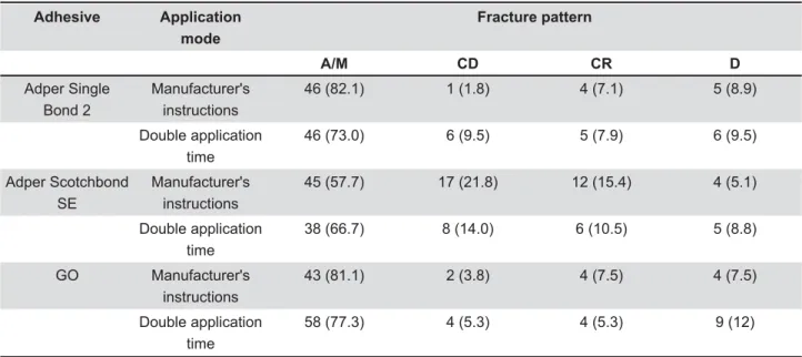
Sclerotic dentin was observed in all specimens used for the μTBS testing and, therefore, no specimen was discarded. The two-way ANOVA revealed that the cross-product interaction Adhesive vs. Application time was statistically " Y' Z \ instructions, the GO adhesive showed the lowest μTBS values. The duplication of the application time yielded the highest μTBS mean only for the GO adhesive (Tukey’s test, p<0.05, Table 1). The fracture pattern of the experimental conditions is !$ H ^ " observed between the groups (data not shown).
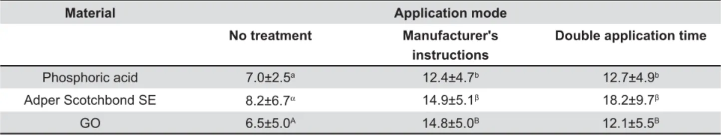
As for the percentage of the tubule area opening, only the main factor, Application time " Y _ (manufacturer’s instructions and duplicating the conditioning time) yielded a similar open tubule `{+ !$ Y+ "# higher than the sclerotic dentin surface (no treatment) (Tukey’s test, p<0.05). Representative
Adhesive Application
mode
Fracture pattern
A/M CD CR D
Adper Single Bond 2
Manufacturer's instructions
46 (82.1) 1 (1.8) 4 (7.1) 5 (8.9)
Double application time
46 (73.0) 6 (9.5) 5 (7.9) 6 (9.5)
Adper Scotchbond SE
Manufacturer's instructions
45 (57.7) 17 (21.8) 12 (15.4) 4 (5.1)
Double application time
38 (66.7) 8 (14.0) 6 (10.5) 5 (8.8)
GO Manufacturer's
instructions
43 (81.1) 2 (3.8) 4 (7.5) 4 (7.5)
Double application time
58 (77.3) 4 (5.3) 4 (5.3) 9 (12)
Table 2- Number and percentage of specimens (%) according to fracture pattern mode and the premature de-bonded specimens from each experimental condition (*)
(*) A/M – adhesive/mixed fracture mode; CD – cohesive fracture mode in dentin; CR – cohesive fracture mode in resin; PF – premature failures.
@!( Application mode
Manufacturer's instructions Double application time
Adper Single Bond 2 31.1±3.0a 30.1±5.1a
Adper Scotchbond SE 34.0±5.0a 31.9±3.2a
GO 18.4±7.0b 30.0±3.3a
Table 1- Micro-tensile bond strength values (MPa) (means ± standard deviations) obtained for each experimental condition
!"!#$"&#
images of each experimental condition can be seen in Figures 4 to 6.
DISCUSSION
Although most in vitro studies that evaluated adhesion to sclerotic dentin used cervical lesions of human dentin as bonding substrates8,19, the
present study employed bovine sclerotic dentin. This substrate is morphologically similar to human dentin3 and, therefore, a suitable substitute for
human teeth in bond strength tests24. Besides that,
bovine specimens are easier to obtain, and the substrate areas for bonding procedures are larger
than sclerotic cervical lesions in human teeth28.
Sclerotic dentin is a common substrate that occurs in response to tooth wear caused by attrition, abrasion, abfraction or erosion29. This substrate
has demonstrated to be a challenge for bonding procedures4,7. The presence of a hyper-mineralized
surface layer, bacteria and sclerotic casts obliterates the dentinal tubules and makes the dentin substrate less susceptible to acid demineralization9.
The results of the present investigation showed that the effect of the experimental treatment on the immediate performance of the adhesive systems is adhesive-dependent, which led us to reject the null hypothesis of this study. The
Material Application mode
No treatment Manufacturer's
instructions
Double application time
Phosphoric acid 7.0±2.5a 12.4±4.7b 12.7±4.9b
Adper Scotchbond SE 8.2±6.7α 14.9±5.1+ 18.2±9.7+
GO 6.5±5.0A 14.8±5.0B 12.1±5.5B
Table 3- Relative percentage of the tubule area opening (means ± standard deviations) obtained for each experimental condition (*)
<$=>"!"!#$"&? used per group
Figure 4- Scanning electron micrographs of sclerotic dentin (A - with no treatment) and after phosphoric acid etching (B and C). In B: sclerotic dentin treated with phosphoric acid for 15 s and in C for 30 s. The number of exposed tubules in B and C is higher than A and similar between them
Figure 5- Scanning electron micrographs of sclerotic dentin (A - with no treatment) and after application of adhesive system Adper Scotchbond SE (B-C). In B, sclerotic dentin treated for 20 s and in C, sclerotic dentin treated duplicating the conditioning time. The number of exposed tubules in B and C is higher than A and similar between them
duplication of phosphoric acid etching was not capable of increasing the removal of the sclerotic casts presented in the hyper-mineralized surface layer of the sclerotic dentin. The recommended and double etching with 35% phosphoric acid resulted in a similar open tubule area and μTBS values.
Cervical sclerotic dentin, unlike sound dentine, exhibit extensive variations in the hybrid layer thickness within the cervical sclerotic lesions26.
Using the recommended etching times, the thickness of the hybrid layer may change abruptly due to an uneven etching26.This fact may be even
worse when the phosphoric acid etching time is duplicated and may account for the controversial results observed when the etching time is duplicated in sclerotic dentin9,26,29. Besides that, the duplication
of the phosphoric acid etching may cause the deepest demineralization of some intertubular and peritubular dentin, which may not be thoroughly
" $# 10.
With regard to the SE adhesives, it has been reported that the additional layers of un-polymerized acidic monomers from SE adhesives may improve their etching potential by increasing the concentration of acidic reagents and counteracting the buffering capacity of hydroxyapatite5. The
application of a single coat of a SE adhesive (Adper Prompt L-Pop, 3MESPE) was reported to $ = "# = #$ and adhesive layers in sound dentin1,22. These
" #> duplication of the SE application could produce a higher dissolution of the sclerotic casts, which was not observed in the present investigation.
Therefore, increased dissolution of sclerotic cast does not explain the increased μTBS for the GO adhesive when applied by double the recommended time. Therefore other mechanisms, operating simultaneously, may explain such findings. For instance, it is known that as the solvent is evaporated between each coat, the concentration of co-monomers after each coating increases12,
thereby improving the quality of the polymer inside the hybrid layer10. This was indirectly demonstrated
by Nakaoki, et al.21 (2005) who observed that the
resin that occupied the area of the inter-tubular dentin of fractured dentin surfaces were much denser when the SE adhesive was applied in multiple coatings.
Based on that, one can argue that this technique # $ $" # weaker polymers. A recent study demonstrated that, among several SE adhesives tested, GO produced the lowest ultimate tensile strength and the lowest μTBS values11. Earlier studies reported that the
ultimate tensile strength of the adhesive systems is positively correlated with the μTBS values11,23.
Therefore, any effort to improve the strength of
the adhesive itself may lead to improvements in the resin-dentin μTBS of the adhesives.
It is likely that the double application of the GO # " into the hybrid layer, contributing for the increase in the ultimate tensile strength of the polymer. This led to the achievement of resin-dentin μTBS values similar to that obtained for ASE and SB under control and experimental conditions.
Besides that, one cannot rule out the fact that the mode of adhesive application might have played a role in the differences between the GO and ASE. In the present study, the materials were applied according to the manufacturer's instructions, varying only the etching time. The recommended application time of the ASE is higher than the GO (Figure 2). Additionally, the former is recommended to be scrubbed on the surface while the latter is recommended to be only slightly applied. Several recent studies have reported that active application produces the highest immediate and long-term μTBS values2,18, due to the formation of a polymer
with increased cross-linking and greater solvent/ water evaporation.
There are other features of the adhesive ASE that may have accounted for this difference. Contrary to GO, which is a 1-step self-etch adhesive, ASE is a 2-step self-etch adhesive that takes the additional advantage of having a more #$ " !+ when applied under manufacturer's instructions, the hybridized complex produced by ASE is richer in #$ " = supply of acidic resin by double application useless. The advantages of such hydrophobic resin coating were demonstrated recently by some studies. The application of one coat of a non-solvent containing resin, used to replace the subsequent coat of the hydrophilic adhesives supplied by the manufacturer, was able to increase the μTBS of a SE adhesive to sound dentin1,15.
}#+ " + $ B from the ASE, produced the thickest adhesive layers, even with a single coat application. Materials similar to ASE produce adhesive layers less sensitive to oxygen inhibition14, ensuring adequate coverage
of the etched dentin and reducing the harmful effects of oxygen inhibition1,12,22.
CONCLUSIONS
REFERENCES
1- Albuquerque M, Pegoraro M, Mattei G, Reis A, Loguercio AD. Effect of double-application or the application of a hydrophobic # "# * * # and dentin. Oper Dent. 2008;33:564-70.
2- Amaral RC, Stanislawczuk R, Zander-Grande C, Gagler D, Reis A, Loguercio AD. Bond strength and quality of the hybrid layer of one-step self-etch adhesives applied with agitation on dentin. Oper Dent. 2010;35:211-9.
3- Camargo MA, Marques MM, Cara AA. Morphological analysis of human and bovine dentine by scanning electron microscope investigation. Arch Oral Biol. 2008;53:105-8.
4- Camargo MA, Roda MI, Marques MM, Cara AA. Micro-tensile $ $ treatment. J Dent. 2008;36:922-7.
5- Camps J, Pashley DH. Buffering action of human dentin in vitro. J Adhes Dent. 2000;2:39-50.
6- De Munck J, Van Landuyt K, Peumans M, Poitevin A, Lambrechts P, Braem M, et al. A critical review of the durability of adhesion to tooth tissue: methods and results. J Dent Res. 2005;84:118-32. 7- El-din AK, Miller BH, Griggs JA. Resin bonding to sclerotic, noncarious, cervical lesions. Quintessence Int. 2004;35:529-40. 8- Eliguzeloglu E, Omurlu H, Eskitascioglu G, Belli S. Effect of surface treatments and different adhesives on the hybrid layer thickness of non-carious cervical lesions. Oper Dent. 2008;33:338-45.
9- Georgescu A, Iovan G, Stoleriu S, Topoliceanu C, Andrian S. } # # on affected and sclerotic dentine. Rom J Morphol Embryol. 2010;51:299-302.
10- Hashimoto M, De Munck J, Ito S, Sano H, Kaga M, Oguchi H, et al. In vitro effect of nanoleakage expression on resin-dentin bond strengths analyzed by microtensile bond test, SEM/EDX and TEM. Biomaterials. 2004;25:5565-74.
''* + = + }+ } adhesive properties on resin-dentin bond strength of one-step self-etching adhesives. J Adhes Dent. 2011;13:417-24.
12- Ito S, Tay FR, Hashimoto M, Yoshiyama M, Saito T, Brackett WW, et al. Effects of multiple coatings of two all-in-one adhesives on dentin bonding. J Adhes Dent. 2005;7:133-41.
13- Kenshima S, Francci C, Reis A, Loguercio AD, Filho LE. Conditioning effect on dentin, resin tags and hybrid layer of different acidity self-etch adhesives applied to thick and thin smear layer. J Dent. 2006;34:775-83.
14- Kim JS, Choi YH, Cho BH, Son HH, Lee IB, Um CM, et al. Effect of light-cure time of adhesive resin on the thickness of the oxygen-inhibited layer and the microtensile bond strength to dentin. J Biomed Mater Res B Appl Biomater. 2006;78:115-23.
15- King NM, Tay FR, Pashley DH, Hashimoto M, Ito S, Brackett WW, et al. Conversion of one-step to two-step self-etch adhesives "# } 2005;18:126-34.
16- Koase K, Inoue S, Noda M, Tanaka T, Kawamoto C, Takahashi A, et al. Effect of bur-cut dentin on bond strength using two all-in-one and one two-step adhesive systems. J Adhes Dent. 2004;6:97-104.
17- Kwong SM, Cheung GS, Kei LH, Itthagarun A, Smales RJ, Tay FR, et al. Micro-tensile bond strengths to sclerotic dentin using a self-etching and a total-etching technique. Dent Mater. 2002;18:359-69.
18- Loguercio AD, Stanislawczuk R, Mena-Serrano A, Reis A. Effect of 3-year water storage on the performance of one-step self-etch adhesives applied actively on dentine. J Dent. 2011;39:578-87. 19- Lopes GC, Baratieri CM, Baratieri LN, Monteiro S Jr, Cardoso Vieira LC. Bonding to cervical sclerotic dentin: effect of acid etching time. J Adhes Dent. 2004;6:19-23.
20- Lopes GC, Vieira LC, Monteiro S Jr, Caldeira de Andrada MA, Baratieri CM. Dentin bonding: effect of degree of mineralization and acid etching time. Oper Dent. 2003;28:429-39.
21- Nakaoki Y, Sasakawa W, Horiuchi S, Nagano F, Ikeda T, Tanaka T, et al. Effect of double-application of all-in-one adhesives on dentin bonding. J Dent. 2005;33:765-72.
22- Pashley EL, Agee KA, Pashley DH, Tay FR. Effects of one versus "+ ** bonding. J Dent. 2002;30:83-90.
23- Reis A, Albuquerque M, Pegoraro M, Mattei G, Bauer JR, Grande RH, et al. Can the durability of one-step self-etch adhesives be improved by double application or by an extra layer of hydrophobic resin? J Dent. 2008;36:309-15.
24- Reis AF, Giannini M, Kavaguchi A, Soares CJ, Line SR. Comparison of microtensile bond strength to enamel and dentin of human, bovine, and porcine teeth. J Adhes Dent. 2004;6:117-21. 25- Senawongse P, Srihanon A, Muangmingsuk A, Harnirattisai C. Effect of dentine smear layer on the performance of self-etching adhesive systems: A micro-tensile bond strength study. J Biomed Mater Res B Appl Biomater. 2010;94:212-21.
26- Tay FR, Pashley DH. Resin bonding to cervical sclerotic dentin: a review. J Dent. 2004;32:173-96.
27- Van Dijken JW. Durability of three simplified adhesive systems in Class V non-carious cervical dentin lesions. Am J Dent. 2004;17:27-32.
28- Wegehaupt F, Gries D, Wiegand A, Attin T. Is bovine dentine an appropriate substitute for human dentine in erosion/abrasion tests? J Oral Rehabil. 2008;35:390-4.