Brazilian Journal of Microbiology (2011) 42: 1220-1226 ISSN 1517-8382
EVALUATION OF INNO-LIPA MYCOBACTERIA V2 ASSAY FOR IDENTIFICATION OF RAPIDLY GROWING
MYCOBACTERIA
Lidia García-Agudo1*, Iría Jesús1, Manuel Rodríguez-Iglesias1, Pedro García-Martos2
1
Microbiology Service. Hospital de Puerto Real, Carretera Nacional IV km 665, 11510 Puerto Real, Cádiz, Spain; 2Microbiology
Service. Hospital Puerta del Mar, Ana de Viya 21, 11009 Cádiz, Spain.
Submitted: August 25, 2010; Returned to authors for corrections: October 25, 2010;Approved: May 16, 2011.
ABSTRACT
A total of 54 rapidly growing mycobacteria (RGM) isolated from patients attended in the two hospitals of
Cádiz Bay (Spain) were selected during a seven-year-period (2000-2006) in order to evaluate the
INNO-LiPA Mycobacteria v2 assay for mycobacterial identification, based on the reverse hybridization principle.
The strains were cultured in Löwenstein-Jensen and Middlebrook 7H9 media and identified to the species
level by sequencing of the 16S rRNA, PCR-restriction enzyme analysis of the hsp65 gene, conventional tests and INNO-LiPA Mycobacteria v2 assay. By the molecular methods we identified a total of 12 different
species: 23 Mycobacterium fortuitum, 11 M. chelonae, 10 M. abscessus, 2 M. senegalense, 1 M. alvei, 1 M. brumae, 1 M. mageritense, 1 M. mucogenicum, 1 M. neoaurum, 1 M. peregrinum, 1 M. septicum and 1 M. smegmatis. Fifty two strains (96.3%) were correctly identified by conventional techniques and 47 strains (87.0%) by INNO-LiPA Mycobacteria v2 assay. We find INNO-LiPA Mycobacteria v2 assay simple to
perform but it provides few advantages in comparison with conventional methods and sometimes needs
complementary tests to identify Mycobacterium fortuitum complex, M. chelonae complex and specific species due to the great heterogeneity in the RGM group.
Key words: Mycobacteria; rapidly growing mycobacteria; M. fortuitum; M. chelonae; M. abscessus.
INTRODUCTION
Rapidly growing mycobacteria (RGM) are ubiquitous in
nature and widespread in water, soil, poultry and animals.
Interest in RGM is rising in the last three decades due to a
notorious increase of post-traumatic and post-surgical
infections, localized and disseminated, and outbreaks of
infection by contaminated medical equipment. RGM have been
described in pulmonary infections, keratitis, endophthalmitis,
suppurative arthritis, osteomyelitis, endocarditis, meningitis,
peritonitis, chronic urinary tract infection, otitis media related
to tympanostomy tube insertion and catheter-associated
bacteremia. The members of the Mycobacterium fortuitum complex, M. chelonae and M. abscessus are the species most frequently associated with human infections; the rest of species
are minority and occasionally reported (2, 3, 6, 13, 14).
García-Agudo, L. et al. Evaluation of INNO-LiPA Mycobacteria
Identification of new species of RGM producing
nosocomial infections and serious infections in oncological
and immunocompromised patients has been possible thanks to
the development of modern microbiological diagnosis systems
and genetic methods, such as the sequencing of bacterial 16S
rRNA region or the restriction enzyme pattern analysis of the
hsp65 gene (PRA), which encodes for the 65-kDa heat shock protein (1, 8, 10). In spite of several target genes have been
used for the mycobacterial identification, the most common is
the 16S rRNA, considered the gold standard as it flanks
species-specific sequences (11). The 65-kDa heat shock protein
contains unique epitopes as well as common epitopes to
various species of mycobacteria. Beyond other PCR-based and
hybridization methods for differentiating mycobacterial species
limitation, PRA does not require hybridization to a panel of
species-specific probes. Those and other molecular techniques
have already been proved to be very useful to identify M. tuberculosis complex (12, 13).
INNO-LiPA Mycobacteria v2, designed to amplify the
mycobacterial 16S-23S rRNA internal transcribed spacer
region (ITS), which is more discriminative than the 16S rRNA
region itself, is a new genetic test for the simultaneous
identification of 16 different species within the genus
Mycobacterium on broth or solid cultures (4, 5, 7, 9, 14, 15). Our purpose was to assess the diagnostic value of this new
molecular method for the identification of RGM to the species
level from positive broth cultures.
MATERIALS AND METHODS
During a seven-year-period (2000-2006), we studied 54
strains of RGM isolated from patients attended in Puerto Real
University Hospital and Puerta del Mar University Hospital
(Cádiz, Spain). All specimens were cultured on
Löwenstein-Jensen and Middlebrook 7H9 media (Becton-Dickinson, UK).
Non-sterile specimens were previously submitted to digestion
and decontamination with N-acetyl-cysteine and sodium
hydroxide to eliminate the contaminating flora.
Sequencing of the 16S rRNA gene
PCR of the sequence coding for 16S rRNA was performed
with the primers PMyc14bio [5'-GRGRTACTCGAGTGGCGA
AC]and PMyc7 [5'-GGCCGGCTACCCGTCGTC] (11). The
presence of amplified DNA was visualized by agarose gel
electrophoresis and staining with ethidium bromide. Direct
sequencing of the PCR product was determined on the
automated ABI PRISM 310 sequencer (Applied Biosystems,
USA), according to the manufacturer's instructions. The
identification of the strain was completed by comparison of the
nucleotide sequence with a library of known sequences. The
databases for this purpose, which are available in the internet,
are the GenBank, the DNA Data Bank of Japan (DDBJ), that of
the European Molecular Biology Laboratory (EMBL) and the
Ribosomal Differentiation of Medical Microsystems database
(RIDOM). In our study, the nucleotide sequences were aligned
with those available in the GenBank database, provided by the
National Center for Biotechnology Information.
PCR-restriction enzyme analysis
PCR-restriction enzyme analysis included a previous PCR
of a segment of the 65-kDa heat shock protein gene (hsp65), amplified by two specific primers, Tb11 [5'-ACCAACGATGG
TGTGTCCAT] and Tb12 [5'-CTTGTCGAACCGCATACC
CT] (16). The presence of amplified products was confirmed
by agarose gel electrophoresis. Restriction analysis was
performed by BstEII and HaeIII enzymes. After digestion fragments were visualized by ethidium bromide staining and
UV light onto an agarose gel. Restriction patterns were
analyzed with Quantity One computer program (BioRad, USA)
and aligned with the available patterns in the database of this
program.
Sequencing of the 16S rRNA and PCR-restriction enzyme
analysis of the hsp65 gene were carried out in the Centro Nacional de Microbiología (Instituto de Salud Carlos III;
Madrid, Spain).
Conventional methods
García-Agudo, L. et al. Evaluation of INNO-LiPA Mycobacteria
(growth rate and growth temperature, pigmentation, colonial
morphology) and biochemical tests (nitrate reduction,
arylsulfatase and urease production, tolerance to 5% NaCl,
carbohydrate utilization (mannitol, inositol, sorbitol), tween 80
hydrolysis, growth on MacConkey agar without crystal violet).
INNO-LiPA Mycobacteria v2 assay
Identification by INNO-LiPA Mycobacteria v2 assay
(Innogenetics, Belgium) required a previous mycobacterial cell
lysis for releasing nucleic acids. From broth cultures, 2 ml were
centrifuged at 13,000 rpm for 15 minutes, the pellet was
resuspended in TE buffer (10 mM Tris HCl, 1 mM EDTA [pH
8]) and heat inactivated at 95°C for 30 minutes, followed by
further centrifugation at 13,000 rpm for 10 seconds, freezing at
220°C for 30 minutes, vortexing, and final centrifugation as
above. Ten microliters of supernatant was used for
amplification in the presence of biotinylated primers
complementary to the sequences flanking the 16S-23S
ribosomal RNA spacer region, which ✂✁☎✄✝✆✞✂✟✡✠☛✟✌☞☛✍✂✟☛✎☎✏✑✟☎✠
✒✑✒✑✓✑✒☛✔✂✕☛✔☎✕☛✔✂✕✌✕✌✓☎✕✌✕✑✕✌✓✌✒☎✕☛✔ ✁✌✎✂✄ ✕✌✓☛✔☎✒✑✓✑✕✑✕✞✔✑✔☎✓✑✓✑✕☛✔☎✒✑✕
✕☛✔✂✕✑✕✗✖✙✘✛✚✌✜☎✢✣✔
mplification was carried out in an automated
thermocycler (GenAmp PCR System 2700; Applied
Biosystems, USA) following the profile indicated by the
manufacturer. The success of the amplification step was
checked by gel electrophoresis. The amplified product was
then hybridized with specific oligonucleotide probes
immobilized as parallel lines on membrane-based strips, bound
with streptavidin labelled with alkaline phosphatase and
incubated with 5-bromo-4-chloro-3-indolyl phosphate and
nitroblue tetrazolium chromogen, resulting in a purple-brown
precipitate if hybridization had ocurred. Hybridization and
detection of the amplified product were performed in the
automated instrument Auto-LiPA (Innogenetics, Belgium).
Appearance of a clearly visible line was considered as a
positive result. The manufacturer provides an interpreting
diagram which includes a MFO line for the M. fortuitum-peregrinum complex, a MSM line for M. smegmatis and three
more for the M. chelonae complex: MCH-1 (groups I, II, III, IV and M. abscessus), MCH-2 (group III and M. abscessus) and MCH-3 (group I). This procedure has been previously
described (7, 14).
RESULTS
We isolated a total of 54 strains of RGM (9.4%) from 576
specimens received in our laboratories during the study period.
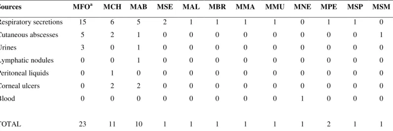
The distribution of specimens were: 34 respiratory secretions, 9
cutaneous abscesses, 4 urines, 4 corneal ulcers, 1 lymphatic
nodule, 1 peritoneal liquid and 1 blood. Strains were identified
by molecular methods as belonging to 12 different species: 23
M. fortuitum, 11 M. chelonae, 10 M. abscessus, 2 M. senegalense, 1 M. alvei, 1 M. brumae, 1 M. mageritense, 1 M. mucogenicum, 1 M. neoaurum, 1 M. peregrinum, 1 M. septicum and 1 M. smegmatis (Table 1).
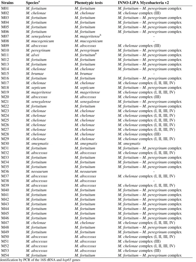
According to the phenotypic characteristics, 53 strains
(98.1%) were recognized as nonchromogen, though one
presented yellowish tonalities (M. brumae) and another produced a late orange pigment (M. smegmatis). Only one strain was considered scotochromogen for producing an orange
pigment (M. neoaurum).
Fifty-two out of 54 strains (96.3%) were correctly
identified by conventional methods. Biochemical tests results
are shown in Table 2. The two misidentified strains through
their biochemical profile were M. senegalense, which did not grow in mannitol and was identified as M. mageritense, and M. alvei, identified as M. fortuitum. INNO-LiPA Mycobacteria v2 assay accurately identified 47 strains (87.0%). The seven
García-Agudo, L. et al. Evaluation of INNO-LiPA Mycobacteria
accuracy for the identification of the rest of species was of
90.4%. Otherwise, two strains of M. chelonae were identified within the MCH-2 complex (III, M. abscessus) and five of M.
abscessus within the MCH-1 complex (I, II, III, IV, M. abscessus). Outcomes by phenotypical tests and by INNO-LiPA Mycobacteria v2 assay are shown in Table 3.
Table 1. Sources of isolation of the 54 rapidly growing mycobacteria strains.
Sources MFOa MCH MAB MSE MAL MBR MMA MMU MNE MPE MSP MSM
Respiratory secretions Cutaneous abscesses Urines Lymphatic nodules Peritoneal liquids Corneal ulcers Blood TOTAL 15 5 3 0 0 0 0 23 6 2 0 0 1 2 0 11 5 1 1 1 0 2 0 10 2 0 0 0 0 0 0 1 1 0 0 0 0 0 0 1 1 0 0 0 0 0 0 1 1 0 0 0 0 0 0 1 1 0 0 0 0 0 0 1 0 0 0 0 0 0 1 1 1 0 0 0 0 0 0 2 1 0 0 0 0 0 0 1 0 1 0 0 0 0 0 1 a
MFO = M. fortuitum, MCH = M. chelonae, MAB = M. abscessus, MSE = M. senegalense, MAL = M. alvei, MBR = M. brumae, MMA = M. mageritense, MMU
= M. mucogenicum, MNE = M. neoaurum, MPE = M. peregrinum, MSP = M. septicum, MSM = M. smegmatis
Table 2. Biochemical profile of the 54 rapidly growing mycobacteria strains.
Species Strains 42ºC NITa ARS URE TW80 NaCl MAC MAN INO SOR
M. abscessus M. alvei M. brumae M. chelonae M. fortuitum M. mageritense M. mucogenicum M. neoaurum M. peregrinum M. senegalense M. septicum M. smegmatis 10 1 1 11 23 1 1 1 1 2 1 1 - - - - vb + - - - + + - - + + - + + - + + + + + + + - + + + + - + + + - + + + + + + + + + + + + - + + vc vd - + + - - - + + - - - + + - - + + + + + - - - - + - - + - + + - - - - - - + + + + + + - - + - - - - + - - - + - - - - - - - - - - - - a NIT = nitrate reduction; ARS = arylsulfatase production; URE = urease production: TW80 = tween 80 hydrolysis; NaCl = tolerance to 5% NaCl; MAC = growth on MacConkey agar without crystal violet; MAN = mannitol utilization; INO = inositol utilization; SOR = sorbitol utilization
García-Agudo, L. et al. Evaluation of INNO-LiPA Mycobacteria
Table 3. Identification of the 54 rapidly growing mycobacteria strains by phenotypic tests and by INNO-LiPA Mycobacteria v2
assay.
Strains Speciesa Phenotypic tests INNO-LiPA Mycobacteria v2
M01 M02 M03 M04 M05 M06 M07 M08 M09 M10 M11 M12 M13 M14 M15 M16 M17 M18 M19 M20 M21 M22 M23 M24 M25 M26 M27 M28 M29 M30 M31 M32 M33 M34 M35 M36 M37 M38 M39 M40 M41 M42 M43 M44 M45 M46 M47 M48 M49 M50 M51 M52 M53 M54 M. fortuitum M. chelonae M. fortuitum M. fortuitum M. fortuitum M. fortuitum M. senegalense M. mucogenicum M. abscessus M. peregrinum M. alvei M. fortuitum M. fortuitum M. chelonae M. brumae M. fortuitum M. chelonae M. septicum M. mageritense M. abscessus M. senegalense M. fortuitum M. chelonae M. chelonae M. chelonae M. chelonae M. chelonae M. chelonae M. chelonae M. smegmatis M. fortuitum M. abscessus M. fortuitum M. fortuitum M. fortuitum M. neoaurum M. abscessus M. abscessus M. abscessus M. fortuitum M. fortuitum M. fortuitum M. fortuitum M. fortuitum M. fortuitum M. fortuitum M. chelonae M. fortuitum M. fortuitum M. abscessus M. abscessus M. abscessus M. abscessus M. fortuitum M. fortuitum M. chelonae M. fortuitum M. fortuitum M. fortuitum M. fortuitum
M. mageritenseb
M. mucogenicum M. abscessus M. peregrinum
M. fortuitumb
M. fortuitum M. fortuitum M. chelonae M. brumae M. fortuitum M. chelonae M. septicum M. mageritense M. abscessus M. senegalense M. fortuitum M. chelonae M. chelonae M. chelonae M. chelonae M. chelonae M. chelonae M. chelonae M. smegmatis M. fortuitum M. abscessus M. fortuitum M. fortuitum M. fortuitum M. neoaurum M. abscessus M. abscessus M. abscessus M. fortuitum M. fortuitum M. fortuitum M. fortuitum M. fortuitum M. fortuitum M. fortuitum M. chelonae M. fortuitum M. fortuitum M. abscessus M. abscessus M. abscessus M. abscessus M. fortuitum
M. fortuitum – M. peregrinum complex
M. chelonae complex (III)
M. fortuitum – M. peregrinum complex
M. fortuitum – M. peregrinum complex
M. fortuitum – M. peregrinum complex
M. fortuitum – M. peregrinum complex
- -
M. chelonae complex (III)
M. fortuitum – M. peregrinum complex
M. fortuitum – M. peregrinum complex
M. fortuitum – M. peregrinum complex
M. fortuitum – M. peregrinum complex
M. fortuitum – M. peregrinum complex
-
M. fortuitum – M. peregrinum complex
M. chelonae complex (I, II, III, IV)
M. fortuitum – M. peregrinum complex
M. chelonae complex (I, II, III, IV)
M. chelonae complex (III)
M. fortuitum – M. peregrinum complex
M. fortuitum – M. peregrinum complex
M. chelonae complex (I, II, III, IV)
M. chelonae complex (I, II, III, IV)
M. chelonae complex (I, II, III, IV)
M. chelonae complex (I, II, III, IV)
M. chelonae complex (I, II, III, IV)
M. chelonae complex (III)
M. chelonae complex (I, II, III, IV)
M. smegmatis
M. fortuitum – M. peregrinum complex
M. chelonae complex (I, II, III, IV)
M. fortuitum – M. peregrinum complex
M. fortuitum – M. peregrinum complex
M. fortuitum – M. peregrinum complex
-
M. chelonae complex (I, II, III, IV)
-
M. chelonae complex (I, II, III, IV)
M. fortuitum – M. peregrinum complex
M. fortuitum – M. peregrinum complex
M. fortuitum – M. peregrinum complex
M. fortuitum – M. peregrinum complex
M. fortuitum – M. peregrinum complex
M. fortuitum – M. peregrinum complex
M. fortuitum – M. peregrinum complex
M. chelonae complex (I, II, III, IV)
M. fortuitum – M. peregrinum complex
M. fortuitum – M. peregrinum complex
M. chelonae complex (I, II, III, IV)
M. chelonae complex (III)
M. chelonae complex (I, II, III, IV)
M. chelonae complex (III)
M. fortuitum – M. peregrinum complex
García-Agudo, L. et al. Evaluation of INNO-LiPA Mycobacteria
DISCUSSION
Rapidly growing mycobacteria are preferably found in the
environment, and rarely in clinic. From our experience, these
mycobacteria are more frequently isolated from respiratory
secretions and cutaneous abscesses (18, 19). The predominant
species of RGM in clinic are Mycobacterium fortuitum, M. chelonae and M. abscessus; the rest are occasionally found. The most of species belong to the nonchromogen group and to
the M. fortuitum complex, including M. fortuitum, M. peregrinum, M. mucogenicum, M. senegalense, M. alvei, M. houstonense, M. boenickei, M. conceptionense, M. porcinum, M. neworleansense and M. brisbanense (8). In our series we identified 12 different species, seven out of them belonged to
this group. We only found a scotochromogen strain of M. neoaurum species and none photochromogen.
The conventional mycobacteria identification according to
their phenotypical characteristics and biochemical behaviour
becomes complex but accessible to any laboratory and of a
great utility for RGM differentiation; however, it is not valid
for some species. For this reason, the use of phenotypic,
biochemical, chromatographic and molecular techniques all
together is recommended for their correct identification.
Sequencing of the ribosomal RNA 16S gene and the study of
the polymorphism of the restriction patterns for the
identification of RGM show an excellent result, although they
are labor-intensive and limited to reference laboratories as they
require sophisticated equipment. Alternative methods as
INNO-LiPA Mycobacteria v2 assay permit simultaneous
identification of 16 species of mycobacteria, including RGM of
M. fortuitum-peregrinum complex, M. chelonae and M. smegmatis, and are reliable for their use in clinical laboratories (4, 5, 7, 9, 14, 15).
In accordance with our results, INNO-LiPA Mycobacteria
v2 assay presents a sensitivity sensibility of 87.0% in the
identification of RGM. In our series there were two strains
uncorrectly identified (M. chelonae and M. mageritense), three strains did not hybridize (M. senegalense, M. mucogenicum
and M. abscessus) and other two could not be identified for not being included in the assay (M. brumae and M. neoaurum). We proved that the technique is well suited with mycobacteria
within the M. fortuitum complex, but it does not differentiate the species inside the group (15); in other respects, the assay
was poor discriminative with M. abscessus strains. Test results are better and sensitivity close to 100% in the identification of
M. tuberculosis complex and other slow growing mycobacteria (7, 9, 14).
In conclusion, we find INNO-LiPA Mycobacteria v2
assay of interest for the identification of the most frequent
RGM in clinic, it is simple to perform but provides few
advantages in comparison with conventional methods and
sometimes needs complementary tests to identify
Mycobacterium fortuitum complex, M. chelonae complex and specific species. Although the ITS has been shown to be more
discriminative than the 16S rRNA itself, the heterogeneity in
the RGM group requires either ITS or hsp65 gene sequencing for a more precise identification of species.
REFERENCES
1. Brunello, F.; Ligozzi, M.; Cristelli, E.; Bonora, S.; Tortoli, E.; Fontana, R. (2001). Identification of 54 mycobacterial species by PCR-restriction fragment length polymorphism analysis of the hsp65 gene. J. Clin. Microbiol.39, 2799-2806.
2. Cramer, J.P.; Sudeck, H.; Burchard, G.D. (2007). Pulmonary infection with rapidly growing mycobacteria in a singer with achalasia: a case report. J. Infect. 54, 219-221.
3. De Groote, M.A.; Uit, G. (2006). Infections due to rapidly growing mycobacteria. Clin. Infect. Dis. 42, 1756-1763.
4. Lebrun, L.; Weill, F.X.; Lafendi, L.; Houriez, F.; Casanova, F.; Gutierrez, C. et al. (2005). Use of the INNO-LiPA-Mycobacteria assay (version 2) for identification of Mycobacterium avium-Mycobacterium intracellulare-Mycobacterium scrofulaceum complex isolates. J. Clin. Microbiol. 43, 2567-2574.
5. Mäkinen, J.; Sarkola, A.; Marjamäki, M.; Viljanen, M.K.; Soini, H. (2002). Evaluation of GenoType and LiPA Mycobacteria assays for identification of finnish mycobacterial isolates. J. Clin. Microbiol. 40, 3478-3481.
García-Agudo, L. et al. Evaluation of INNO-LiPA Mycobacteria
following liposuction. Clin. Infect. Dis. 34, 1500-1507.
7. Padilla, E.; González, V.; Manterola, J.M.; Pérez, A.; Quesada, M.D.; Gordillo, S. et al. (2004). Comparative evaluation of the new version of the INNO-LiPA Mycobacteria and GenoType Mycobacterium assays for identification of Mycobacterium species from MB/BacT liquid cultures artificially inoculated with mycobacterial strains. J. Clin. Microbiol. 42, 3083-3088.
8. Schinsky, M.F.; Morey, R.E.; Steigerwalt, A.G.; Douglas, M.P.; Wilson, R.W.; Floyd, M.M. et al. (2004). Taxonomic variation in the
Mycobacterium fortuitum third-biovariant complex: Description of
Mycobacterium bonickei sp. nov., Mycobacterium houstonense sp. nov.,
Mycobacterium neworleansense sp. nov., Mycobacterium concordense
sp. nov., Mycobacterium brisbanense sp. nov., and recognition of
Mycobacterium porcinum from human clinical isolates. Int. J. Syst. Evol. Microbiol. 54, 1653-1667.
9. Suffys, P.N.; Da Silva Rocha, A.; De Oliveira, M.; Dias Campos, C.E.; Werneck Barreto, A.M.; Portaels, F.; Rigouts, L.; Wouters, G.; Jannes, G.; Van Reybroeck, G.; Mijs, W.; Vanderborght, B. (2001). Rapid identification of mycobacteria to the species level using INNO-LiPA Mycobacteria, a reverse hybridization assay. J. Clin. Microbiol. 39, 4477-4482.
10. Tortoli, E. (2003). Impact of genotypic studies on mycobacterial taxonomy: the new mycobacteria of the 1990s. Clin. Microbiol. Rev. 16, 319-354.
11. Kox, L.F.F.; Leeuwen, J.; Knijper, S.; Jansen, H.M.; Kolk, A.H.J. (1995). PCR assay based on DNA coding for 16S rRNA for detection and identification of mycobacteria in clinical samples. J. Clin. Microbiol. 33, 3225-3233.
12. Figueiredo, E.E.S.; Carvalho, R.C.T.; Silvestre, F.G.; Lilenbaum, W.; Fonseca, L.S.; Silva, J.T.; Paschoalin, V.M.F. (2010). Detection of
Mycobacterium bovis DNA in nasal swabs from tuberculous cattle by a multiplex PCR. Braz. J. Microbiol. 41, 386-390.
13. Marchi, A.M.; Juttel, I.D.; Kawacubo, E.M.; Dalmarco, E.M.; Blatt, S.L.; Cordova, C.M.M. (2008). Evaluation of methods for detection and identification of Mycobacterium species in patients suspected of having pulmonary tuberculosis. Braz. J. Microbiol. 39, 613-618.
14. Tortoli, E.; Mariottini, A.; Mazzarelli, G. (2003). Evaluation of INNO-LiPA Mycobacteria v2: Improved reverse hybridization multiple DNA probe assay for mycobacterial identification. J. Clin. Microbiol. 41, 4418–4420.
15. Trombert-Paolantoni, S.; Poveda, J.D.; Figarella, P. (2004). Comparison de deux techniques d’hybridation moléculaire dans l’identification de mycobactéries en pratique courante. Pathol. Biol. 52, 462-468.
16. Telenti, A.; Marchesi, F.; Balz, M.; Bally, F.; Botrger, E.C.; Bodmer, T. (1993). Rapid identification of mycobacteria to the species level by polymerase chain reaction and restriction enzyme analysis. J. Clin. Microbiol. 31,175-178.
17. Smet, K.A.L.; Brown, I.N.; Yates, M.; Ivanyi, J. (1995). Ribosomal internal transcribed spacer sequences are identical among Mycobacterium avium-intracellulare complex isolates from AIDS patients, but vary among isolates from elderly pulmonary disease patients. Microbiology. 141, 2739-2747.
18. Uslan, D.Z.; Kowalski, T.J.; Wengenack, N.L.; Virk, A.; Wilson, J.W. (2006). Skin and soft tissue infections due to rapidly growing mycobacteria: comparison of clinical features, treatment, and susceptibility. Arch. Dermatol. 142, 1287-1292.
19. Wallace, R.; Brown, B.; Griffith, D. (1998). Nosocomial outbreak/pseudo outbreaks caused by nontuberculous mycobacteria. Annu. Rev. Microbiol. 52, 453-490.