Restriction enzyme analysis of the
hsp
65 gene in
clinical isolates from patients suspected of having
pulmonary tuberculosis in Teresina, Brazil*
,**
Análise de restrição enzimática do gene hsp65 de isolados clínicos de pacientes com suspeita de tuberculose pulmonar em Teresina, Piauí
Maria das Graças Motta e Bona, Maria José Soares Leal, Liline Maria Soares Martins, Raimundo Nonato da Silva, José Adail Fonseca de Castro, Semiramis Jamil Hadad do Monte
Abstract
Objective: To identify mycobacterial species in the sputum of patients suspected of having pulmonary tuberculosis and to determine the impact that the acquisition of this knowledge has on the therapeutic approach. Methods: We evaluated 106 patients suspected of having pulmonary tuberculosis and referred to the pulmonology department of a public hospital in the city of Teresina, Brazil. Morning sputum specimens were evaluated for the presence of mycobacteria by sputum smear microscopy and culture. We used PCR and restriction enzyme analysis of the hsp65 gene (PRA-hsp65) to identify the strains of mycobacteria isolated in culture. Results: A total of 206 sputum samples were analyzed. Patient ages ranged from 15 to 87 years, and 67% were male. There was cough in 100% of the cases. The predominant radiographic pattern was moderate disease, observed in 70%. Smear positivity was 76%, and isolation in culture occurred in 91% of the cultures. Traditional tests identified nontuberculous mycobacteria (NTM) in 9% of the isolates. The PRA-hsp65 method confirmed these data, showing seven band patterns that were able to identify the isolated species of NTM: Mycobacterium kansasii; M. abscessus 1; M. abscessus 2; M. smegmatis; M. flavescens 1; M. gordonae 5; and M. gordonae 7. All of the patients with NTM were over 60 years of age, and bronchiectasis was seen in 88% of the X-rays. There were two cases of reinfection, initially attributed to M. abscessus and M. kansasii. Conclusions: In immunocompetent patients, NTM can infect the lungs. It is important to identify the specific NTM in order to establish the correct diagnosis and choose the most appropriate therapeutic regimen. The PRA-hsp65 method is useful in identifying NTM species and can be implemented in molecular biology laboratories that do not specialize in the identification of mycobacteria.
Keywords: Tuberculosis; Mycobacteria, atypical; Polymerase chain reaction; Brazil.
Resumo
Objetivo: Identificar as espécies de micobactérias encontradas no escarro de pacientes com suspeita de tuberculose pulmonar e analisar o impacto dessas identificações na abordagem terapêutica. Métodos: Foram avaliados 106 pacientes com suspeita de tuberculose pulmonar encaminhados para o serviço de pneumologia de um hospital público em Teresina, Piauí. Espécimes de escarro matinal foram avaliados quanto à presença de micobactérias por baciloscopia e cultura. Foram utilizadas PCR e análise de restrição enzimática do gene hsp65 (PRA-hsp65) para a identificação das cepas de micobactérias isoladas em cultura. Resultados: Foram analisadas 206 amostras de escarro. A idade dos pacientes variou de 15 a 87 anos, sendo 67% do gênero masculino. Tosse ocorreu em 100% dos casos. O padrão radiográfico predominante foi de lesão moderada, observada em 70%. A positividade no esfregaço foi de 76%, e isolamento em cultura ocorreu em 91% das culturas executadas. Testes tradicionais identificaram micobactérias não tuberculosas (MNT) em 9% dos isolados. O método PRA-hsp65 confirmou esses dados, mostrando sete padrões de bandas capazes de identificar as espécies de MNT isoladas: Mycobacterium kansasii; M. abscessus 1; M. abscessus 2; M. smegmatis; M. flavescens 1; M. gordonae 5 e M. gordonae 7. Todos os pacientes com MNT tinham mais de 60 anos, e observaram-se bronquiectasias em 88% das radiografias. Houve dois casos de reinfecção, identificados inicialmente como infecção por M. abscessus e M. kansasii. Conclusões:
As MNT causam infecção pulmonar em pacientes imunocompetentes, e a identificação das MNT é importante para estabelecer o diagnóstico correto e a decisão terapêutica mais adequada. O método PRA-hsp65 é útil para identificar espécies de MNT e pode ser implantado em laboratórios de biologia molecular não especializados em micobactérias.
Descritores: Tuberculose; Micobactérias atípicas; Reação em cadeia da polimerase; Brasil. * Study carried out at the Universidade Federal do Piauí – UFPI, Federal University of Piauí – Teresina, Brazil.
Correspondence to: Semiramis Jamil Hadad do Monte. Campus Ministro Petrônio Portela, Bloco 16, Bairro Ininga, CEP 64048-232, Teresina, PI, Brasil.
Tel. 55 86 32155691. Fax: 55 86 32155690. E-mail: [email protected]
Financial support: This study received financial support from the Federal University of Piauí Immunogenetics and Molecular Biology Laboratory.
Submitted: 6 October 2010. Accepted, after review: 29 April 2011.
essential to improving management and clinical outcomes.(9)
Microbiology is currently the gold standard for diagnosing mycobacterial infections. Identification of AFB in sputum smears is a simple, fast, low-cost technique; however, its sensitivity ranges from 30% to 80% depending on the isolation in culture, the local prevalence, and the methodology adopted.(10) In contrast,
sputum culture is a highly sensitive and specific method, albeit laborious and time-consuming (28-60 days to obtain a result).(11)
Various studies have suggested that molecular techniques are useful for the etiological diagnosis of TB. In 1993, one group of authors proposed a molecular method(12)
based on PCR and restriction enzyme analysis of the hsp65 gene (PRA-hsp65), a gene found in all mycobacterial species with species-specific nucleotide sequences. An effective, widely used, and easily reproducible method that does not require viable microorganisms, PRA-hsp65 allows the identification of various species of NTM in a single reaction.(9,12-14)
The accurate etiological diagnosis of mycobacterial infection at the species level is essential for the appropriate management of mycobacterial infections. Based on the hypothesis that PRA-hsp65 is able to differentiate between M. tuberculosis and NTM for clinical and therapeutic purposes, the objective of the present study was to identify the mycobacterial species isolated from the sputum of patients suspected of having pulmonary TB (PTB). We also attempted to determine the impact that the acquisition of this knowledge has on the therapeutic approach taken.
Methods
Between January and June of 2007, 106 patients suspected of having PTB were referred to the pulmonology department of a public hospital in the city of Teresina, located in the state of Piauí, Brazil. The inclusion criterion was being able to produce sputum spontaneously. The exclusion criteria were being under treatment with antituberculosis drugs at the time of sputum collection and having been diagnosed with extrapulmonary TB. The study project was approved by the Research Ethics Committee of the Federal University of Piauí (Protocol no. CAAE 0124.0.045.000-06),
Introduction
Tuberculosis (TB) remains one of the main public health problems in most countries. It is estimated that one third of the world population is infected with Mycobacterium tuberculosis. Brazil ranks 14th among the 22 countries with the highest incidence of TB, at 48 cases/100,000 population in 2007.(1) The increase in the
number of TB cases is mostly caused by poverty, extreme malnutrition, HIV infection, migration of infected people, inadequate control measures, inaccessibility to effective pharmacological treatment, and difficulty in monitoring the pharmacological treatment.(2)
Those factors have collectively prompted the spread of drug-resistant strains, including the multidrug-resistant forms.(2) The presence of
comorbidities, including HIV infection,(3) and
the delay in diagnosis are important concerns and should be targets of TB control strategies in Brazil because undiagnosed cases of TB are a major source of TB transmission.(4)
Since the advent of effective treatment for TB in the 1950s, the isolation of M. tuberculosis has become routine. Some of the cases initially described as TB infection were subsequently recognized as cases of infection with nontuberculous mycobacteria (NTM).
(5) Because these organisms are ubiquitous,
exposure to them is universal. There is as yet no evidence of animal-to-human transmission of NTM. In humans, NTM disease is assumed to be acquired through exposure to environmental factors, although a specific source has yet to be identified.(6,7) Among immunocompetent
patients, the incidence of pulmonary disease caused by NTM has been increasing worldwide.
(5,6) Prior to the AIDS pandemic, most cases of
lung disease in individuals with bronchiectasis, COPD, sequelae of TB, or pneumoconiosis were attributable to species belonging to the M. avium-intracellulare complex.(8)
step, the supernatant was transferred to a new tube. All centrifugation steps were carried out at 13,000 rpm in microcentrifuge tubes (Eppendorf, Hamburg, Germany).
A total of 5 µL of lysate were added to each reaction tube. The composition of the PCR mixture (50 µL) was 50 mM KCl, 10mM Tris-HCl (pH = 8.3), 1.5 mM MgCl2, 10% glycerol,
200 mM (each) deoxynucleoside triphosphate, 0.5 µM (each) primer, and 1.25 U of Taq polymerase (HotStarTaq Plus DNA Polymerase; QIAGEN, Düsseldorf, Germany). The reaction was subjected to 45 cycles of amplification (1 min at 94°C, 1 min at 60°C, and 1 min at 72°C); this was followed by 10 min of extension at 72°C. The primers Tb11 (5’-ACCAACGATGGTGTGTCCAT) and Tb12 (5’-CTTGTCGAACCGCATACCCT) amplified a 439-bp fragment between nucleotide positions 398 and 836 of the published gene sequence.(16)
Restriction analysis was performed using 15 µL of PCR products digested separately with 5 U of the restriction enzymes BstEII and HaeIII, in accordance with the manufacturer instructions (Promega Corporation, Madison, WI, USA). BstEII was digested by incubation at 60°C for 1.5 h, and HaeIII was digested by incubation at 37°C for 1.5 h.
Digestion products were mixed with 4 µL of gel loading buffer (0.25% bromophenol blue and 40% sucrose in water) and loaded onto gels prepared with 4% agarose in Tris-borate-EDTA buffer (89 mM Tris, 89 mM boric acid, and 2 mM EDTA; pH = 8.0). DNA size markers (50- and 25-bp DNA ladders) were applied in three lanes (two 50-bp lanes at the extremities of the gel and one 25-bp lane in the center). After electrophoresis, the gels were stained with ethidium bromide, visualized on a UV transilluminator, and photographed. The PRA-hsp65 patterns were interpreted with the aid of previously published tables.(17) Negative
and positive controls—water and M. tuberculosis H37Rv DNA (ATCC 27294; American Type Culture Collection, Manassas, VA, USA), respectively—were included in each experiment. The medical charts of the patients were reviewed in order to obtain information regarding previous diagnoses of TB, positive AFB results, and previous isolations of M. tuberculosis, NTM, or both.
in accordance with Brazilian National Health Council Resolution 196/1996. All of the patients gave written informed consent.
After performing clinical and radiographic evaluations, we collected spontaneous morning sputum samples (two from each patient). The samples were sent to the Maria José Leal Laboratory for Microbiological Diagnosis. Laboratory techniques (sputum smear microscopy and culture) were standardized in accordance with the norms and guidelines described in the Guidebook for Tuberculosis Bacteriology published by the Brazilian National Ministry of Health.(15) Ziehl-Neelsen-stained
smears were examined under microscopy for the semi-quantitative AFB counts and cord factor detection.(12,15) For the isolation of mycobacteria,
culture was performed on Löwenstein-Jensen medium.(15) When two cultures were positive,
the one with the most abundant growth of mycobacterial strains was selected for macroscopic, microscopic, and biochemical analyses, as well as for genotyping. Phenotyping of strains, for presumptive identification, included macroscopic screening (colony morphology and pigmentation), microscopic analysis (cord factor detection and identification of contaminating microorganisms), and biochemical analysis, the last performed with an M. tuberculosis identification kit (MTBAC; Probac do Brasil Produtos Bacteriológicos Ltda., São Paulo, Brazil). Strains with pigmented or smooth, fast-growing, nonphotochromogenic colonies that showed no cord factor under microscopy were presumptively classified as NTM.
Genotyping was performed by PRA-hsp65, as previously described,(12) in the Molecular Biology
and Immunogenetics Laboratory of the Federal University of Piauí.
Samples were prepared as follows: a loop of mycobacteria grown on Löwenstein-Jensen medium was suspended in 1 mL of Tris-EDTA buffer (10 mM Tris and 1 mM EDTA; pH = 8) and was heat-inactivated for 10 min at 80°C. After inactivation, the bacteria were centrifuged for 15 min. The pellet was resuspended in 100 µL of Tris-EDTA and transferred to a tube containing glass beads (Precellys®24; Bertin Technologies,
Montigny-le-Bretonneux, France). The suspended cells were mechanically disintegrated for 2 min in a Precellys®24 apparatus (Bertin
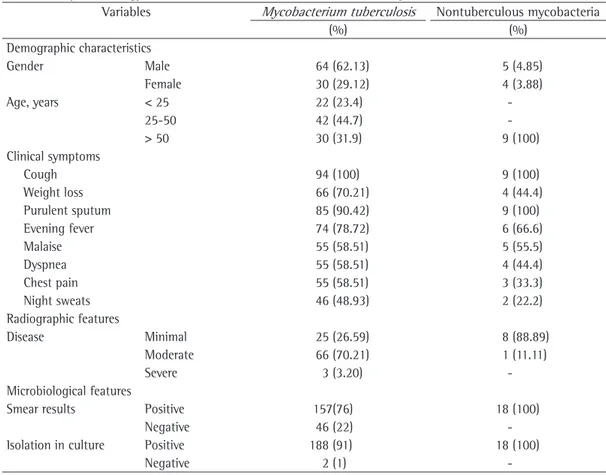
Only 2 patients were classified as cases of reinfection. The mean age was 46.8 years (range, 15-87 years), and males predominated (67%). The two most common symptoms were cough and purulent sputum. Table 1 shows the clinical, radiographic, and microbiological features of the patients.
The biochemical tests employed proved capable of identifying M. tuberculosis and NTM. Mean age was significantly higher among the patients in whom NTM were identified than among those in whom M. tuberculosis was identified (64.1 vs. 46.8 years; p < 0.05). There were no significant differences between the patients with M. tuberculosis and those with NTM regarding clinical symptoms or gender. However, all of the patients who presented with NTM also presented with radiological findings of bronchiectasis, AFB-positive smear, and isolation in culture. Of the isolates submitted to PRA-hsp65, 91.0% showed a BstEII or HaeIII For data analysis, we compared the
fragments obtained after enzyme treatment of amplicons with the restriction patterns stored in a PRA electronic database,(17) as well as with
the HaeIII and BstEII patterns, thus creating an algorithm for differentiating among NTM strains at the species level. After obtaining the clinical, radiographic, microbiological, and molecular results, we analyzed the data using the Statistical Package for the Social Sciences (SPSS Inc., Chicago, IL, USA).
Results
Of the 106 patients evaluated, 3 were excluded: 2 because the sputum samples were insufficient for analysis and 1 because of culture contamination. Of the 103 remaining patients, 94 were diagnosed with PTB on the basis of isolation of M. tuberculosis from sputum samples. Strains of NTM were isolated from the sputum samples of the remaining 9 patients.
Table 1 - Clinical, radiographic, and microbiological features of patients who were clinically suspected of having pulmonary infection with Mycobacterium tuberculosis or nontuberculous mycobacteria and were treated at a pulmonology referral center in Teresina, Brazil, from January to June of 2007.
Variables Mycobacterium tuberculosis Nontuberculous mycobacteria
(%) (%)
Demographic characteristics
Gender Male 64 (62.13) 5 (4.85)
Female 30 (29.12) 4 (3.88)
Age, years < 25 22 (23.4)
-25-50 42 (44.7)
-> 50 30 (31.9) 9 (100)
Clinical symptoms
Cough 94 (100) 9 (100)
Weight loss 66 (70.21) 4 (44.4)
Purulent sputum 85 (90.42) 9 (100)
Evening fever 74 (78.72) 6 (66.6)
Malaise 55 (58.51) 5 (55.5)
Dyspnea 55 (58.51) 4 (44.4)
Chest pain 55 (58.51) 3 (33.3)
Night sweats 46 (48.93) 2 (22.2)
Radiographic features
Disease Minimal 25 (26.59) 8 (88.89)
Moderate 66 (70.21) 1 (11.11)
Severe 3 (3.20)
-Microbiological features
Smear results Positive 157(76) 18 (100)
Negative 46 (22)
-Isolation in culture Positive 188 (91) 18 (100)
-Although the patient was not hospitalized, empirical treatment with the rifampin-isoniazid-pyrazinamide combination was administered for 2 months while we awaited the results of the molecular identification. Because the patient remained symptomatic, the rifampin-isoniazid-pyrazinamide combination was replaced by clarithromycin (500 mg/12 h for 18 months) and amikacin (15 mg • kg−1 • day−1 administered
intravenously every 12 h for 15 days), followed by the same doses of clarithromycin and amikacin administered on Mondays, Wednesdays, and Fridays for 2 months. After 18 months, the patient was asymptomatic and the culture results were negative. Therefore, the treatment was discontinued. Because of socioeconomic factors, the remaining 7 patients suspected of having TB were hospitalized and treated with the rifampin-isoniazid-pyrazinamide combination while we awaited the results of the sputum culture. Those patients also remained symptomatic. The results of molecular identification by the PRA-hsp65 method were as follows: M. gordonae, in 3 patients; M. flavescens, in 1; M. smegmatis, in 1; M. kansasii, in 1; and M. abscessus 2, in 1. Because of operational problems, it was impossible to carry out a sensitivity test; nevertheless, the therapeutic regimen was changed. For the 3 patients in whom M. gordonae was isolated in culture, the antibiotic regimen was changed to clarithromycin (500 mg/12 h). In the first month, the patients showed significant clinical improvement. Culture results were negative by the sixth month. The treatment lasted 12 months, after which the patients were completely asymptomatic. The patient in whom M. flavescens was isolated in culture showed no improvement during the first pattern for M. tuberculosis. The PRA-hsp65
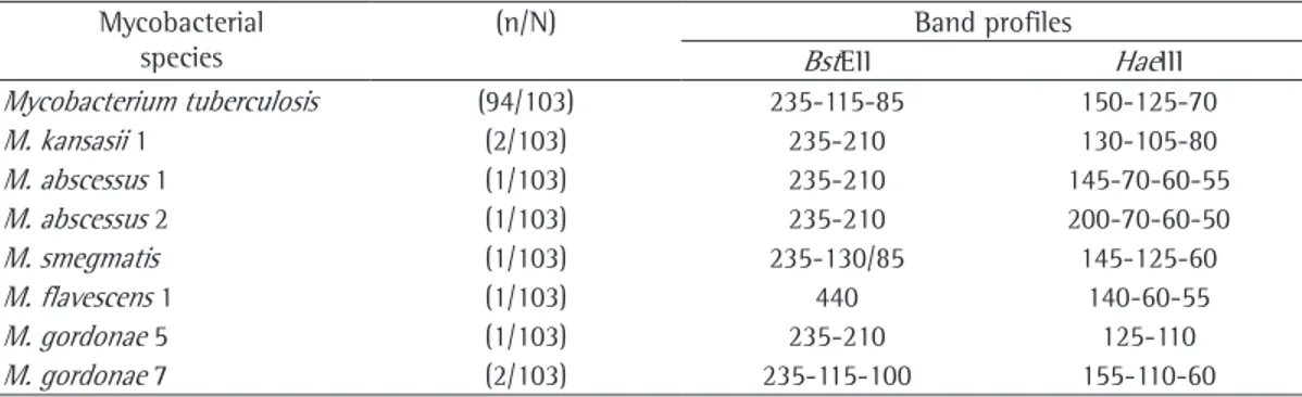
identified seven NTM strains: M. kansasii; M. abscessus 1; M. abscessus 2; M. smegmatis; M. flavescens 1; M. gordonae 5; and M. gordonae 7.(17) The band patterns are shown in Table 2. As
in the cases of primary infection, M. abscessus 1 and M. kansasii were identified in the sputum samples from the 2 patients with reinfection. All of the NTM strains identified by PRA-hsp65 were also identified as NTM by the p-nitrobenzoic acid test and the thiophene-2-carboxylic acid hydrazide test.
The standard treatment regimen (the rifampin-isoniazid-pyrazinamide combination) failed in 2 of the 103 patients under study. Those 2 patients had previously experienced treatment failure with ethionamide, pyrazinamide, ethambutol, and streptomycin. M. kansasii and M. abscessus 1 were isolated from the sputum samples collected from those patients. In addition, both species had been isolated from earlier sputum samples from those patients, supporting the diagnosis. The patient in whom M. kansasii was isolated in culture was treated with clarithromycin (500 mg p.o. every 12 h) and ethambutol for 12 months. Follow-up evaluation showed complete clinical remission. The patient became asymptomatic 1 year after treatment initiation and remained so after clarithromycin discontinuation. The patient in whom M. abscessus 1 was isolated showed no clinical response to any of the various therapeutic regimens that were initiated on the basis of susceptibility test results. The patient remained symptomatic until death, which occurred 5 years after the diagnosis of pulmonary disease caused by NTM. M. abscessus 2 was isolated from 1 patient.
Table 2 - Molecular identification, by PCR and restriction enzyme analysis of the hsp65 gene, of species of mycobacteria isolated in culture of clinical samples collected from patients with pulmonary tuberculosis treated at a pulmonology referral center in Teresina, Brazil, from January to June of 2007.
Mycobacterial species
(n/N) Band profiles
BstEII HaeIII
Mycobacterium tuberculosis (94/103) 235-115-85 150-125-70
M. kansasii 1 (2/103) 235-210 130-105-80
M. abscessus 1 (1/103) 235-210 145-70-60-55
M. abscessus 2 (1/103) 235-210 200-70-60-50
M. smegmatis (1/103) 235-130/85 145-125-60
M. flavescens 1 (1/103) 440 140-60-55
M. gordonae 5 (1/103) 235-210 125-110
The actual prevalence of NTM in Brazil has yet to be determined.(18-20) It is known
that NTM can be isolated from environmental sources, can colonize the mucosal surfaces of immunocompetent individuals, and can cause infection.(20) Consequently, isolating NTM only
once might not indicate infection. Therefore, it is mandatory to use criteria in order to differentiate between colonization and infection.(21)
A substantial increase in the occurrence of NTM as etiological agents of both pulmonary and cutaneous infections is expected, mainly among elderly individuals with COPD(7,22) and
cross infection. Studies have shown that NTM not only colonize but also pervade tissues, eventually causing infection.(23-25) However, it
is not easy to make this differentiation. The criteria for making this distinction have been standardized by the American Thoracic Society.
(7,26)
In our study, species that had previously been identified in the same patients (M. abscessus 1 and M. kansasii) were identified in only 2 cases, which were therefore characterized as cases of reinfection. It is of note that those patients were female, had not undergone any endoscopic procedure, and presented with bronchiectasis.
Good epidemiological control requires rapid and precise identification of the etiological agent. Therefore, in order to complement the conventional methods, it is crucial to develop new biochemical strategies for the identification of mycobacteria. In this context, PRA-hsp65 is an attractive option because it is fast, accurate, and reproducible. In addition, it can be performed in laboratories set up for microbiology and molecular biology. These features hasten test results and allow better management. Therefore, PRA-hsp65 can be a valuable addition to the array of diagnostic tests for mycobacterial infections. In this scenario, the incorporation of a molecular biology method to be performed in clinical diagnostic laboratories becomes increasingly relevant. The choice of therapeutic regimen for the treatment of TB should not be based exclusively on AFB smear results, because TB treatment is long and requires microorganism eradication in order to avoid the development and dissemination of multidrug-resistant forms deriving from an inappropriate initial treatment. In Brazil and other developing countries, in which the incidence of TB is higher, it is common 2 months of therapy. When PRA-hsp65 revealed
the presence of M. flavescens, the standard therapy was discontinued, and clarithromycin (500 mg p.o. every 12 h) was administered for 1 year. The patient achieved complete clinical remission within 1 month after treatment initiation and remained asymptomatic after the discontinuation of clarithromycin. The patients in whom M. smegmatis and M. kansasii were isolated in culture were treated with clarithromycin (500 mg p.o. every 12 h) and ethambutol (1,000 mg/day p.o.) for 12 months. All 7 patients achieved complete clinical remission within 1 year after treatment initiation. We can therefore affirm that all of the patients were infected, rather than colonized, given that they satisfactorily responded to the changes in the therapeutic regimen.
Discussion
In the present study, NTM accounted for 9% of the isolates, M. gordonae and M. kansasii being the most common strains. The maximum time elapsed between isolation in culture and species identification by PRA-hsp65 was 24 h, which is impossible when growth and chemical tests are used. The time required for those tests to provide definitive species identification ranges from several weeks to as long as 2 months, given that several of those tests require macroscopic growth of colonies in culture in order to be read.(9-11,13-15) In order to discriminate between
M. tuberculosis complex and NTM, we used the conventional methods (i.e., the p-nitrobenzoic acid test and the thiophene-2-carboxylic acid hydrazide test). The time required in order to obtain the results was 4 weeks.
Studies have identified new species of NTM, and more than 140 species are currently known, the identification of species by conventional techniques being therefore an arduous, time-consuming task that requires several laboratory tests and highly qualified human resources.(9,13)
References
1. World Health Organization. Global Tuberculosis Control: Epidemiology, Strategy, Financing: WHO Report 2009. Geneva: World Health Organization; 2009.
2. Maher D, Raviglione M. Global epidemiology of tuberculosis. Clin Chest Med. 2005;26(2):167-82, v. 3. Rao VK, Iademarco EP, Fraser VJ, Kollef MH. The impact
of comorbidity on mortality following in-hospital diagnosis of tuberculosis. Chest. 1998;114(5):1244-52. 4. Belo MT, Luiz RR, Hanson C, Selig L, Teixeira EG,
Chalfoun T, et al. Tuberculosis and gender in a priority city in the state of Rio de Janeiro, Brazil. J Bras Pneumol. 2010;36(5):621-5.
5. Runyon EH. Anonymous mycobacteria in pulmonary disease. Med Clin North Am. 1959;43(1):273-90. 6. Falkinham JO 3rd. Nontuberculous mycobacteria in the
environment. Clin Chest Med. 2002;23(3):529-51. 7. Griffith DE, Aksamit T, Brown-Elliott BA, Catanzaro
A, Daley C, Gordin F. An official ATS/IDSA statement: diagnosis, treatment, and prevention of nontuberculous mycobacterial diseases. Am J Respir Crit Care Med. 2007;175(4):367-416. Erratum in: Am J Respir Crit Care Med. 2007;175(7):744-5.
8. Hadad DJ, Palhares MC, Placco AL, Domingues CS, Castelo Filho A, Ferrazoli L, et al. Mycobacterium avium complex (MAC) isolated from AIDS patients and the criteria required for its implication in disease. Rev Inst Med Trop Sao Paulo. 1995;37(5):375-83.
9. Tortoli E. Impact of genotypic studies on mycobacterial taxonomy: the new mycobacteria of the 1990s. Clin Microbiol Rev. 2003;16(2):319-54.
10. Sommers HM, McClatchy JK. Laboratory Diagnosis of the Mycobacterioses. Washington, D.C.: American Society for Microbiology, 1983.
11. Kent PT, Kubica GP. Public Health Mycobacteriology: A Guide for the Level III Laboratory. Atlanta: U.S. Dept. of Health and Human Services, Public Health Service, Centers for Disease Control; 1985.
12. Telenti A, Marchesi F, Balz M, Bally F, Böttger EC, Bodmer T. Rapid identification of mycobacteria to the species level by polymerase chain reaction and restriction enzyme analysis. J Clin Microbiol. 1993;31(2):175-8. 13. Leão SC, Bernardelli A, Cataldi A, Zumarraga M, Robledo
J, Realpe T, et al. Multicenter evaluation of mycobacteria identification by PCR restriction enzyme analysis in laboratories from Latin America and the Caribbean. J Microbiol Methods. 2005;61(2):193-9.
14. Devallois A, Goh KS, Rastogi N. Rapid identification of mycobacteria to species level by PCR-restriction fragment length polymorphism analysis of the hsp65 gene and proposition of an algorithm to differentiate 34 mycobacterial species. J Clin Microbiol. 1997;35(11):2969-73.
15. Brasil. Ministério da Saúde. Centro de Vigilância em Saúde. Centro de Referência Prof. Hélio Fraga. Manual de Bacteriologia da Tuberculose. Rio de Janeiro: Ministério da Saúde, Centro de Vigilância em Saúde, Centro de Referência Prof. Hélio Fraga, Departamento de Vigilância Epidemiológica, Coordenação Geral de Laboratórios de Saúde Pública; 2005.
16. Shinnick TM. The 65-kilodalton antigen of Mycobacterium tuberculosis. J Bacteriol. 1987;169(3):1080-8.
practice to start patients on antituberculosis drugs before the identification of mycobacteria, particularly in cases of positive sputum smear. However, physicians should be alert to the possibility that there might be no clinical response, given that NTM are common, as shown in the present study. The correct identification of NTM species is crucial, because other, specific antimicrobials will have to be chosen on the basis of identification and susceptibility test results.(19,27) In this context, PRA-hsp65 is the
most appropriate method because it can identify the NTM species within 24 h after their isolation in culture. In contrast, the time required for the conventional method of identifying the NTM species (which is based on growth and chemical properties) is over 30 days. Because PRA-hsp65 yields results more rapidly, the appropriate therapeutic regimen can be started more rapidly.
In conclusion, the present study showed that the implementation of PRA-hsp65 for the identification of mycobacteria in a nonspecialized molecular biology laboratory is feasible. Although it is recognized that PRA-hsp65 should not be considered a replacement for other tests that are used in order to diagnose TB, PRA-hsp65 constitutes an additional diagnostic tool to investigate the etiology of the disease, especially in individuals with no clinical response to antituberculosis drugs. The method can contribute to a faster identification of mycobacteria in public laboratories, thus optimizing management.
Acknowledgments
from: ftp://ftp.cve.saude.sp.gov.br/doc_tec/tb/MNT_ Final_9-12-05a.pdf
22. Guide SV, Holland SM. Host susceptibility factors in mycobacterial infection. Genetics and body morphotype. Infect Dis Clin North Am. 2002;16(1):163-86.
23. Tanaka E, Amitani R, Niimi A, Suzuki K, Murayama T, Kuze F. Yield of computed tomography and bronchoscopy for the diagnosis of Mycobacterium avium complex pulmonary disease. Am J Respir Crit Care Med. 1997;155(6):2041-6.
24. Moore EH. Atypical mycobacterial infection in the lung: CT appearance. Radiology. 1993;187(3):777-82. 25. Jeong YJ, Lee KS, Koh WJ, Han J, Kim TS, Kwon OJ.
Nontuberculous mycobacterial pulmonary infection in immunocompetent patients: comparison of thin-section CT and histopathologic findings. Radiology. 2004;231(3):880-6.
26. Ueki SY, Martins MC, Telles MA, Virgilio MC, Giampaglia CM, Chimara E, et al. Micobactérias não-tuberculosas: diversidade das espécies no estado de São Paulo. J Bras Patol Med Lab. 2005;41(1):1-8.
27. Koh WJ, Kwon OJ, Lee KS. Nontuberculous mycobacterial pulmonary diseases in immunocompetent patients. Korean J Radiol. 2002;3(3):145-57.
17. Prasite [homepage on the Internet]. Lausanne: Hospices Cantonaux; c1999 [updated 2007 Sep 15; cited 2010 Aug 12]. Available from: http://app.chuv.ch/prasite/ index.html.
18. Matos ED, Santana MA, de Santana MC, Mamede P, de Lira Bezerra B, Panão ED, et al. Nontuberculosis mycobacteria at a multiresistant tuberculosis reference center in Bahia: clinical epidemiological aspects. Braz J Infect Dis. 2004;8(4):296-304.
19. Pedro HS, Pereira MI, Goloni MR, Ueki SY, Chimara E. Nontuberculous mycobacteria isolated in São José do Rio Preto, Brazil between 1996 and 2005. J Bras Pneumol. 2008; 34(11):950-5.
20. Zamarioli LA, Coelho AG, Pereira CM, Nascimento AC, Ueki SY, Chimara E. Descriptive study of the frequency of nontuberculous mycobacteria in the Baixada Santista region of the state of São Paulo, Brazil. J Bras Pneumol. 2008;34(8):590-4.
21. Centro de Vigilância Epidemiológica “Alexandre Vranjac” [homepage on the Internet]. São Paulo: Secretaria da Saúde do Estado de São Paulo. [cited 2006 Jul 15]. Micobacterioses: recomendações para o diagnóstico e tratamento. [Adobe Acrobat document, 29p.] Available
About the authors
Maria das Graças Motta e Bona
Adjunct Professor. Universidade Federal do Piauí – UFPI, Federal University of Piauí – Teresina, Brazil.
Maria José Soares Leal
Professor. Universidade Federal do Piauí – UFPI, Federal University of Piauí – Teresina, Brazil.
Liline Maria Soares Martins
Assistant Professor. Universidade Estadual do Piauí – UESPI, Piauí State University – Teresina, Brazil.
Raimundo Nonato da Silva
Molecular Biologist. Immunogenetics and Molecular Biology Laboratory, Universidade Federal do Piauí – UFPI, Federal University of Piauí – Teresina, Brazil.
José Adail Fonseca de Castro
Researcher, Immunogenetics and Molecular Biology Laboratory, Universidade Federal do Piauí – UFPI, Federal University of Piauí – and Adjunct Professor II, Universidade Estadual do Piauí – UESPI, Piauí State University – Teresina, Brazil.
Semiramis Jamil Hadad do Monte