RBCCV 44205-1619
DOI 10.5935/1678-9741.20140113
Mycobacterial endocarditis: a comprehensive review
Endocardite micobacteriana: uma revisão abrangente
Shi-Min Yuan
1, MMed, PhD
1The First Hospital of Putian, Teaching Hospital, Fujian Medical University,
Putian, People’s Republic of China.
This study was carried out at First Hospital of Putian, Teaching Hospital, Fujian Medical University, Putian, Fujian Province, People’s Republic of China.
No inancial support.
Correspondence address: Shi-Min Yuan
Longdejing Street, 389 - Chengxian District, Putian, Fujian Province, Peo-ple’s Republic of China
E-mail: [email protected]
Article received on April 24th, 2014
Article accepted on September 30th,2014 Abstract
Objective: A systematic analysis was made in view of the epidemiology, clinical features, diagnosis, treatment and main outcomes of mycobacterial endocarditis.
Methods: The data source of the present study was based on a comprehensive literature search in MEDLINE, Highwire Press and Google search engine for publications on mycobacte-rial endocarditis published between 2000 and 2013.
Results: The rapidly growing mycobacteria become the pre-dominant pathogens with Mycobacterium chelonae being the most common. This condition has changed signiicantly in terms of epidemiology since the 21st century, with more broad patient age range, longer latency, prevailed mitral valve infections and better prognosis.
Conclusion: Mycobacterial endocarditis is rare and the causative pathogens are predominantly the rapidly growing mycobacteria. Amikacin, ciproloxacin and clarithromycin are the most frequently used targeted antimicrobial agents but often show poor responses. Patients with deep infections may warrant a surgical operation or line withdrawal. With period-ic multidrug therapy guided by drug susceptibility testing, and surgical managements, patients may achieve good therapeutic results.
Descriptors: Heart Valves. Endocarditis. Mycobacterium.
Resumo
Objetivo: Uma análise sistemática foi feita considerando epi-demiologia, quadro clínico, diagnóstico, tratamento e principais resultados da endocardite micobacteriana.
Métodos: Foi realizada uma pesquisa bibliográica abran -gente no MEDLINE, Highwire Press e no Google para publi-cações sobre endocardite micobacteriana, publicados entre 2000 e 2013.
Resultados: As micobactérias de crescimento rápido tor-nam-se os patógenos predominantes, com Mycobacterium che-lonae sendo a mais comum. Essa condição se alterou signiica -tivamente em termos de epidemiologia, desde o início do século 21, abrangendo faixa etária mais ampla, maior latência, preva -lecendo infecções da valva mitral e melhor prognóstico.
Conclusão: Endocardite micobacteriana é rara e os pató-genos causadores são predominantemente as micobactérias de crescimento rápido. Amicacina, ciproloxacina e claritromicina são os agentes antimicrobianos mais frequentemente utilizados, mas muitas vezes apresentam respostas pobres. Pacientes com infecções profundas podem justiicar um procedimento cirúr -gico ou retirada de linha. Com a poliquimioterapia periódica guiada por testes de sensibilidade às drogas, e abordagens cirúr -gicas, os pacientes podem obter bons resultados terapêuticos.
INTRODUCTION
Cardiac disorders, pregnancy and other surgical maneu-vers can be risk factors of bacterial infective endocardi-tis[1-4]. Increasingly utilization of foreign medical materials,
indwelling catheter insertions and intravenous drug uses are recognized risk factors predisposing to bacterial infec-tive endocarditis of the present era[5]. Continuous changes
in terms of epidemiology and management strategies of the bacterial infective endocarditis have been elucidated[5].
Staphylococcus aureus has become the most common mi-croorganism of the bacterial infective endocarditis particu-larly associated with increasing foreign material implant[6],
while Streptococcus viridans infections reduced[5].
How-ever, there is no updated elaboration on recent changes of mycobacterial endocarditis.
Mycobacterial endocarditis is rare. It showed a sig-niicant predilection of non-tuberculous over tuberculous mycobacteria in terms of infective endocarditis. Due to more resistant to antimicrobial therapies than other patho-gens, mycobacteria are often refractory to antimicrobial treatments and are associated with a very high mortality[7].
Rapid-growth non-tuberculous mycobacteria including Mycobacterium (M.) chelonae, M. abscessus and M. fortu-itum accounted for 68% of the isolates[8] and thus being the
predominant mycobacteria for the infections. There have been systemic reviews on infective endocarditis caused by M. fortuitum[7,9] in 2002, and by M. abscessus[10] and
M. chelonae[11] in recent years. Due to the rarity, regular
management strategies are still scanty. As for the dificulty of pathogen identiications, poor responses to antimicro-bial therapy and poor prognosis, this condition remains a challenge with regard to diagnosis and treatment. However, mycobacterial endocarditis has not been suficiently elabo-rated. The present study is designed to highlight the clinical pictures of mycobacterial endocarditis based on relevant literature information published since 2000.
METHODS
MEDLINE, Highwire Press and Google search engine were searched for publications in the English language on mycobacterial endocarditis from 2000 to 2013. The terms “mycobacteria”, “heart valve”, “heart valve prosthesis”, “tu-berculous”, “non-tuberculous” and “endocarditis” were used for the searches. All the articles, titles and subject headings were screened carefully for potential relevance. Articles were
Abbreviations, acronyms & symbols
AVR Aortic valve replacement M. Mycobacterium MVR Mitral valve replacement
included if the patient had an established diagnosis of myco-bacteria endocarditis on current admission and outcome data were reported.
Due to the rarity of the condition, all the discovered ar-ticles reported only sporadic single or small series; no large population, comparative studies were retrievable. Therefore, data from this systematic review were qualitatively analyzed as suggested in the Quality of Reporting of Meta-Analyses recommendations.
The search identiied 31 relevant studies from 2000 to 2013[7,9-38], including 24 case reports[7,9,10,12,13,15,17-22,24-28,30-33,36-38],
2 case series[14,23], 2 original articles[11,34], 1 medical
imag-ing[16], 1 poster abstract session[29], and 1 “letter to the
edi-tor”[35]. After reviewing selected articles, all 31 articles were
included and no one was excluded. Data were extracted from the text, igures, or tables and included details of the study population, demographics, types of mycobacteria, sites of infections, locations of vegetations, latency, sensitivity, anti-microbial spectrum, management strategies, clearance time, follow-up length and main outcomes (survivals, complica-tions, relapses, reinterventions and mortality).
Quantitative data were presented as mean±standard de-viation, and intergroup differences were compared by un-paired t-test. Comparisons of frequencies were performed by Fisher’s exact test. P<0.05 was considered statistically signiicant.
RESULTS
Demographics
The patient setting included 50 patients with mycobacte-rial endocarditis. There were 29 males and 21 females with a male-to-female ration of 1.38:1. Their ages were 45.9±19.8 (range, 0.5-78; median 50) years (n=50). Age distribution of the patients conformed to the normal distribution by proba-bility–probability plot.
Clinical features
The major symptoms on admission were described in 46 patients including fever in 35 (76.1%)[7,9-29,33,37,38],
dys-pnea in 10 (21.7%)[10,11,31,32] and chest pain in 1 (2.1%)
pa-tient[30] (χ2=60.7, P=0.000). The duration of the symptoms
was 4.0±4.4 (range, 0.17-18; median, 2) months (n=31). The temperature of the febrile patients were 38.9±0.8℃ (n=12). Of the febrile patients, fever grade was not indicated in 22[11, 14,18,23,24,26,28,29,33,35,36,38]. In the remaining 13 patients, 5 (38.5%)
had a high fever[12,17,19,20,22,29], 5 (38.5%) had a moderate
fe-ver[13,15,21,25,34] and 3 (23.1%) had a low-grade fever[9,27,37]
(χ2=0.9, P=0.630). Of them, 4 were prolonged fever[24,35,36]
and 2 were fever of unknown origin[14,16].
in 2[13,18], a systolic murmur in the aortic region[36], a
pansys-tolic murmur at the apex + a diaspansys-tolic murmur in the aortic region[25], a pansystolic murmur at the apex + a systolic
mur-mur in the aortic region[15], a systolic murmur at the lower left
sternal boarder[37] and an unspeciied cardiac murmur[32] in 1
patient each) and an absence of a cardiac murmur in 3 (20%) patients[20,22,24] (χ2=10.8, P=0.001).
Laboratory examinations revealed their hemoglobin was 9.9±1.8 (range, 5.9-12.5; median, 10) g/L (n=10)[9,10,12,13,15,18,20, 27,30,36,38], white blood cell count was 8.9±4.4 (range, 3.3-16.8;
median, 8.2) ×109/L (n=13)[9,10,13,15,20-22,24,27,28,30,33,38], platelet
count was 165.6±128.7 (range, 67-450; median, 124)×109/L
(n=7)[10,13,15,18,20,27,38], C-reaction protein was 6.8±4.7 (range,
0.14-11; median, 8) mg/dL (n=4)[10,20,27,28], and CD4 was
240.5 ± 206.3 (range, 44-587; median 196)/mm3 (n=6) [9,10,13,14,19,37]. CD4 was normal (>500/mm3) in 1 (16.7%)
pa-tient, and was abnormal (<500/mm3) in 5 (83.3%) patients
(χ2=5.3, P=0.021).
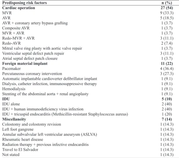
Predisposing risk factors
Cardiac surgery, foreign material implant, intravenous drug use and miscellaneous risk factors were the underly-ing etiologies for the development of infective endocarditis (χ2=31.9, P=0.000) (Table 1). Of the causative cardiac
opera-tions, 23 (85.2%) were heart valve operations and 4 (14.8%) were congenital atrial/ventricular defect patch repairs (χ2=26.7, P=0.000). There were 22 (95.7%) valve
replace-ments and 1 (4.3%) valve repair (χ2 = 38.4, P=0.000).
Eigh-teen (81.8%) were single valve replacements, and 4 (18.2%) were double valve replacements (χ2=17.8, P=0.000); and 17
(77.3%) were irst time valve replacements and 5 (22.7%) were redo operations (χ2=13.1, P=0.001). A total of 26 valves
were replaced including 13 (50%) aortic and 13 (50%) mitral valve replacements; 20 (80%) were biological, and 5 (20%) were mechanical valve prostheses (χ2 = 18, P=0.000), except
the one whose detail of aortic valve prosthesis was not given.
Table 1. Predisposing risk factors. Predisposing risk factors Cardiac operation MVR
AVR
AVR + coronary artery bypass grafting Composite AVR
MVR + AVR Redo-MVR + AVR Redo-AVR
Mitral valve ring plasty with aortic valve repair Ventricular septal defect patch repair
Atrial septal defect patch closure Foreign material implant Pacemaker
Percutaneous coronary intervention
Automatic implantable cardioverter deibrillator implant
Dialysis, catheter infection, immunosuppressive therapy Hemodialysis
Stenting of the abdominal aorta + renal angioplasty IDU
IDU alone
IDU + human immunodeiciency virus infection
IDU + tricuspid endocarditis (Methicillin-resistant Staphylococcus aureus) Miscellaneity
Colostomy and colostomy revision Left foot gangrene
Annular subvalvular left ventricular aneurysm (ASLVA) Rheumatic heart disease
Radiation therapy + previous infective endocarditis Travel to El Salvador
Not stated
n (%) 27 (54) 9 (33.3) 5 (18.5) 1 (3.7) 1 (3.7) 1 (3.7) 3 (11.1)
2 (7.4) 1 (3.7) 3 (11.1)
1 (3.7) 11 (22) 4 (36.4) 3 (27.3) 1 (9.1) 1 (9.1) 1 (9.1) 1 (9.1) 5 (10) 2 (40) 2 (40) 1 (20) 7 (14) 1 (14.3) 1 (14.3) 1 (14.3) 1 (14.3) 1 (14.3) 1 (14.3) 1 (14.3)
The latency from the presence of the predisposing risk fac-tors to symptom onset was 21.9±25.9 (range, 0.067-96; medi-an, 12) months (n=31)[7,9,11,12,14-18,20,21,23,26,27,31-33,37]. There were 9
(29.0%) early onsets (latency <8 weeks), 2 (6.5%) intermedi-ate onsets (lintermedi-atency was between 8 weeks and 8 months) and 20 (64.5%) late onsets (latency >8 months) (χ2=23.9, P=0.000).
The latency of the patients with cardiac operations was much shorter than that of the patients with a foreign material im-plant, but did not reach a statistical signiicance (16.4±20.1 months vs. 34.8±39.0 months, P=0.136). It was incompati-ble with intravenous drug use patients, of which latency was reported in only one patient, and was much shorter than that of the miscellaneity reaching a quasi-statistical difference (16.4±20.1 months vs. 44±34.6 months, P=0.051) (Figure 1). Bioprosthetic valve endocarditis was associated with a longer latency than mechanical without showing a signiicant differ-ence (17.5±22.5 months vs. 10.8±7.1 months, P=0.569). Four patients had a delayed diagnosis for 0.81±0.24 (range, 0.5-1; median, 0.88) months (n=4)[12,13,20,26].
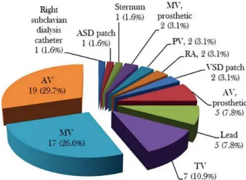
Infection sites
The infection sites could be divided into 5 types according the location and number of the mycobacterial infections: sin-gle intracardiac infection in 38 (76%), two intracardiac infec-tions in 9 (18%), and triple valve infecinfec-tions, single intracardiac + single extracardiac infections, and double intracardiac + sin-gle extracardiac infections in 1 (2%) patient, each (χ2=128.5,
P=0.000). Including extracardiac infections associated with the endocarditis, totally 64 sites were affected with a mean of 1.28±0.54 (range, 1-3; median, 1) infection sites per patient. There were 1.24±0.48 infection sites in the non-tuberculous and 1.75±0.96 infection sites in the tuberculous endocarditis
patients (P=0.067). Native aortic, mitral and tricuspid valves were the most commonly affected sites of mycobacterial en-docarditis, representing 29.7%, 26.6% and 10.9%, respec-tively (Figure 2). No difference was found in the prevalence of infection sites between non-tuberculous and tuberculous mycobacterial endocarditis (Table 2), or in the strain distribu-tions between aortic and mitral valves (Table 3).
Vegetations were detected in 44 patients with 43 detected by echocardiography and 1 patient detected by positron emis-sion tomography with 18F-luorodeoxyglucose uptake[16]. Of
them, 12 (27.3%) patients did not have a visualized vegeta-tion[7,9,11,14,17,28,31], but one of them with an abscess along the
in-ferior and septal walls, instead[31] and 32 (72.7%) patients had
(χ2=18.2, P=0.000). The vegetation locations of the
remain-ing 32 patients were mitral valve in 10 (31.3%)[11,20,21,30,36,37]
(two of them were in the prosthetic mitral valve)[20,21],
aor-tic valve in 7 (21.9%)[11,13,18,32,33,38] (one was in the prosthetic
aortic valve)[33], pacemaker lead in 3 (12.5%)[16,26,27], tricuspid
valve in 3 (9.4%)[10,19,35], both mitral valve and right
subclavi-an catheter in 1 (3.1%)[36], both mitral and aortic valves in 2
(6.3%)[12,15], both aortic and tricuspid valves in 2 (6.3%)[22,24],
mitral, aortic and tricuspid valves in 1 (3.1%)[25], tricuspid
valve and ventricular septal defect patch in 2 (6.3%)[35] and
atrial septal patch in 1 (3.1%) patient[23], respectively. The
de-tection time for a positive vegetation was described in 3 pa-tients, which was 5[13], 21[20] and 105 days[15] after admission,
respectively. Dimensions of the vegetations were recorded in 16 patients for 17 vegetations. Five vegetations of 5 patients from a single report[11] were recorded as “minimal”, which
were excluded from the calculation of the vegetation size. The size of the remaining 12 vegetations of 11 patients was 19.7±18.4 (range, 5-70; median, 15.5) mm[10,15,18,20,23-26,30,32,37].
Fig. 2 - Distribution of infection sites.
ASD=atrial septal defect; AV=aortic valve; MV=mitral valve; RA=right atrium; PV=pulmonary valve; TV=tricuspid valve; VSD=ventricular septal defect
Valve insuficiency was present in 23 (46%) patients: mitral valve regurgitation in 9 (39.1%)[11,30,31,37] (6 were prosthetic
mitral valve leaks[11]), aortic valve regurgitation in 5 (21.7%) [11,28,32] (4 were prosthetic aortic valve leaks[11,32]), aortic and
mitral valve regurgitation in 5 (21.7%)[11,12,14,38] (2 were
pros-thetic aortic and mitral valve leaks[11]) and tricuspid valve
regurgitation in 4 (17.4%) patients[10,27,35]. There were totally
11 (47.8%) native valve regurgitations and 12 (52.2%) pros-thetic valve leaks (χ2=0.1, P=0.768). Besides, one patient had
inferior and septal wall abscess associated with mitral valve regurgitation[31] and one patient had ventricular septal patch
dehiscence[35].
Pathogens
The initial blood culture results were not indicated in 5 patients[29,34]. In the remaining 45 patients, a negative culture
prevailed, followed by an acid-fast bacterium (Table 4). Thirty-seven patients had more investigations performed for strain identiications. Fourteen patients had more sam-ples than blood for cultures: 7 (50%) patients had one more sample, 4 (28.6%) patients had 2 more samples, 2 (14.3%) patients had 3 more samples and 1 (7.1%) patient had 4 more samples for cultures, respectively. There were totally 25 ad-ditional samples for cultures including 4 (16%) intraopera-tive excised valves[12,18], or valve prosthesis[7], or prosthetic
valve ring[14], 4 (16%) resected vegetations[27,30,35], 3 (12%)
sputum[27,28,34], 2 (8%) bronchoalveolar lavage[14,27], 2 (8%)
urine[14,27], 2 (8%) bone marrow[14,37], 1 (4%) removed patch[35],
removed pacing lead[26], intraoperative specimens (with no
details available)[34], pacemaker generator pocket site[26],
as-pirated luid[26], sternum[7], cerebrospinal luid[37] and tracheal
swab[15] for each, respectively. Lowenstein–Jensen medium
was once used for valve, sternum and blood cultures[7]. Three
(12%) were negative and 22 (88%) were positive (χ2=28.8,
P=0.000). The 22 positive cultures identiied the strains to be
M. fortuitum in 7 (31.8%), M. chimaera in 4 (18.2%), M. ab-scessus in 3 (13.6%), M. chelonae in 2 (9.1%), M. neoaurum
in 1 (4.5%), acid-fast bacilli in 2 (9.1%), rapidly growing mycobacteria in 1 (4.5%) and tubercule bacilli in 2 (9.1%), respectively. At least 15 (68.2%) cases were rapid growing mycobacteria infections.
Seventeen patients (four of them had additional samples for cultures) had a positive histological staining results by Ziehl-Neelsen and auramine-rhodamine stains[12,17,18,27] with
acid-fast organisms shown on luorescence microscopy. Ten patients had a molecular analysis of the mycobacte-ria, where 16s ribosomal deoxyribonucleic acid sequencing was applied in 4 (40%)[14,20,21,23], reverse line blot
hybridiza-tion in 4 (40%)[29] and a polymerase chain reaction-restriction
fragment length polymorphism analysis in 2 (20%) patients
[10,33], respectively. All patients had fresh samples, and one
of them had additional cryopreserved samples for investiga-tions. However, analyses on the cryopreserved samples dis-closed negative results, while all fresh specimens displayed positive results. In addition, two patients had mycolic acid analysis by biochemical and chromatographic techniques[7,9].
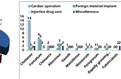
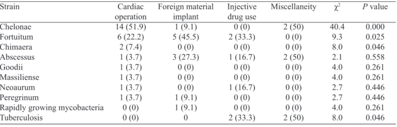
By preliminary blood cultures, histological staining, mo-lecular analyses and chromatographic techniques, the eventu-al mycobacterieventu-al strains were identiied in eventu-all but one patient (Figure 3). Distributions of the mycobacteria responding to the four predisposing risk factors showed cardiac operation was associated with more, prevailed rapidly growing myco-bacteria (M. chelonae, M. fortuitum and M. chimaera) en-docarditis, foreign material implant was associated with M. fortuitum, and intravenous drug use and miscellaneity were prone to be of tuberculous endocarditis (Figure 4, Table 5).
According to the antimicrobial spectrum, intravenous ami-kacin 300 mg twice daily, ciproloxacin 400-500 mg twice daily and clarithromycin 500 mg twice daily were the most frequently used targeted antimicrobial agents, and imipenem
Table 2. A comparison of infection sites between non-tuberculous and tuberculous mycobacterial infective endocarditis
Infection site
Aortic valve Mitral valve
Aortic valve, prosthetic Lead
Tricuspid valve Mitral valve, prosthetic Pulmonary valve VSD patch ASD patch Right atrium
Right subclavian dialysis catheter Sternum
Tuberculous (n = 8) 3 (37.5)
2 (25) 0 (0) 0 (0) 2 (25)
0 (0) 0 (0) 0 (0) 0 (0) 1 (12.5)
0 (0) 0 (0)
ASD=atrial septal defect; VSD=ventricular septal defect
Non-tuberculous (n = 56) 16 (28.6) 15 (26.8) 5 (8.9) 5 (8.9) 5 (8.9) 2 (3.6) 2 (3.6) 2 (3.6) 1 (1.8) 1 (1.8) 1 (1.8) 1 (1.8)
χ2
0.3 0.0 1.4 1.4 1.9 0.5 0.5 0.5 0.3 2.7 0.3 0.3
P value
Fig. 4 - Distributions of the mycobacteria responding to the four predisposing risk factors.
Fig. 3 - Eventual mycobacterial strains.
Table 3. A comparison of pathogens between aortic and mitral valve endocarditis.
Pathogen
Chelonae Fortuitum Abscessus Chimaera Peregrinum
Rapidly growing mycobacteria Goodii
Neoaurum Tuberculous
Mitral valve (n=20) 11 (55) 1 (5) 3 (15)
1 (5) 0 (0) 0 (0) 1 (5) 1 (5) 2 (10) Aortic valve
(n=22) 8 (36.4) 6 (27.3) 3 (13.6) 1 (4.5) 1 (4.5) 1 (4.5) 0 (0) 0 (0) 2 (9.1)
χ2
1.5 3.7 0.0 0.0 1.4 1.4 1.7 1.7 0.0
P value
0.226 0.053 0.900 0.945 0.235 0.235 0.199 0.199 0.920
Table 4. Initial blood culture results.
Initial blood culture Negative
Acid-fast bacteria Gram-positive bacilli/rod Atypical mycobacterial infection Non-tuberculous mycobacteria Rapidly growing mycobacteria Mycobacterium species Fortuitum
Abscessus Neoaurum Chimaera Peregrinum Tuberculous
n (%) 18 (40)
9 (20) 5 (11.1)
1 (2.2) 1 (2.2) 1 (2.2) 1 (2.2) 4 (8.9) 1 (2.2) 1 (2.2) 1 (2.2) 1 (2.2) 1 (2.2)
References [11,14,25,27,28,30] [7,10,13,22,23,35,36]
[15,20,26,31,33] [37] [38] [34] [21] [9,12,17,24]
(500 mg/6 hours), linezolid, rifampicin and trimethoprim/sul-famethoxazole 160 mg/800 mg (p.o., thrice daily) were more frequently used for non-tuberculous mycobacterial infection; while ethambutol (20 mg/kg/day), isoniazid (10 mg/kg/day), pyrazinamide (25 mg/kg/day) and rifampicin (10 mg/kg/day) were for tuberculous mycobacterial infection. The antimyco-bacterial course was life in 2 patients[18,35]. In another 19 patie
nts[10-13,22-24,26,28,33,34], the treatment course was indicated, which
was 2.7±2.3 (range, 0.83-9; median, 1.67) months. The clear-ance time interval was 31.8±58.4 (range, 2-210; median, 12.5) days (n=12)[12,13,15,19-21,23,34,35].
Prognosis
Patients were at a follow-up of 48.7±39.8 (range, 3-116; median, 32) months (n=23)[9,11,17,19-21,23,26,30,32,34]. Totally 27 (50%)
patients were event-free survivals[12,16,17,19,20,22-24,26,28,30,33-35], 3
(6.25%) patients relapsed at 0.5, 5 and 10 months, respective-ly[9,10,18], 4 (6.25%) patients were complicated[21,25,29,32] and one
of them required reintervention[32], which constituted the only
reintervention of the whole setting, and 17 (34%) patients died[11-15,18,27,29,31,36-38] at 83.9±85.9 (range, 1-270; median, 64)
days (n=11). The death causes were described in 8 patients, which were multiorgan failure in 2[12,15], and candidaemia and
hospital-acquired pneumonia[36], persistent mycobacteremia
and stroke[29], progressive heart failure[14], respiratory distress[37],
splenic rupture[14] and variceal bleeding[31] in 1 patient, each.
Two patients did not receive either medical or surgical treatment, but had a good prognosis in each. Of the remain-ing 48 patients, 32 (66.7%) patients received an antimicrobi-al therapy antimicrobi-alone, 10 (20.8%) patients had a cardiac operation and 6 (12.5%) patients had an intervention for removal of catheter/lead/deibrillator (χ2=36.8, P=0.000).
In the patients with medical treatment, there were 17 event-free survivals[11,19,22,30,34,35], 11 deaths[11,13,14,29,31,37,38],
2 complicated (spleen infarct, renal infarct and cerebral abscess on day 10, and prosthetic valve endocarditis due to coagulase negative staphylococcal species at 1 year in one patient[21]; and periaortic abscess in another[29]) and 2
Table 5. Pathogens corresponding to predisposing risk factors.
Strain Chelonae Fortuitum Chimaera Abscessus Goodii Massiliense Neoaurum Peregrinum
Rapidly growing mycobacteria Tuberculosis Cardiac operation 14 (51.9) 6 (22.2) 2 (7.4) 1 (3.7) 1 (3.7) 1 (3.7) 1 (3.7) 1 (3.7) 0 (0) 0 (0) Injective drug use 0 (0) 2 (33.3) 0 (0) 1 (16.7) 0 (0) 0 (0) 1 (16.7) 0 (0) 0 (0) 2 (33.3) Foreign material implant 1 (9.1) 5 (45.5) 0 (0) 3 (27.3) 0 (0) 0 (0) 0 (0) 1 (9.1) 1 (9.1) 0 Miscellaneity 2 (50) 0 (0) 0 (0) 2 (50) 0 (0) 0 (0) 0 (0) 0 (0) 0 (0) 2 (50) χ2 40.4 9.3 8.0 2.1 4.0 4.0 2.7 2.7 4.0 8.0
P value
0.000 0.025 0.046 0.558 0.261 0.261 0.446 0.446 0.261 0.046
relapses[9,10]. In the surgical treatment group, all patients
received a valve replacement operation, including 2 aor-tic valve replacements (AVRs), 1 AVR with root replace-ment, 1 redo-AVR, 1 2nd redo-AVR, 2 AVRs + mitral valve
replacements (MVRs), 1 AVR + MVR + tricuspid valvu-loplasy, 1 2nd redo-AVR + MVR and 1 redo-MVR. There
were 5 event-free survivals[12,17,20,28,33], 3 deaths[12,18,29], 2
complicated (1 preoperative stroke and 1 complication re-lated with his previous ascending aortic replacement)[25,32],
1 relapse[18] and 1 reintervention[32]. In the patients with a
catheter/lead/deibrillator removal, there were 3 event-free survivals[16,24,26] and 3 deaths[15,27,36]. Time of deaths was
de-scribed in 11 patients. There were 4 (36.4%) early deaths and 7 (63.6%) late deaths (χ2=1.6, P=0.201).
DISCUSSION
In 1959, Runyon[39], according to pigment of colony and
rate of growth, divided mycobacteria into 4 types: photo-chromogen, scotophoto-chromogen, nonphotochromogen and rapid grower. The latter one, rapidly growing mycobacterium, was deined so because the mycobacterial colonies form at 25-45˚C on solid agar in 5-7 days. Both slowly and rapidly growing mycobacteria are environmental opportunistic mycobacteria that are normal inhabitants of natural waters, drinking water and soils[40]. The most important slowly growing species are
M. avium and M. intracellulare, called the M.avium complex; and rapidly growing mycobacteria (M. abscessus, M. chelonae
and M. fortuitum), which are opportunistic pathogens[40].
Mycobacterial endocarditis is rare. In a recent report of infective endocarditis incorporating information from 13 tertiary hospitals in Turkey, no myocabacterial endocarditis was reported[41]. Wallace et al.[42] stated that the incidence of
mycobacterial endocarditis was 33.3% (4/12) out of M. for-tuitum or M. chelonae bacteria and was 3.2% of rapid growth mycobacterial blood infections. Olalla et al.[7] reviewed 19
native and 15 prosthetic valve endocarditis, with a biological valve prosthesis in 6 and a mechanical valve prosthesis in 9 patients.
The degree of fever may not correspond to the severity of the illness[43]. As it was illustrated in the present article, no
dominance of fever grade was associated with mycobacterial endocarditis. Strabelli et al.[11] reported that the latent for the
febrile onset and development of valve dysfunction was few days and months to years, respectively. In 5 of 13 patients with prosthetic mycobacterial endocarditis, the latent peri-od for the diagnosis of bioprosthetic endocarditis was 1-4 years[11]. Olalla et al.[7] reported a latency of 12 weeks from
infection to symptom onset with no difference found between biological and mechanical prosthetic valve endocarditis. The present study revealed a more broad patient age range from infant to 78 years old with a normal age distribution, equality of affected aortic and mitral valves, more native than pros-thetic valve and more bioprospros-thetic than mechanical biopros-thetic endocarditis. Moreover, cardiac operations caused a shorter latency to endocarditis occurrence than foreign ma-terial implant and miscellaneous risk factors. Bioprosthetic valve endocarditis was associated with a longer latency than mechanical but lack of a signiicant difference. Kunin et al.[9]
reported a prevailed early onset of mycobacterial endocar-ditis. Valve dysfunction was noted in 10 (76.9%) patients, while only 3 (23.1%) patients were free of valve dysfunc-tion (χ2=12.5, P=0.001). The present study demonstrated a
predominant late onset and a decreased incidence of valve dysfunction.
The most common predisposing risk factors for myco-bacterial infections include medical procedures (central ve-nous access, hemodialysis catheter indwelling, various sur-gical operations including mammoplasty, arthroplasty and cardiothoracic operations)[9], immunocompromised,
particu-larly patients with human immunodeiciency virus/acquired immunodeiciency syndrome, hematological malignancies, or those treated with immunosuppressive drugs after solid or-gan transplantation[44]. More recently, the use of tumor
necro-sis factor-α blockers has become an additional risk factor[45].
Line-related non-tuberculous infections did not have any pre-dominant organisms including M. abscessus, M. chelonae,
M. neoaurum, M. fortuitum and M. mucogenicum[34]. Further,
possible manufacturer contamination of bioprosthesis by M. chelonea has been recognized[46]. In addition, nosocomial
in-fections were once evidenced from sources like cardioplegic solutions[47], water supply[48], antiseptic solutions[49] and valve
prosthesis preservation solutions[50].
Positivity of blood cultures varied according to types of affected valves. It was 75% in mechanical, 20% in biologi-cal and 100% in native valves[7]. Removed valve prostheses
showed a high positivity of mycobacterial cultures[48]. Upon
colonial growth, gram and acid-fast stains should be per-formed, and then when blood cultures are negative, acid-fast
bacilli stains on histological examination of the removed prosthetic valves[11] and Ziehl-Neelsen stain should be
em-ployed, followed by subcultures[40].
Mycobacteria are more resistant to chemical disinfec-tion than other pathogens[11]. Infective endocarditis caused
by rapidly growing mycobacteria is always refractory due to extensive drug resistance and substantially delayed diagno-sis[36]. The poor response to therapy can be a result of
var-ious factors that may lead to drug resistance, including the presence of bioilms, type of antimicrobial agent used and presence of a novel inducible erm(41) gene[51]. With
peri-odic multidrug therapy guided by drug susceptibility testing, patients may achieve good therapeutic results[51]. Therefore,
antimicrobial susceptibility tests of the isolates are of im-portant clinical signiicance[36]. The management of rapidly
growing mycobacteria is usually an empiric therapy with two agents and a successive targeted regimen according to drug sensitivity tests[9]. Amikacin was the most reliable agent for
the treatment of non-tuberculous mycobacteria and alterna-tive effecalterna-tive agents included ciproloxacin, clarithromycin, imipenem and linezolid[34]. Non-tuberculous mycobacteria are
more sensitive to some antibiotics than other rapidly growing mycobacteria[32]. Clarithromycin was proved to be sensitive
to M. abscessus/chelonae. Moxiloxacin was discovered to be the best to treat M. fortuitum infections, very active against M. chelonae when used alone and more effective against all the strains when combined with clarithromycin and amika-cin[52]. A combined use of clarithromycin with moxiloxacin
or linezolid at a high concentration (16 μg/ml) also displayed activity against M. abscessus[51]. Disseminated infections with
atypical mycobacteria often develop in immunocompromised patients with reduced CD4 count and complement levels[37]. A
combined antimicrobial therapy with prolonged treatment du-ration is often the regimen of choice. Amikacin and imipenem have been proved to be effective agents for the disseminated cases[32].
Patients with deep infections of rapidly growing myco-bacteria often warrant a surgical intervention including line removal, debridement, or removal of the foreign material[34].
An improved survival was advocated to be associated with surgical interventions[33], whereas some patients with
conser-vative managements failed to survive[13]. In the patient series
of Olalla et al.[7] reported in 2002, 15 of 19 cases of M.
for-tuitum complex (M. fortuitum and M. chelonae) endocarditis were prosthetic valve endocarditis and valve replacement was performed in 8 (42.1%) patients with an overall mortality of 88%. Recently, Strabelli et al.[11] reported 13 patients with M.
chelonae endocarditis received a valve replacement and an overall mortality was decreased to 23.1% (3/13) including 1 (7.7%) early and 2 (15.4%) late deaths. Exceptionally, a few patients did not receive any antimicrobial treatment and pa-tients were at long-term event-free survival[11]. These results
Authors’ roles & responsibilities
SMY Main Author
risk factor for infective endocarditis, and usage of higher dose and combined antimicrobial regimens with reference to the mycobacterial species and surgical treatment can lead to improved outcomes.
The rarity of this condition as well as the relevant pub-lished materials made this study a limited patient population. Abundant valuable date in the future may replenish the re-search with more accurate results.
CONCLUSION
Mycobacterial endocarditis is rare but dreadful. The rap-idly growing mycobacteria become the predominant patho-gens with chelonae being the most common. This condition has changed signiicantly in terms of epidemiology since the 21st century, with more broad patient age range, longer
la-tency, prevailed mitral valve infections and better prognosis. The better prognoses than before might be attributed to the faster strain identiication molecular techniques, higher dose of antimicrobial agents, periodic multidrug therapy guided by drug susceptibility testing and more requirements of sur-gical interventions.
REFERENCES
1. Yuan SM, Demesthenous E, Coman V. Long QT syndrome in extensive infective endocarditis complicating hypertrophic
obstructive cardiomyopathy. Kardiol Pol. 2009;67(1):53-7.
2. Yuan SM, Jing H. The bicuspid aortic valve and related disorders. Sao Paulo Med J. 2010;128(5):296-301.
3. Yuan SM, Wu B, Hu XN, Jing H. Axillary artery thrombus and infective endocarditis in lupus. J Chin Med Assoc. 2011;74(1):40-3.
4. Al Jama PE. Obstetric outcome of teenage pregnancies: a 5-year
experience in a university hospital. Kuwait Med J. 2012;44:195-9.
5. Yuan SM. Right-sided infective endocarditis: recent epidemiologic changes. Int J Clin Exp Med. 2014;7(1):199-218.
6. Vaccarino GN, Nacinovich F, Piccinini F, Mazzetti H, Segura E, Navia D. Pacemaker endocarditis: approach for lead extraction
in endocarditis with large vegetations. Rev Bras Cir Cardiovasc. 2009;24(4):570-3.
7. Olalla J, Pombo M, Aguado JM, Rodríguez E, Palenque E, Costa JR, et al. Mycobacterium fortuitum complex endocarditis-case report and literature review. Clin Microbiol Infect. 2002;8(2):125-9.
8. Matos ED, Santana MA, de Santana MC, Mamede P, de Lira Bezerra B, Panão ED, et al. Nontuberculosis mycobacteria at a multiresistant tuberculosis reference center in Bahia: clinical epidemiological aspects. Braz J Infect Dis. 2004;8(4):296-304.
9. Kunin M, Salamon F, Weinberger M, Genkin I, Sagie A, Tur-Kaspa R. Conservative treatment of prosthetic valve endocarditis
due to Mycobacterium fortuitum. Eur J Clin Microbiol Infect Dis. 2002;21(7):539-41.
10. Tsai WC, Hsieh HC, Su HM, Lu PL, Lin TH, Sheu SH, et al. Mycobacterium abscessus endocarditis: a case report and
literature review. Kaohsiung J Med Sci. 2008;24(9):481-6.
11. Strabelli TM, Siciliano RF, Castelli JB, Demarchi LM, Leão SC, Viana-Niero C, et al. Mycobacterium chelonae valve endocarditis resulting from contaminated biological prostheses. J Infect. 2010;60(6):467-73.
12. Collison SP, Trehan N. Native double-valve endocarditis by Mycobacterium fortuitum following percutaneous coronary intervention. J Heart Valve Dis. 2006;15(6):836-8.
13. Spell DW, Szurgot JG, Greer RW, Brown JW 3rd. Native valve endocarditis due to Mycobacterium fortuitum biovar fortuitum: case report and review. Clin Infect Dis. 2000;30(3):605-6.
14. Achermann Y, Rössle M, Hoffmann M, Deggim V, Kuster
S, Zimmermann DR, et al. Prosthetic valve endocarditis and bloodstream infection due to Mycobacterium chimaera. J Clin Microbiol. 2013;51(6):1769-73.
15. Al-Benwan K, Ahmad S, Mokaddas E, Johny M, Kapoor MM.
Diagnosis of endocarditis caused by Mycobacterium abscessus. Ann Saudi Med. 2010;30(5):408-11.
16. Amraoui S, Texier-Maugein J, Bordachar P. PET scan in suspected but unproven pacemaker endocarditis. Arch Cardiovasc Dis. 2012;105(2):125-6.
17. Bosio S, Leekha S, Gamb SI, Wright AJ, Terrell CL, Miller DV. Mycobacterium fortuitum prosthetic valve endocarditis: a
case for the pathogenetic role of bioilms. Cardiovasc Pathol.
2012;21(4):361-4.
18. Corrales-Medina V, Concha R, Simkins J, Sanchez M, Baracco G. Native valve endocarditis caused by rapidly growing mycobacteria: case report and review of the literature. Scand J Infect Dis. 2007;39(6-7):639-41.
Bacterial endocarditis: a role for Mycobacterium tuberculosis? AIDS. 2002;16(13):1845-6.
20. Jönsson G, Rydberg J, Sturegård E, Christensson B. A case of Mycobacterium goodii prosthetic valve endocarditis in a non-immunocompromised patient: use of 16S rDNA analysis for rapid diagnosis. BMC Infect Dis. 2012;12:301.
21. Kumar A, Pazhayattil GS, Das A, Conte HA. Mycobacterium
neoaurum causing prosthetic valve endocarditis: a case report and review of the literature. Braz J Infect Dis. 2014;18(2):235-7.
22. Natsag J, Min Z, Hamad Y, Alkhalil B, Rahman A, Williams R. A mysterious gram-positive rods. Case Rep Infect Dis. 2012;2012:841834.
23. Sadanandan R, Kuriakose KM, Ramanarayanan PV, Philomina
B. Mycobacterium massiliense: an emerging pathogen in cardiovascular infections. Asian Cardiovasc Thorac Ann. 2013;21(1):77-8.
24. Shah P, Vishnevsky A. Mycobacterium fortuitum device infection with subsequent endocarditis. Med Forum 2012;13:Article 13 [Accessed Apr 5 2014]. Available at: http://jdc.jefferson.edu/tmf/ vol13/iss1/13
25. Shaikh Q, Mahmood F. Triple valve endocarditis by mycobacterium tuberculosis: a case report. BMC Infect Dis. 2012;12:231.
26. Sharma S, Tleyjeh IM, Espinosa RE, Costello BA, Baddour LM. Pacemaker infection due to Mycobacterium fortuitum. Scand J Infect Dis. 2005;37(1):66-7.
27. Sharma H, Keshavan A, Little MA, Cross J, Lipman
MC, Talukdar S, et al. Fortuitous vasculitis. Ren Fail. 2012;34(3):378-82.
28. Sogabe O, Ohya T. A case of tuberculous endocarditis with acute
aortic valve insuficiency and annular subvalvular left ventricular
aneurysm. Gen Thorac Cardiovasc Surg. 2007;55(2):61-4.
29. Soman R, Gupta N, Shetty A, Rodrigues C. 1732. Prosthetic endocarditis due to nontuberculous Mycobacteria: a new problem in the developing world. Session: Poster Abstract Session: Tuberculosis and other Mycobacterial Infections. Saturday, October 20, 2012. Room: SDCC Poster Hall F-H [Accessed Apr 5 2014]. Available at: https://idsa.confex.com/idsa/2012/ webprogram/Paper35271.html
30. Sultan FA, Fatimi S, Jamil B, Moustafa SE, Mookadam F. Tuberculous endocarditis: valvular and right atrial involvement. Eur J Echocardiogr. 2010;11(4):E13.
31. Takekoshi D, Al-Heeti O, Belvitch P, Schraufnagel DE. Native-valve endocarditis caused by Mycobacterium chelonae,
misidentiied as polymicrobial gram-positive bacillus infection.
J Infect Chemother. 2013;19(4):754-6.
32. Torres-Duque CA, Díaz C, Vargas L, Serpa EM, Mosquera W, Garzón MC, et al. Disseminated mycobacteriosis affecting a
prosthetic aortic valve: irst case of Mycobacterium peregrinum
type III reported in Colombia. Biomedica. 2010;30(3):332-7.
33. Vail G, Kohler R, Steiner F, Donepudi R. Successful treatment
of Mycobacterium fortuitum prosthetic valve endocarditis: case report. Clin Infect Dis. 2000;30(3):629-30.
34. van Duin D, Goldfarb J, Schmitt SK, Tomford JW, Tuohy MJ,
Hall GS. Nontuberculous mycobacterial blood stream and cardiac infections in patients without HIV infection. Diagn Microbiol Infect Dis. 2010;67(3):286-90.
35. Vuković D, Parezanović V, Savić B, Dakić I, Laban-Nestorović S, Ilić S, et al. Mycobacterium fortuitum endocarditis
associated with cardiac surgery, Serbia. Emerg Infect Dis. 2013;19(3):517-9.
36. Williamson JC, Miano TA, Morgan MR, Palavecino EL.
Fatal Mycobacterium abscessus endocarditis misidentiied as
Corynebacterium spp. Scand J Infect Dis. 2010;42(3):222-4.
37. Liebeskind DS, Ostrzega N, Wasterlain CG, Buttner EA. Neurologic manifestations of disseminated infection with Mycobacterium abscessus. Neurology. 2001;56(6):810-3.
38. Jagadeesan N, Patra S, Singh AP, Nagesh CM, Reddy B, Badnur SC, et al. Spontaneous endocarditis caused by rapidly growing non-tuberculous Mycobacterium chelonae in an immunocompetent patient with rheumatic heart disease. J Cardiovasc Dis Res. 2013;4(4):254-6.
39. Runyon EH. Anonymous mycobacteria in pulmonary disease. Med Clin North Am. 1959;43(1):273-90.
40. Falkinham JO 3rd. Nontuberculous mycobacteria in the environment. Clin Chest Med. 2002;23(3):529-51.
41. Elbey MA, Akdağ S, Kalkan ME, Kaya MG, Sayın MR, Karapınar H, et al. A multicenter study on experience of 13
tertiary hospitals in Turkey in patients with infective endocarditis.
Anadolu Kardiyol Derg. 2013;13(6):523-7.
42. Wallace RJ Jr, Swenson JM, Silcox VA, Good RC, Tschen JA, Stone MS. Spectrum of disease due to rapidly growing mycobacteria. Rev Infect Dis. 1983;5(4):657-79.
43. Tunkel AR. Fever in adults [Accessed Apr 5 2014]. Available at: http://www.merckmanuals.com/home/infections/biology_of_ infectious_disease/fever_in_adults.html
44. van Ingen J. Diagnosis of nontuberculous mycobacterial infections. Semin Respir Crit Care Med. 2013;34(1):103-9.
46. Tyras DH, Kaiser GC, Barner HB, Laskowski LF, Marr JJ.
Atypical mycobacteria and the xenograft valve. J Thorac Cardiovasc Surg. 1978;75(3):331-7.
47. Wallace RJ Jr, Musser JM, Hull SI, Silcox VA, Steele LC, Forrester GD, et al. Diversity and sources of rapidly growing mycobacteria associated with infections following cardiac surgery. J Infect Dis. 1989;159(4):708-16.
48. Kuritsky JN, Bullen MG, Broome CV, Silcox VA, Good RC,
Wallace RJ Jr. Sternal wound infections and endocarditis due to organisms of the Mycobacterium fortuitum complex. Ann Intern Med. 1983;98(6):938-9.
49. Fraser DW. Bacteria newly recognized as nosocomial pathogens.
Am J Med. 1981;70(2):432-8.
50. Laskowski LF, Marr JJ, Spernoga JF, Frank NJ, Barner HB, Kaiser
G, et al. Fastidious mycobacteria grown from porcine prosthetic-heart-valve cultures. N Engl J Med. 1977;297(2):101-2.
51. Jarand J, Levin A, Zhang L, Huitt G, Mitchell JD, Daley CL. Clinical and microbiologic outcomes in patients receiving treatment for Mycobacterium abscessus pulmonary disease. Clin Infect Dis. 2011;52(5):565-71.