Mafalda X. Henriques
Dissertation presented to obtain the Ph.D degree in Biology
Instituto de Tecnologia Química e Biológica | Universidade Nova de Lisboa
Oeiras, December, 2012
Studies of the assembly of the Streptococcus
pneumoniae capsular polysaccharide
Mafalda X. Henriques
First edition, December 2012
Cover image
Left, upper panel: Localization of Wze-Citrine, expressed from the
natural chromosomal locus of the
wze gene, in encapsulated
Streptococcus pneumoniae cells.
Right, lower panel: Detection of the capsular polysaccharide, by
immunofluorescence, on the Wze null mutant.
Back image: Detection of the capsular polysaccharide, by
Aos meus pilares,
From left to right: Professor Mário Ramirez, Professor Leonilde
Moreira, Professor Carlos Romão, Doctor Rut Carballido-López, Mafalda
X. Henriques, Doctor Sérgio Filipe, Professor Adriano Henriques
December, the 17th of 2012
Supervisor: Professor Carlos Romão
Main opponents: Professor Mário Ramirez
Doctor Rut Carballido-López
Acknowledgments
The development of a PhD thesis is never an individual process.
Many people contribute to its construction, either by suggesting
experiments, discussing ideas, keeping us focused on the problem at hand
or simply by listening to our complaints after a bad day of work. To all the
people that accompanied me in this journey, I would like to declare my
acknowledgments.
I will start by thanking my supervisor, Dr. Sérgio Filipe. When I
joined the Bacterial Cell Surfaces and Pathogenesis laboratory I was not
sure I wanted, or had the necessary characteristics, to enroll in a PhD. I
was lucky to be given a very interesting and exciting project, and soon I
was completely involved with it. For that opportunity and for all his
guidance and ideas throughout these years, I would like to thank him.
I would like to thank the members of my Thesis Committee, Dr.
Mariana Pinho and Dr. Ana Rute Neves. Dr. Mariana Pinho has
accompanied my work very closely and her suggestions have no doubt
been extremely helpful, as well as the different perspective and
suggestions of Dr. Ana Rute Neves.
I deeply thank my lab colleagues at the Bacterial Cell Surfaces
and Pathogenesis laboratory. To Magda Atilano, for her sincere
friendship, advices and help in the lab. A month after we met I was going
to be away from the lab for a couple of days and you said you would miss
having me around. That meant a lot to me and I still remember it. I miss
you already! I thank Tatiana Rodrigues for all her help in the lab but
mostly for her friendship, her joy and the good moments and laughs we
shared. I truly think that without you I would not been able to endure part
of this thesis. Thank you very much. The lab is not the same since you
present, for providing a good work environment.
I also thank my colleagues from the Bacterial Cell Biology
laboratory. Ana Jorge, thank you for your friendship, for sharing your
knowledge with me and for teaching me new protocols. I think we hit it of
very quickly and I truly miss having you around. I thank Pedro Matos for
his ideas, willingness to help and sense of humor. I thank Patricia Reed,
for sharing all her knowledge and tips with me when I was lost in
unexpected results. I also thank all other members of the Bacterial Cell
Biology group, past and present.
I thank all my friends and colleagues from other labs. It was a
pleasure and an honor to share this time with you.
I greatly thank Teresa Baptista da Silva for all her help in the lab.
You have made our life so much easy, thank you very much!
I thank Instituto Tecnologia Química e Biológica for the
ooportunity to work and develop my project in an institute with such
excellent conditions. ITQB is truly a good place to work. To Fundação
para a Ciência e Tecnologia I thank for providing the funding for my PhD.
Finalmente, quero agradecer às pessoas mais importantes para o
desenrolar deste percurso. Aos meus pais, por toda a formação, educação,
amizade, carinho e amor que sempre me deram. A vocês devo tudo o que
sou e agradeço-vos profundamente. Não há palavras para descrever o
quanto vos adoro. Ao Ivo, por me acompanhar há mais de um terço da
minha vida. Obrigada pelo teu carinho, companheirismo e compreensão. E
pela tua paciência para os meus longos e frequentes desabafos...
agradeço-te do fundo do meu coração. Considero-me uma pessoa priveligiada por
Table of Contents
Acknowledgments
v
Abbreviations and Acronyms
1
Abstract
3
Resumo
6
Chapter I: General Introduction
9
Structure of the S. pneumoniae capsular polysaccharide 11 Role of the S. pneumoniae capsular polysaccharide 12
Genetic organization of the S. pneumoniae capsular polysaccharide
locus 15
S. pneumoniae capsular polysaccharide synthesis 18
Regulation of the S. pneumoniae capsular polysaccharide
synthesis 22
S. pneumoniae capsule synthesis as a potential drug target 28
State-of-the-art of S. pneumoniae cell biology 31
S. pneumoniae cell division 32
References 39
Chapter II: Construction of improved tools for protein localization
in Streptococcus pneumoniae
53
Abstract 55 Introduction 56
Materials and Methods 58 Results and Discussion 64 Final Remarks 79
Acknowledgments 80 References 81
septum of
Streptococcus pneumoniae is dependent on a bacterial
tyrosine kinase
93
Abstract 95 Introduction 96
Materials and Methods 100 Results 111
Discussion 130
Acknowledgments 137 References 138
Supplementary Information 144
Chapter IV: Wzd, a spatial regulator of capsule synthesis, interacts
with and recruits to the septum the capsular polysaccharide ligase
Wzg of Streptococcus pneumoniae
159
Abstract 161 Introduction 163
Materials and Methods 165 Results 170
Discussion 178 Acknowledgments 183 References 184
Supplementary Information 188
Chapter V: General Discussion and Future Perspectives
195
How does the Wzd/Wze complex localize to the septum? 197 How does Wzd/Wze control CPS synthesis at midcell? 200 Are there two CPS synthesis modes in S. pneumoniae 201 Concluding remarks 204
Abbreviations and Acronyms
Amp Ampicillin
ATP Adenosine triphosphate
BY-kinase Bacterial tyrosine kinase
CFP Cyan fluorescent protein
CPS Capsular polysaccharide
CW Cell wall
CWPS Cell wall polysaccharide
DTT Dithiothreitol
EDTA Ethylenediaminetetraacetic acid
Em Erythromycin
Gal Galactose
GFP Green fluorescent protein
Glc Glucose
GlcNAc N-acetylglucosamine
GlcUA Glucoronic acid
HMW High molecular weight
HPLC High pressure liquid chromatography
IPD Invasive pneumococcal disease
IPTG Isopropyl--D-thiogalactopyranoside
Kan Kanamycin
LB Luria-Bertani broth
LMW Low molecular weight
NDP Nucleotide diphosphate
NETs Neutrophil extracellular traps
PCP Polysaccharide co-polymerase
PGN Peptidoglycan
PBP Peptidoglycan binding protein
Rha Rhamnose
Rib Ribitol
SDS Sodium dodecyl sulfate
SDS-PAGE Sodium dodecyl sylfate polyacrylamide gel electrophoresis
Tet Tetracycline
TSA Tryptic soy agar
UDP Uracil diphosphate
Und-P Undecaprenyl phosphate
Abstract
The capsular polysaccharide (CPS), or capsule, is one of the main
virulence factors of Streptococcus pneumoniae. This gram-positive
bacterium can asymptomatically colonize the nasopharynx of healthy
individuals, but it also has the capacity to spread to other parts of the host’s body and cause disease. The ability of pneumococcal bacteria to cause invasive disease is dependent upon the presence of the CPS, which
surrounds the entire cell surface, protecting it from the host immune
system.
There are more than 90 different types of capsule currently
known. Importantly, in almost all serotypes the regulation of
polymerization and cell wall attachment of the CPS seems to be
dependent upon two proteins belonging to the Bacterial Tyrosine Kinase
family. These proteins, Wzd and Wze, are proposed to function as main
regulators of CPS synthesis in S. pneumoniae. In this work, the study of
the dependence of CPS synthesis upon these two proteins was performed.
Investigation of how capsule synthesis proceeds and its coordination with
other essential cellular processes, such as cell division and cell wall
synthesis, is described in this thesis.
Chapter II of this dissertation describes the construction and
improvement of tools for S. pneumoniae cell biology studies. The
developed tools allow the in vivo localization of proteins fused, either at
the N- or C- terminus, to fluorescent proteins mCherry, Citrine, CFP and
sfGFP. These new cell biology tools were subsequently used to study the
cellular localization of Wzd and Wze. As described in chapter III, Wzd, a
membrane protein, and Wze, a cytoplasmic protein, localized at the
division septum when in the presence of each other. This process did not
Bacterial Two-Hybrid assay, Wzd and Wze were found to interact and the
roles of the two conserved motifs of Wze, a Walker A ATP-binding motif
and a C-terminal tyrosine cluster, in this interaction were determined.
Mutation of conserved residues in the Wze Walker A ATP-binding motif,
which prevents ATP binding to Wze, was shown to eliminate the
interaction with Wzd, while substitution of the C-terminal tyrosines for
phenylalanines had no effect. Moreover, Wze containing a mutated
Walker A ATP-binding motif no longer localized to the septum of
encapsulated S. pneumoniae cells, but became dispersed throughout the
cytoplasm. In the absence of Wzd and Wze, capsular polysaccharide was
produced and attached to the cell wall, but it was found to be absent from
the division septum, the site of cell wall synthesis. These results suggest
that Wzd and Wze are spatial regulators of capsule synthesis, ensuring
that it occurs at the correct cellular localization.
In chapter IV the investigation of how Wzd and Wze spatially
regulate capsule synthesis is described. Using a Bacterial Two-Hybrid
assay, screens for interactions between Wzd and Wze, and other
components of the CPS synthesis machinery, were performed.
Interestingly, a positive interaction between Wzd and the CPS-cell wall
ligase, Wzg, was detected. Fluorescent derivatives of Wzg were found to
localize at the division septum of encapsulated bacteria in the presence of
Wzd and Wze. This suggests that one role of the Wzd and Wze proteins
may be to activate the attachment of the CPS to the cell wall at the
division septum, ensuring that the whole surface is protected by the
capsule.
The work described in this thesis has contributed to the
elucidation of the mechanism of capsule synthesis in S. pneumoniae. A
new role for the main regulators of this process, Wzd and Wze, was
machinery were found. The findings described here may have important
implications in the design of new strategies against pneumococcal
Resumo
O polissacárido capsular (CPS), ou cápsula, é um dos principais
factores de virulência de Streptococcus pneumoniae. Esta bactéria
Gram-positiva pode colonizar assintomaticamente a nasofaringe de indivíduos
saudáveis, possuindo também a capacidade de se espalhar para outras
partes do corpo do hospedeiro e causar doença. A aptidão das bactérias
pneumocócicas de causar doença invasiva é dependente da presença de
CPS, que rodeia toda a superfície celular, protegendo-a do sistema
imunitário do hospedeiro.
Existem mais de 90 tipos diferentes de cápsula actualmente
conhecidos. No entano, em quase todos os serótipos, a regulação da
polimerização e ligação da cápsula à parede celular parece ser dependente
de duas proteínas que pertencem à família das Tirosinas Cinases
Bacterianas. Estas proteínas, Wzd e Wze, são propostas funcionar como
os principais reguladores da síntese de CPS em S. pneumoniae. Neste
trabalho, estudou-se a dependência da síntese de CPS destas proteínas. A
investigação de como decorre a síntese da cápsula e de como esta é
coordenada com outros processos celulares essenciais, como a divisão
celular e a síntese de parede celular, é descrita nesta tese.
O capítulo II desta dissertação descreve a construção e
optimização de ferramentas para estudos de biologia celular de S.
pneumoniae. As ferramentas desenvolvidas permitem a localização in vivo
de proteínas fundidas, quer no seu N- ou C-terminal, às proteínas
fluorescentes mCherry, Citrine, CFP e sfGFP. Estas novas ferramentas de
biologia celular foram subsequentemente utilizadas no estudo da
localização celular do Wzd e do Wze. Como descrito no capítulo III, o
Wzd, uma proteína de membrana, e o Wze, uma proteína citoplasmática,
processo não requereu nenhuma proteína adicional codificada no operão
cps. Usando um ensaio de Bacterial Two-Hybrid, demonstrou-se que o
Wzd e o Wze interagem e determinou-se o papel dos dois motivos
conservados do Wze, um motivo Walker A de ligação de ATP e um grupo
de tirosinas no seu C-terminal, nesta interacção. A mutação dos resíduos
conservados no motivo Walker A de ligação de ATP do Wze, impedindo
a ligação de ATP, eliminou a interacção com o Wzd, enquanto que a
substituição das tirosinas do C-terminal por fenilalaninas não teve
qualquer efeito. Além disso, o Wze contendo um motivo Walker A de
ligação de ATP mutado deixou de se localizar no septo de células
encapsuladas de S. pneumoniae, encontrando-se disperso pelo citoplasma.
Na ausência do Wzd ou do Wze, o polissacárido da cápsula era produzido
e ligado à parede celular, mas encontrava-se ausente do septo de divisão, o
local da síntese de parede. Estes resultados sugerem que o Wzd e o Wze
são reguladores espaciais da síntese da cápsula, garantindo que esta ocorre
na sua correcta localização celular.
No capítulo IV descreve-se a investigação de como o Wzd e o
Wze regulam espacialmente a síntese de cápsula. Usando um ensaio de
Bacterial Two-Hybrid, testaram-se interacções entre o Wzd e o Wze e
outros componentes da maquinaria de síntese de CPS. Curiosamente,
detectou-se uma interacção positiva entre o Wzd e o Wzg, a proteína que
liga o CPS à parede celular. Derivados fluorescentes do Wzg
localizaram-se no localizaram-septo de divisão de bactérias encapsuladas na prelocalizaram-sença do Wzd e do
Wze. Isto sugere que um dos papéis das proteínas Wzd e Wze poderá ser a
activação da ligação do CPS à parede celular no septo de divisão,
assegurando que toda a superfície celular é protegida pela cápsula.
O trabalho descrito nesta tese contribuiu para a elucidação do
mecanismo da síntese de cápsula em S. pneumoniae. Um novo papel para
e interacções com outros membros da maquinaria de síntese do CPS
foram encontradas. As descobertas aqui descritas poderão ter importantes
implicações no desenvolvimento de novas estratégias contra a infecção
Chapter I
When, in 1880, Pasteur described the isolation of Streptococcus
pneumoniae, he noted: “Each of these little particles is surrounded at a
certain focus with a sort of aureole which corresponds perhaps to a
material substance.” [5]. This structure, observed by Pasteur more than a century ago, was in fact the capsule or capsular polysaccharide (CPS) that
surrounds pneumococcal bacteria. Since then, the CPS of S. pneumoniae
has been at the center of major scientific observations in the fields of
genetics, immunology and pathogenesis. Its study elucidated the role of
capsular polysaccharides in bacterial virulence [6] and in protective
immunity observed in the infected host [56], but perhaps the more
important contribution to biology was the discovery of the capsule
transformation reaction [34], which ultimately led to the identification of
DNA as the genetic material [7]. Today, the study of pneumococcal
capsule remains an area of active research, particularly with the aim of
developing strategies to fight S. pneumoniae infections.
Structure of the
S. pneumoniae
capsular polysaccharide
The capsular polysaccharide forms the outermost layer of all S.
pneumoniae fresh clinical isolates. The CPS is approximately 200 – 400
nm thick [96] and is usually found covalently attached to the outer surface
of the cell wall peptidoglycan [95]. Currently, a total of 94 capsular
serotypes have been reported in S. pneumoniae [14, 16, 82], and it is
expected that additional serotypes will probably be identified in the future
[114].
Capsular polysaccharides differ in their sugar composition and in
the linkages between each of their components. They are complex
structures containing different sugars, linkages and sometimes side chains
a simpler structure, being composed of only one or two sugars, such as
types 3 and 37 [112].
Figure I-1: S. pneumoniae capsular polysaccharide repeating units. The capsule repeating units of specific S. pneumoniae serotypes are shown to demonstrate their diversity. Glc, glucose; Gal, galactose; GlcNAc, N-acetylglucosamine; Rha, rhamnose; GlcUA, glucuronic acid; Rib, ribitol. Adapted from references [83, 112].
Role of the
S. pneumoniae
capsular polysaccharide
The essential role of the capsule in pneumococcal virulence was
found by Avery and co-workers, when they discovered that enzymatic
removal of the capsule protected mice from a pneumococcal infection [6].
Since then, the essential role of capsule in S. pneumoniae virulence has
been demonstrated with unencapsulated mutants or with mutants that
produce reduced amounts of capsule. These strains contained mutations in
essential capsule genes, which render them avirulent in mice models
[11-12, 36, 66], or mutations that impaired the activity of cellular enzymes
virulence requires not only the expression of capsule, but also its
attachment to the cell wall [70].
Cell surface protection
Although structurally diverse, capsules perform the same major
role: to confer resistance to opsonophagocytosis by reducing access of
phagocytic receptors to complement bound to the cell wall of S.
pneumoniae.
Figure I-2: Scheme of the S. pneumoniae cell surface. The membrane (light orange) is surrounded by the cell wall (dark orange). Teichoic acids and lipoteichoic acids (blue lines), choline (red circles) and Choline-binding proteins (green shapes) are shown. The capsule surrounds the entire surface of the cell (yellow). Modified from reference [81].
The S. pneumoniae cell wall can activate the complement system.
In the absence of capsule, antibodies and C3b molecules, bound to the
surface of the cells, are easily recognized by specific receptors produced
by the host’s phagocytes. In encapsulated bacteria, the CPS acts as a shield or mechanical barrier (Figure I-2) that prevents a successful
interaction. This renders the pneumococcal cells resistant to phagocytosis
of S. pneumoniae by neutrophil extracellular traps (NETs), but not to be
required for resistance to NET-mediated killing [103].
Contribution to the colonization of the host
Although a thick capsule prevents the exposure of surface
molecules that might be important for the establishment of the
colonization stage, the total absence of capsular polysaccharide is
nevertheless deleterious for pneumococcal bacteria even at this stage. In
fact, unencapsulated mutants are not able to colonize mouse models,
requiring at least ~20% of the amount of capsular polysaccharide found in
the parental encapsulated strains to be able to colonize at parental levels
[58]. The reason for this might be the reduced clearance of the
pneumococcus from the mucus, in the absence of CPS. Unencapsulated
strains remain agglutinated in the luminal mucus and are less likely to
migrate to the epithelial surface, where stable colonization occurs. The
capsule inhibits mucus binding, in a process possibly mediated by
electrostatic repulsion [74].
Importance of capsule type
The capsular type that is expressed by a particular pneumococcal
strain has a direct role in the virulence and in the accessibility of surface
adhesins and antigens to antibodies, although CPS structure does not seem
to be the only factor involved in these mechanisms [1, 44, 87]. Also,
isolates of the serotypes more commonly found in invasive disease resist
better to complement deposition [62].
Importantly, the occurrence of spontaneous capsular
transformation events, i. e. the exchange of genes that determine capsule
which S. pneumoniae avoids opsonophagocytosis by serotype-specific
antibodies during infection. Most importantly, it can lead to the spreading
of a multiresistant phenotype into new serotypes, and to the increase in
blood virulence and invasive phenotype of multiresistant strains [75].
Despite the differences between capsular types, the virulence
observed with different strains within the same serotype is correlated with
the amount of capsular polysaccharide that is expressed at the surface of
the cells [57].
Why certain serotypes are more prevalent than others is a question
that remains to be answered. It has been proposed that the serotypes
producing a less metabolically costly CPS will be more heavily
encapsulated, and hence be able to resist phagocytosis and persist in
carriage. However, many serotypes with low metabolic cost remain rare,
indicating that this is not sufficient for high prevalence [104].
Genetic organization of the
S. pneumoniae
capsular
polysaccharide locus
In all the S. pneumoniae serotypes, with the exception of serotype
37, the genes that encode the enzymes responsible for the capsular
polysaccharide synthesis are located in the same chromosomal region, the
cps operon, located between the dexB and aliA genes [13, 113]. Although
some of the nucleotide diphospho-sugars necessary for CPS production
that are common to other metabolic pathways may be obtained from
cellular pools [37, 113], most of the enzymes required for capsule
synthesis are encoded in this region.
The 5’ region of the cps operon is highly conserved between serotypes and comprises four genes: wzg, wzh, wzd and wze (also known
ter
I
-
and encodes the initiating glycosyltransferase. This is constituted by
WchA (CpsE) in the 68 serotypes in which glucose (Glc) is the first sugar
of the capsule repeat unit [13, 113]. The sequence of gene wzg is the most
conserved. It falls into only one sequence cluster, whereas the sequences
of wzh through wze or wchA can be divided into two different clusters [61,
69, 101]. Interestingly, these two clusters have been associated with
ancestral forms of the capsular regulatory genes or with sequences
probably acquired by lateral gene transfer, which in turn correlated with a
predominance of carriage serotypes or invasive serotypes, respectively
[101].
The downstream region of the cps operon is considered to be
serotype specific (Figure I-3). This sequence is not conserved as the 5’
region and it encodes the enzymes responsible for the assembly of the
specific sugar repeating unit of each serotype [13, 48]. This includes
genes encoding enzymes responsible for the synthesis of NDP-sugars that
are unique to the CPS, glycosyltransferases and sugar modification
enzymes. Also in this region is encoded the enzyme responsible for the
transport of the repeat unit across the membrane, the Wzx flippase, as well
as the enzyme required for its polymerization, the Wzy polymerase [13,
113].
The cps locus of the S. pneumoniae serotype 3 is also located
between genes dexB and aliA and it includes the common region
described above. However, this is not transcribed and all the genes except
wzd are mutated. The 3’ end of the type 3 locus is also constituted by
truncated sequences and only two genes, ugd (cps3D) and wchE (cps3S)
are required for serotype 3 capsular polysaccharide synthesis [4, 23, 113].
As mentioned above, serotype 37 is the only exception to this
this is mutated. A single gene, tts, located elsewhere in the pneumococcal
genome, is responsible for the synthesis of the type 37 capsule [55].
S. pneumoniae
capsular polysaccharide synthesis
The majority of the pneumococcal capsular polysaccharide types
are synthesized through the Wzy-dependent mechanism [13]. In this mode
of synthesis, which proceeds in a similar way as the synthesis of
peptidoglycan, the repeating units of the polymer are assembled in the
inner face of the cytoplasmic membrane, transported across it to the outer
face by a Wzx flippase and then polymerized in a nonprocessive manner
by a Wzy polymerase. The defining enzymes of this pathway are the Wzx
flippase and the Wzy polymerase [113]. Only in serotypes 3 and 37 the
CPS synthesis occurs by a different mechanism, the synthase-dependent
mechanism [13]. In this case, a single enzyme is responsible for the
initiation, polymerization and transport of the capsular polysaccharide
polymer [113]. The polysaccharides synthesized through this mechanism
are simpler than those synthesized by the Wzy-dependent manner,
consisting of only one or two repeating sugars. Also, defined repeating
units are not formed, instead single sugars are added directly to the
growing chain while it is being synthesized and transported across the
cytoplasmic membrane.
Synthase dependent synthesis
Synthesis of the capsular type 3 is the paradigm for the synthase
dependent capsule mode of synthesis in S. pneumoniae. It requires only
one membrane associated enzyme, WchE (Cps3S), to transport and link
the two sugar residues, from glucose (Glc) and
Synthesis initiates with the addition of one of the sugar residues, the
majority of the times from UDP-Glc, to phosphatidylglycerol [20].
Subsequently, alternating UDP-Glc and UDP-GlcUA units are used until
the oligosaccharide reaches a critical length of ~8 monosaccharides [31].
This causes the oligosaccharylphosphatidylglycerol to become tightly
associated with the synthase and shifts the enzyme to a highly processive
phase, leading to the production of polymer of high molecular weight
[31].
The rate of capsule synthesis in serotype 3 is regulated by the
concentration of the UDP-sugars. In the absence or presence of
insufficient levels of one of the UDP-sugars, synthesis is stopped and the
polysaccharide is ejected from the enzyme in an abortive translocation
reaction [30]. The presence of the second UDP-sugar prevents the release
of the polysaccharide and, in a concentration-dependent manner, leads to
the processive addition of sugars to the polymer [18]. Since cellular levels
of Glc are usually sufficient to inhibit the ejection reaction,
UDP-GlcUA levels are the limiting factor in this process [31]. Because capsule
production correlates with capsule chain length, the concentration of
UDP-GlcUA also modulates this property [102].
Wzy-dependent synthesis
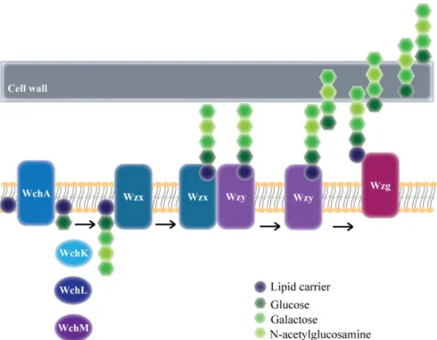
Synthesis of the S. pneumoniae capsule by the Wzy-dependent
mechanism (Figure I-4) begins with reversible transfer of an activated
sugar, the majority of the times glucose (Glc) [13], to the C55
undecaprenyl-phosphate (Und-P) lipid carrier, leading to the formation of
Und-PP-Glc [17]. This transfer was shown to be catalyzed by WchA, the
first glycosyltransferase encoded in the cps locus, in serotypes 2, 8, 9V
Once the formation of the repeating unit is initiated, subsequent
sugars are added by additional glycosyltransferases. These are usually
identified by homology from the genes encoded in the cps locus, but for
serotype 14 the activities of the putative enzymes have been confirmed
[46, 49]. WchK is a -1, 4-galactosyltransferase that transfers galactose to lipid-linked glucose, the second step in the assembly of the type 14
repeating unit. WchL is a -1, 3-N-acetylglucosaminyltransferase that adds the third sugar of the repeating unit, N-acetylglucosamine. Finally,
WchM, a -1, 4-galactosyltransferase, adds the last sugar to the repeating unit (Figure I-4).
linkage of the capsule to the cell wall peptidoglycan, a reaction catalyzed by protein Wzg. See text for more details.
Following the assembly of the repeating unit in the inner face of
the cytoplasmic membrane, the polymer is “flipped” to the external side by protein Wzx. At the outer face of the cytoplasmic membrane, repeating
units are added and high molecular weight capsular polysaccharide is
formed in a reaction catalyzed by the Wzy polymerase [113] (Figure I-4).
The Wzy polymerases are generally predicted by their putative membrane
topologies. Only for the Escherichia coli O-polysaccharide biosynthetic
pathway (in an in vitro system) has unambiguous biochemical evidence
that Wzy acts as a sugar polymerase been presented [107].
The final step in capsular polysaccharide synthesis is the covalent
linkage of the capsule to the cell wall peptidoglycan [95]. For a long time
the enzyme responsible for this step has remained unknown. Recently,
Kawai and colleagues [43] have identified the Wzg protein, the product of
the first gene of the cps operon, as the long searched capsule-cell wall
ligase. Wzg belongs to a family of proteins, named LCP (LytR –Cps2A –
Psr), that carry out the transfer of anionic cell wall polymers, such has
teichoic acids and acidic capsular polysaccharides, from their lipid-linked
precursors to peptidoglycan. Crystallographic studies have demonstrated
the phosphotransferase activity of the serotype 2 Wzg protein, supporting
the proposed enzymatic function of the protein [25]. Importantly, the LCP
proteins apparently do not recognize the repeating chain of the anionic
polymer, recognizing instead the lipid and pyrophosphorylated part [25].
Therefore, these proteins are not specific and have somewhat redundant
roles. The precise linkage of the CPS to peptidoglycan is not known.
In the last years, protein WchA has gained importance has a
central player in the control of polymer assembly. Knockout mutants of
dehydrogenase necessary for side chain synthesis in this serotype) were
only obtained in the presence of suppressor mutations [111]. In the
majority of the cases, the suppressor mutations were mapped in wchA. It
was proposed that the lethality of the wzx, wzy and ugd mutations was due
to sequestration of Und-P in the capsule synthesis pathway, preventing its
turnover for utilization in other essential cellular pathways, like
peptidoglycan synthesis, or desestabilization of the membrane due to the
accumulation of lipid-linked intermediates. The suppressors led to a
reduction in the amount of capsule, presumably to a level that did not
seriously affect the levels of available Und-P. The mutations in WchA
occurred in the non-conserved extracytoplasmic and in the conserved
cytoplasmic glyosyltransferase domains of the protein, indicating both
have important roles [111]. It is possible that these mutations disrupt
interactions with other proteins of the pathway, causing a reduction of its
global activity.
Regulation of capsular polysaccharide synthesis in the
Wzy-dependent mode occurs through a complex phosphoregulatory system,
which is discussed in the following section.
Regulation of the
S. pneumoniae
capsular polysaccharide
synthesis
The production of capsule and the length of the CPS chain in the
Wzy-dependent synthesis are regulated by proteins from the Bacterial
Tyrosine Kinase (BY-kinase) family. The members of this family are
often involved in the regulation of the synthesis of polysaccharides, such
as exopolysaccharides and capsular polysaccharides [33].
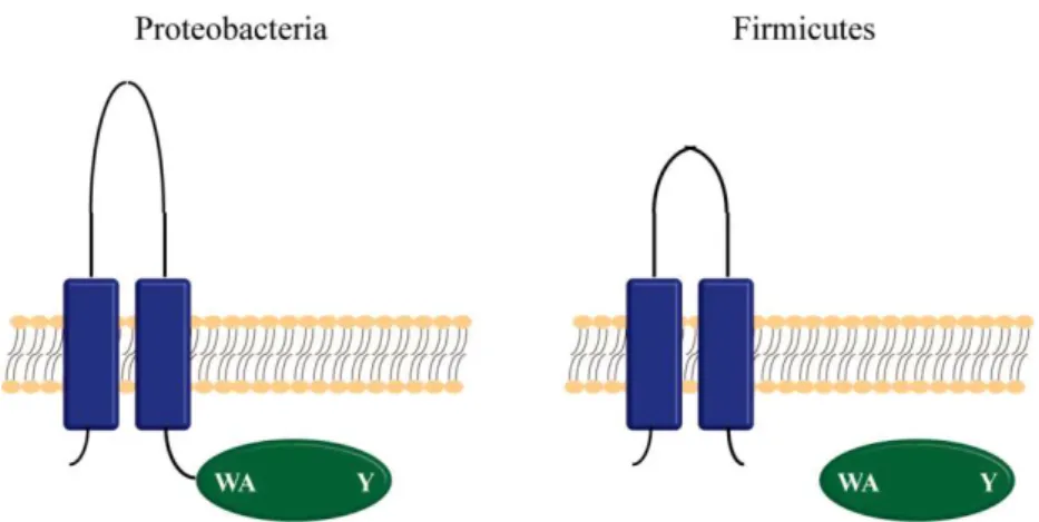
The BY-kinase regulatory systems consist of a membrane sensor
dephosphorylates the latter. Two transmembrane domains and an
extracytoplasmic loop between them, which is proposed to participate in
the sensing of signals from the outside, constitute the membrane sensor
domain. The catalytic cytoplasmic domain is the kinase part of the system.
It possesses conserved Walker A and B ATP binding motifs and a tyrosine
cluster at its C-terminal end. In Gram-negative bacteria and in
Actinobacteria, these two domains are present in the same protein. On the
other hand, in the firmicutes, such as S. pneumoniae, the two domains
exist as two separate, interacting proteins [33] (Figure I-5).
Figure I-5: Organization of Bacterial Tyrosine Kinases. BY-kinases are constituted by a transmembrane “sensory” domain (blue) and an intracellular catalytic domain (green). The cytoplasmic catalytic domain possesses conserved motifs: a Walker A ATP binding (WA) and a C-terminal tyrosine cluster (Y). In proteobacteria, the BY-kinases exist as a single protein, whereas in firmicutes they exist as two separate interacting polypeptides. The loop between the two transmembrane -helices is shorter in firmicutes than in proteobacteria.
Protein Wzd is the sensor transmembrane domain in S.
pneumoniae CPS synthesis. It belongs to the Polysaccharide
Co-Polymerase (PCP) family [72] and it was shown to be required for the
[71]. However, immunoblot analysis revealed the production of
short-chain CPS polymers in these mutants [11]. Also, immunofluorescence
experiments detected the presence of low levels of CPS-related material at
the cell poles of some of the cells [71].
Protein Wze is the catalytic cytoplasmic domain part of the
BY-kinase system involved in S. pneumoniae CPS synthesis. As mentioned
above for these types of proteins, it possesses the conserved motifs
characteristic of the group: the Walker A and B ATP binding motif and
the C-terminal tyrosine cluster [71]. Wze autophosphorylates at this
cluster and this requires the binding of ATP and the presence of protein
Wzd [12, 71]. Null mutants of wze produced the same phenotype as
described above for wzd [11, 66, 71]. Interestingly, mutation of the two
conserved motifs of Wze had different effects on capsule synthesis.
Mutation of the Walker A ATP binding motif eliminated capsule
production and prevented tyrosine phosphorylation, whereas mutation of
the C-terminal tyrosines to phenylalanines still allowed capsule
production [71]. However, deletion of the whole tyrosine cluster produces
a phenotype indistinguishable from a wze null mutant and at least two
functional tyrosine repeats are required for the normal function of the
protein [67], indicating that this motif is involved in CPS production.
As mentioned above, the third member of the BY-kinase
regulatory system is a phosphatase, responsible for dephosphorylating the
cytoplasmic kinase. In S. pneumoniae CPS synthesis, the phosphatase is
protein Wzh, a manganese-dependent phosphotyrosine-protein
phosphatase that belongs to the Polymerase and Histidinol Phosphatase
(PHP) family [68]. Wzh was shown to bind and dephosphorylate Wze, but
also to inhibit its phosphorylation [12]. Pneumococcal wzh null mutant
colonies have a rough phenotype, but immunobloting and
immunofluorescence experiments have shown that, although at lower
cells [66, 71]. Also, the capsule produced has a normal chain size, as seen
by the CPS ladder pattern observed in immunoblots, which is identical to
the WT [11]. Point mutations in conserved aspartate and histidine residues
of wzh, predicted to be involved in catalysis and metal binding,
respectively, inactivate the protein. This leads to an increase in the levels
of phosphorylated Wze and produces the same phenotype as a wzh null
mutant [68].
For a long time, the role of the tyrosine phosphorylation in
capsular polysaccharide synthesis has been the subject of debate, since
apparently conflicting results were obtained by different authors. Weiser
et al. observed that changes in the availability of environmental oxygen
had an effect in the amount of capsule produced by transparent (T
variants, producing less CPS) and opaque (O variants, producing more
CPS) pneumococci [105]. Oxygen had an inhibitory effect in capsule
production and this correlated with a decrease in the levels of tyrosine
phosphorylation of Wze. Therefore, it seemed that tyrosine
phosphorylation positively regulated capsule synthesis. This was also
observed by Bender and colleagues, who found that the amounts of
capsule and tyrosine-phosphorylated Wze correlated directly [11].
However, Morona and colleagues observed the opposite trend: tyrosine
phosphorylation appeared to reduce capsule synthesis [67, 71]. It was only
when the distinction between capsule synthesized and capsule attached to
the cell wall was made that this question was elucidated. Mutants in which
capsule was synthesized at normal levels but was defectively attached to
the cell wall expressed mutated Wzd proteins, whose specific point
mutations allowed the protein to function independently of Wze tyrosine
phosphorylation [70]. Also, wzh mutants produced less CPS than the WT
strain, but the proportion of attached CPS versus total amount produced
of CPS biosynthesis and increase the rate of cell wall-CPS attachment
[70].
Currently, the model for S. pneumoniae capsular polysaccharide
synthesis regulation is the following [70] (Figure I-6): nonphosphorylated
Wze interacts with Wzd, and ATP binds to the Walker A motif of Wze.
This promotes the interaction with other proteins of the machinery such
that CPS biosynthesis/polymerization proceeds at a maximal level (Figure
I-6A). At some point, due to an as yet unknown signal, Wze becomes
phosphorylated. This changes the interactions between the proteins in the
assembly machinery, slowing down CPS polymerization and promoting
instead the transfer of the polymer to the cell wall ligase and consequent
linkage to peptidoglycan. When Wzh dephosphorylates Wze, the cycle
can be repeated (Figure I-6B).
Structural studies of the Wzd and Wze Staphylococcus aureus
homologues, CapA and CapB, have provided a model for the molecular
mechanism of polysaccharide synthesis regulation. Olivares-Illana et al.
solved the structure of the S.aureus tyrosine kinase CapB fused at its
N-terminus with the carboxyl-terminal cytoplasmic part of its cognate
transmembrane modulator, CapA [80]. The unphosphorylated form of the
protein was found to associate in a ring-shaped octamer and this led to the
proposal of a structural-mechanical cycling process.
Phosphorylation/dephosphorylation of the tyrosine kinase would induce
cyclic dissociation-association of the octamer. These conformational
changes would probably be transmitted to the other members of the
polysaccharide synthesis machinery, affecting their interactions, this way
S. pneumoniae
capsule synthesis as a potential drug target
Importance of S. pneumoniae as a human pathogen
S. pneumoniae is the leading cause of otitis media,
community-acquired pneumonia and bacterial meningitis. The pneumococcus spreads
from person-to-person by aerosols and nasopharynx colonization is
generally asymptomatic, with carriage rates ranging from 5 – 10% of
healthy adults to 20 – 40% of healthy children. When the bacteria present at the nasopharynx migrates to other parts of the human body, spreading
into the inner ear, lungs, bloodstream or brain, invasive pneumococcal
disease (IPD) develops. IPD risk groups include infants, the elderly and
immunocompromised individuals [81].
Although the rates of colonized individuals which develop IPD
are low, the morbidity associated with S. pneumoniae is very high, due to
its high colonization levels. In 2005, it was estimated that pneumococcus
was the cause of 1.6 million deaths annually, including 0.7 – 1 million
deaths of children under the age of 5 [110]. The situation is of most
concern in developing countries, in which the majority of the latter occur.
In 2000, 19% of the deaths of children under the age of five, which were
HIV negative, due to pneumococcal disease, occurred solely in India and
more than half occurred in only six countries (India, Nigeria, Ethiopia,
Democratic Republic of the Congo, Afghanistan and China) [108]. In
Europe and the United States, S. pneumoniae is the most common cause
of community-acquired bacterial pneumonia in adults. In these
economically developed regions, IPD is also responsible for high
mortality levels. The mortality rates average 10 – 20% for adults with
pneumococcal pneumonia, reaching as much as 50% in high-risk groups
[109]. Furthermore, pneumococcal disease is also responsible for a high
direct medical costs reached as much as 3.5 billion US dollars [41]. When
one considers the work loss and productivity, the value is even higher.
Pneumococcal disease still remains a substantial cause of morbidity and
mortality in developed countries nowadays.
Current strategies to fight S. pneumoniae infections
Given the figures presented above, it is clear that effective
strategies against S. pneumoniae disease are required. In the search for
strategies to fight pneumococcal infections, the capsule has been a subject
of interest for a long time.
Despite the existence of several S. pneumoniae serotypes, the
majority of the disease is caused by only a small subset [8]. Therefore,
polysaccharide vaccines (containing capsular polysaccharide) and
conjugated vaccines (containing capsular polysaccharides conjugated with
a highly immunogenic protein) against these CPS types have been
developed [86]. Unfortunately, they only confer protection to the
particular serotypes contained in their formulation. This has led to a
phenomenon of replacement, in the population, of the vaccine serotypes
by other non-vaccine serotypes [3, 38, 42]. New approaches, preferably
directed at several serotypes simultaneously, are therefore required and
would be a major breakthrough in the field. Vaccines against non-variable
pneumococcal surface structures are being developed and will possibly be
of great use in the future [59].
BY-kinases as novel drug targets
Since proteins Wzd, Wze and Wzh, as seen above, are key
designed against pneumococcal infections. Moreover, the fact that they
belong to the BY-kinase family is another important factor, since this
family of proteins has been raising attention as novel drug targets.
Several features of BY-kinases meet the criteria for an
antibacterial target [21]. These enzymes control the metabolism of
polysaccharides that are present at the surface of the bacterial cells, but
not of the host cells, and they are embedded in the membrane, allowing
relatively easy access. Also, through the phosphorylation of other cellular
targets, they are involved in a broad spectrum of essential metabolic
pathways. Most importantly, the structures of two BY-kinases, Etk from
the Gram-negative E. coli [54] and CapB from the Gram-positive S.
aureus [80], have been solved and they revealed some important features.
Besides the absence of sequence similarity with tyrosine kinases from
eukaryotes, the fold of BY-kinases is also entirely different from their
eukaryotic counterparts, as well as their mechanism of action [53].
Not only BY-kinases serve as good candidates for drug
development, their cognate phosphatases are also promising targets. The
first structure of a tyrosine phosphatase from a Gram-positive, Wzh from
S. pneumoniae serotype 4, was solved and shown to have a completely
different structure from its E. coli functional homologue Wzb [35].
Recently, a screening for inhibitors of the phosphatase Wzh led to the
finding of a metabolite produced by a marine sponge that inhibited the
activity of the enzyme, causing a decrease in capsule synthesis [97].
Interestingly, despite their different structures, this metabolite also
inhibited the phosphatase activity of the E. coli Wzb, demonstrating the
State-of-the-art of
S. pneumoniae
cell biology
In order to fully comprehend how important and essential
mechanisms for the survival of bacteria occur and how they are regulated,
it is mandatory to determine where the proteins involved in those
mechanisms are localized in the cell and what factors control that
localization. In S. pneumoniae, fundamental processes such as cell wall
synthesis [64-65] and cell division [27, 32, 51, 78] have been studied
through the use of immunofluorescence techniques. However, these types
of techniques present several disadvantages. They require the fixation of
the cells and their lysis in order to allow access of antibodies, therefore
they are prone to the production of artifacts. Importantly, they also do not
allow the visualization of live cells, making it impossible to follow the
dynamics of a particular protein throughout the cell cycle of the bacteria
[90].
The discovery of the GFP protein and its application to the
construction of protein fusions that allow their in vivo visualization has
revolutionized the cell biology field. Several characteristics of GFP make
it an attractive reporter, in particular the fact that no external co-factors
are necessary for its fluorescence and its self-contained domain structure.
This reduces the chance of perturbations of the fluorescence by the protein
to which GFP is fused, while simultaneously allowing that protein to
retain its function [60, 100]. However, GFP maturation requires
post-translational oxidation of the protein, which has hindered the application
of this technology in microaerophiles such as S. pneumoniae [100].
In 2000, Acebo and colleagues provided the first report on the
expression of GFP in S. pneumoniae [2]. They were able to measure
directly the fluorescence of GFP in cells expressing single or multiple
strategies in the same direction and allowing tighter control of GFP
expression followed [76-77].
It was only in 2009 that the first study of protein localization
through GFP fusions, in live pneumococcal cells, was reported [26].
Eberhardt and colleagues successfully localized proteins involved in
choline decoration of teichoic acids, and since then several proteins
involved in processes such as chromosome segregation [63], cell division
[10, 29] or capsule synthesis [25] have been studied.
The use of GFP protein fusions in S. pneumoniae has no doubt
been an important breakthrough in the cell biology of this important
human pathogen, and the constant laboratory evolution and improvement
of the GFP proteins [28, 94] is constantly increasing its range of
applications [24]. Despite this, pneumococcal processes are still being
studied through immunofluorescence techniques [9, 50, 92]. This
indicates that the field of pneumococcal cell biology is still in need of
more and better tools for the in vivo study of protein localization.
S. pneumoniae
cell division
S. pneumoniae cells have the shape of an elongated ellipsoid,
similar to a rugby ball. Cocci bacteria which have this shape have been
designated as ovococci, to distinguish from the truly spherical cocci such
as Staphylococcus aureus. Pneumococcal cells can be visualized as
isolated cells, as diplococcic or as small chains, a characteristic which is
the result of their mode of division which proceeds in successive parallel
S. pneumoniae possesses two modes of cell wall synthesis
In rod-shaped cells, cell wall growth occurs in two phases,
elongation and division. On the contrary, cocci do not possess a cell wall
elongation machinery, so that all cell wall growth is associated with
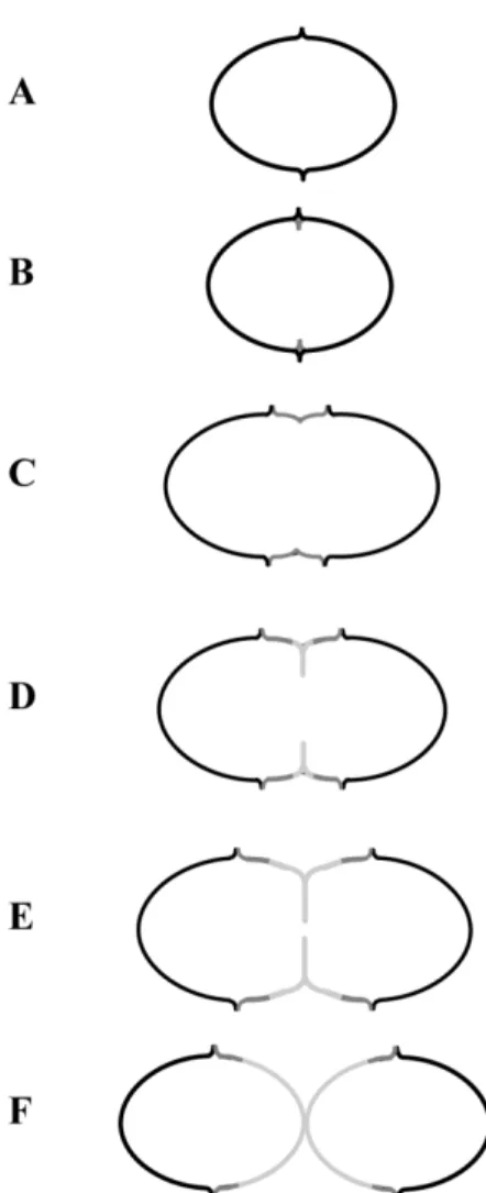
division. Ovococcal cells possess an annular outgrowth of peptidoglycan
which encircles the cells at their middle [39, 98], termed the equatorial
ring (Figure I-7A), and this is the only site where new cell wall material is
incorporated [15, 99]. In these bacteria, peptidoglycan is synthesized in
two ways: peripheral synthesis and septal synthesis [40].
In ovococci, division initiates with the appearance of an ingrowth
below the equatorial ring (Figure I-7B). When the equatorial ring splits at
its middle, peripheral cell wall synthesis takes place as new material is
incorporated between the two separating equatorial rings (Figure I-7C).
Septal cell wall synthesis begins when the ingrowth, which has remained
equidistant from the two rings, starts to grow centripetally, leading to the
formation of the cross-wall, perpendicular to the main axis of the cell
(Figure I-7D-E) [40, 115]. It is important to note that both types of cell
wall synthesis in ovococci are division specific, since peripheral synthesis
is different from the elongation, or lateral wall synthesis, that occurs in
rod-shaped bacteria.
PBPs (Penicillin-binding proteins) are enzymes responsible for
the last steps of peptidoglycan synthesis: the transglycosylation of
disaccharide units that form the glycan strands; and the transpeptidation
that cross-links the glycans through peptide bridges [89]. These proteins
can be classified into high-molecular-weight (HMW) and
low-molecular-weight (LMW) PBPs. HMW PBPs can be further divided into class A,
which includes bifunctional proteins with both transglycosylase and
endopeptidases [89]. S. pneumoniae has 3 class A HMW PBPs (PBP1a,
PBP1b and PBP2a), two class B HMW PBPs (PBP2b and PBP2x) and
one LMW PBP (PBP3) [115].
Figure I-7: Schematic representation of the ovococci mode of division.
Initial immunofluorescence studies on the localization of the
pneumococcal HMW PBPs led to the identification of two distinct
localization patterns, septal and equatorial, which seemed to relate with
the organization of the PBPs in two cell wall synthesis machineries, septal
and peripheral [65]. Importantly, subsequent studies have shown that this
patterns were an artifact and that all PBPs have the same localization at
the middle of the cells [115]. However this does not rule out the existence
of two cell wall synthesis machineries in ovococci, which is supported by
recent data.
First, Lactococcus lactis, an ovococcus, was found to have the
ability to become rod-shaped. Studies on the mechanism that allows this
morphological transition led to the identification of PBPs associated with
septal growth (PBP2x) and peripheral growth (PBP2b), which function
independently of each other. This allows elongation to occur during
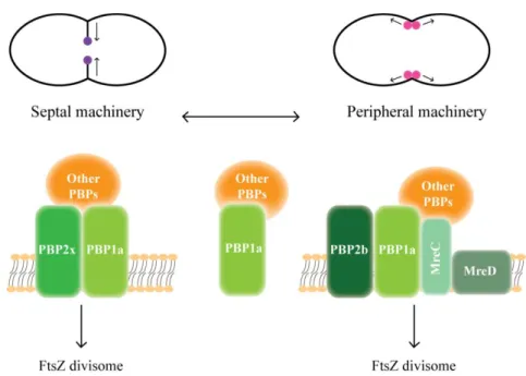
inhibition of division and vice versa [85]. Second, proteins MreC and
MreD, which are required to maintain the shape of rod-shaped bacteria,
were found to direct peripheral peptidoglycan synthesis in S. pneumoniae.
MreCD achieved this through the control of PBP1a localization or activity
[50]. Based on these results and on other studies from Bacillus subtilis,
which showed that PBP1 shuttles between peripheral and septal
peptidoglycan synthesis, a model for the composition of the septal and
peripheral peptidoglycan synthesis machineries has been proposed (Figure
I-8) [93].
Interestingly, dual labeling of PBPs showed that different
populations of PBPs are differentially localized within the same region of
a cell. The observation that the activities of PBPs are not uniform in the
midcell regions is in accordance with the model that two cell wall
synthesis machineries, septal and peripheral, exist and operate at that site
[45].
Coordination of cell wall synthesis with cell division
The first event in cell division is the recruitment of the
tubulin-like protein FtsZ to the division site, where it assembles into a ring
structure at the inner face of the cytoplasmic membrane. Following the
formation of the Z-ring other division proteins are recruited to the division
site, including the PBPs [115]. These were initially thought to localize
through protein-protein interactions, but increasing evidence has been
in this process. The S. pneumoniae LMW PBP3, a carboxypeptidase that
hydrolyses the D-Ala-D-Ala terminus of the peptidoglycan precursor, was
found to localize all over the cell surface of the pneumococcal cells, but
absent from the future division site. This implies that pentapetides are
only available at the equator, making the localization of the HMW PBPs
at that site dependent on the availability of their substrate. In the absence
of PBP3, the HMW PBPs rings were no longer co-localized with the FtsZ
ring [64]. Also, in L. lactis the substitution of the terminal D-Ala of the
peptidoglycan precursors for D-Lac led to the production of spherical
cells, instead of ovococcal, that remained attached in chains [22]. The
changes in the structure of the peptidoglycan precursor were postulated to
affect the activity of the carboxypeptidase peptidoglycan processing
enzymes, which in turn affected the recruitment of other PBPs.
Other proteins seem to be important in S. pneumoniae cell
division. DivIVA, a protein that in B. subtilis is involved in division site
selection, localizes as a ring at the division septum and as dots at the poles
of S. pneumoniae cells. In the absence of DivIVA, the percentage of cells
with constricted FtsZ rings is reduced, indicating a possible role in Z-ring
progression. Using a Bacterial Two-Hybrid assay, DivIVA was found to
interact with itself and with FtsZ, as well as with several other division
proteins [27]. Protein DivIB seems to have a role in the early and late
stages of the division process, since in its absence cells form chains and a
small fraction are enlarged and have defective septa [52]. DivIB forms a
trimeric complex with proteins DivIC and FtsL, the function of which has
been proposed to be related to septal peptidoglycan synthesis [78].
Recently, protein StkP has emerged as a main regulator of
pneumococcal cell division. StkP is the only eukaryotic-type Ser/Thr
protein kinase in S. pneumoniae and it was shown to affect the
round or elongated and form chains, with non-functional septa and with
delocalized sites of peptidoglycan synthesis [10, 29]. Importantly, the cell
division defects observed in the absence of StkP are the result of the lack
of phosphorylation of its targets [10], among which is DivIVA [79]. StkP
localizes to the division site [10, 32], and this localization occurs through
the sensing of uncross-linked peptidoglycan, since the protein is
delocalized in the presence of antibiotics that target the late stages of
peptidoglycan synthesis, as well as in cells that stopped dividing [10]. It
seems that cell wall synthesis and uncross-linked peptidoglycan are
sensed by StkP, leading to its recruitment to the cell division sites and to
the stimulation of its autophosphorylation activity. StkP, trough the
phosphorylation of key division substrates, would in this way regulate cell
division and control the shift from peripheral to septal cell wall synthesis
References
1. Abeyta, M., Hardy, G. G., and Yother, J., 2003. Genetic Alteration of
Capsule Type but Not PspA Type Affects Accessibility of Surface-Bound
Complement and Surface Antigens of Streptococcus pneumoniae.
Infection and Immunity, 71(1):218-225.
2. Acebo, P., et al., 2000. Quantitative Detection of Streptococcus
pneumoniae Cells Harbouring Single or Multiple Copies of the Gene
Encoding the Green Fluorescent Protein.Microbiology, 146:1267-1273.
3. Aguiar, S. I., et al., 2010. Serotypes 1, 7F and 19A Became the
Leading Causes of Pediatric Invasive Pneumococcal Infections in Portugal
after 7 Years of Heptavalent Conjugate Vaccine Use. Vaccine,
28(32):5167-73.
4. Arrecubieta, C., López, R., and García, E., 1994. Molecular
Characterization of cap3A , a Gene from the Operon Required for the
Synthesis of the Capsule of Streptococcus pneumoniae Type 3:
Sequencing of Mutations Responsible for the Unencapsulated Phenotype
and Localization of the Capsular Cluster on the Pneumococcal
Chromosome.Microbiology, 176(20):6375-6383.
5. Austrian, R., 1981. Pneumococcus : The First One Hundred Years.
Reviews Of Infectious Diseases, 3(2):183-189.
6. Avery, O. T. and Dubos, R., 1931. The Protective Action of a Specific
Enzyme against Type III Pneumococcus Infection in Mice. Journal of
Experimental Medicine, 54(1):73-90.
7. Avery, O. T., Macleod, C. M., and McCarty, M., 1944. Studies on the
Chemical Nature of the Substance Inducing Transformation of
Pneumococcal Types: Induction of Transformation by a
Desoxyribonucleic Acid Fraction Isolated from Pneumococcus Type III.
8. Babl, F. E., et al., 2001. Constancy of Distribution of Serogroups of
Invasive Pneumococcal Isolates among Children: Experience During 4
Decades.Clinical Infectious Diseases, 32(8):1155-61.
9. Barendt, S. M., Sham, L.-T., and Winkler, M. E., 2011.
Characterization of Mutants Deficient in the L, D -Carboxypeptidase
(DacB and WalRK (VicRK) Regulon, Involved in Peptidoglycan
Maturation of Streptococcus pneumoniae Serotype 2 Strain D39. Journal
of Bacteriology, 193(9):2290-2300.
10. Beilharz, K., et al., 2012. Control of Cell Division in Streptococcus
pneumoniae by the Conserved Ser/Thr Protein Kinase StkP. Proceedings
of the National Academy os Sciences of the USA, 109(15):E905-13.
11. Bender, M. H., Cartee, R. T., and Yother, J., 2003. Positive
Correlation between Tyrosine Phosphorylation of CpsD and Capsular
Polysaccharide Production in Streptococcus pneumoniae. Journal of
Bacteriology, 185(20):6057-6066.
12. Bender, M. H. and Yother, J., 2001. CpsB Is a Modulator of
Capsule-Associated Tyrosine Kinase Activity in Streptococcus pneumoniae.
Journal of Biological Chemistry, 276(51):47966-74.
13. Bentley, S. D., et al., 2006. Genetic Analysis of the Capsular
Biosynthetic Locus from All 90 Pneumococcal Serotypes.PLoS Genetics,
2(3):e31.
14. Bratcher, P. E., et al., 2010. Identification of Natural Pneumococcal
Isolates Expressing Serotype 6D by Genetic, Biochemical and Serological
Characterization.Microbiology, 156(Pt2):555-560.
15. Briles, E. B. and Tomasz, A., 1970. Segregation of
Choline-3H-Labeled Teichoic Acid.The Journal of Cell Biology, 47(3):786-790.
16. Calix, J. J., et al., 2012. Biochemical, Genetic and Serological
Characterization of Two Capsule Subtypes among Streptococcus
pneumoniae Serotype 20 Strains: Discovery of a New Pneumococcal
17. Cartee, R. T., et al., 2005. CpsE from Type 2 Streptococcus
pneumoniae Catalyzes the Reversible Addition of Glucose-1-Phosphate to
a Polyprenyl Phosphate Acceptor, Initiating Type 2 Capsule Repeat Unit
Formation.Journal of Bacteriology, 187(21):7425-7433.
18. Cartee, R. T., et al., 2001. Expression of the Streptococcus
pneumoniae Type 3 Synthase in Escherichia coli. Journal of Biological
Chemistry, 276(52):48831-48839.
19. Cartee, R. T., et al., 2000. Mechanism of Type 3 Capsular
Polysaccharide Synthesis in Streptococcus pneumoniae. Journal of
Biological Chemistry, 275(6):3907-3914.
20. Cartee, R. T., Forsee, W. T., and Yother, J., 2005. Initiation and
Synthesis of the Streptococcus pneumoniae Type 3 Capsule on a
Phosphatidylglycerol Membrane Anchor. Journal of Bacteriology,
187(13):4470-4479.
21. Cozzone, A. J., 2009. Bacterial Tyrosine Kinases: Novel Targets for
Antibacterial Therapy? Trends in Microbiology, 17(12):536-43.
22. Deghorain, M., et al., 2010. Functional and Morphological
Adaptation to Peptidoglycan Precursor Alteration in Lactococcus lactis.
Journal of Biological Chemistry, 285(31):24003-24013.
23. Dillard, J. P., Vandersea, M. W., and Yother, J., 1995.
Characterization of the Cassette Containing Genes for Type 3 Capsular
Polysaccharide Biosynthesis in Streptococcus pneumoniae. Journal of
Experimental Medicine, 181(3):973-983.
24. Dinh, T. and Bernhardt, T. G., 2011. Using Superfolder GFP for
Periplasmic Protein Localization Studies. Journal of Bacteriology,
193(18):4984-4987.
25. Eberhardt, A., et al., 2012. Attachment of Capsular Polysaccharide to
the Cell Wall in Streptococcus pneumoniae. Microbial Drug Resistance,
