Ana Laura Paixão
Dissertation presented to obtain the Ph.D degree in Biochemistry
Instituto de Tecnologia Química e Biológica António Xavier | Universidade Nova de Lisboa
The role of pneumococcal carbon
metabolism in colonisation and
invasive disease
Ana Laura Paixão
Supervisor:
Dr. Ana Rute Neves
Co-Supervisors:
Professor Peter William Andrew and Dr.
Hasan Yesilkaya
Interim Co-Supervisor:
Professor Adriano Oliveira Henriques
(2012-2015)
Dissertation presented to obtain the Ph.D degree in Biochemistry Instituto de Tecnologia Química e Biológica António Xavier | Universidade Nova de Lisboa
From left to right: Patrick Maria Franciscus Derkx, Raquel Sá Leão
Domingues da Silva, Karina de Bívar Xavier, Cecília Maria Pais de Faria de
Andrade Arraiano, Ana Laura Paixão, Ana Rute Neves, Adriano Oliveira
Henriques and Sofia Rocha Pauleta.
Oeiras, March 27th
, 2015
Apoio financeiro da Fundação para a Ciência e Tecnologia e do FSE no âmbito do Quadro Comunitário de apoio, Bolsa de Doutoramento com a referência SFRH/BD/46997/2008.
Acknowledgments
It was a long journey since I started my PhD, in the end of 2009. A full life experience. A path full of mixed feelings. The energy and motivation of a new beginning and the fear of failing such a challenge. The happiness of achieving the goals of this work, the surprise of reaching unexpected results and the frustration of failed experiments. The winnings and losses, laughs and tears, and the discovery of different worlds. And now? A new beginning. But before, I would like to express my gratitude to those that enriched this work and personal experience.
First, I would like to thank my supervisor, Dr. Rute Neves, for the opportunity I was given to conduct my PhD in the LAB & in vivo NMR
laboratory. Thank you for giving me the conditions to accomplish this work until the end, for the guidance, the scientific mentoring and the critical support in reviewing and writing this thesis. I thank the final effort to conclude this thesis in time. The encouragement to share my results in several European meetings and the opportunity to travel to amazing places.
I thank my co-supervisors, Prof. Peter William Andrew and Dr. Hasan Yesilkaya, for accepting me as a PhD student. For the openness to discuss my work and reviewing the first manuscript, contributing to a fruitful collaboration.
troublesome times. I am thankful for being always open to discuss my work and for teach me to do one thing at a time.
To Dr. Jan-Willem Veening who received me in his lab (Molecular Genetics - Groningen Institute of Biomolecular Sciences & Biotechnology) and supervised my work during my stay in Groningen. The best work experience I ever had. Thank you for running such a nice lab, full of interesting people, who received me so well.
To my dearest post-doc, Morten Kjos, the kindest person I have ever met. Always concerned with my work and my well-being while I was in Groningen. Thank you for all the support and help in the complementation assays, especially when I left Groningen and there were still experiments to be concluded.
To Prof Helena Santos for the valuable discussions held in her lab meeting presentations during the first years of my PhD.
To Ana Lúcia Carvalho for her tireless help. For her support and effort to conclude all the in vivo NMR experiments with me before leaving to her
new professional challenge. For teaching me everything she knew about HPLC and in vivo NMR. And most importantly, for her friendship. Thank
you for all the good moments in and outside the lab.
To José Caldas I thank all the help, dedication and interest in the microarrays analysis. Thank you for such a fruitful collaboration.
To Luis Gafeira, for being such an enthusiastic, passionate about science, who contributes with creative ideas to the work of everyone, including mine. Always willing to help. I will remember him for being a good listener and a friend.
To Paula Gaspar for sharing her knowledge and technical support.
To all the past and present members of Cell Physiology and NMR group (because you are so many I won’t enumerate – I could fail someone), for being always available to help and for sharing good parties with the LAB & in vivo NMR laboratory.
To Joana Oliveira for doubling my hands in the growth experiments. Thank you for the effort.
To all my colleagues in the in the LAB & in vivo NMR that contributed
to this experience in so many ways.
To Dr. Susana Vinga and André Veríssimo for the modelling of the growth curves and statistical analysis.
To Dr. Rita Ventura and Eva Lourenço for the synthesis of phosphorylated compounds.
To all the other collaborators that contributed in different ways to this thesis: Dr. Tomas Kloosterman, Dr. Vitor Fernandes and Prof. Dr. Oscar Kuipers.
To Teresa Maio, Ana Mingote, Sónia Estêvão, Teresa Ferreira, Tiago Pais and Marta Rodrigues for contributing to the most hilarious times in and outside the lab.
To all my colleagues from the 2010 PhD classes with whom I shared good moments.
To Fundação para a Ciência e a Tecnologia for the financial support that made this doctoral work possible (SFRH / BD / 46997 / 2008).
With the greatest gratitude, I dedicate this thesis to my parents and my husband, Gonçalo. This experience was also yours. I am grateful for your unconditional support, comprehension, love, and advices. Without them, it would have been much more difficult. Thank you for being always there when I needed the most.
Abstract
Streptococcus pneumoniae is a common asymptomatic commensal of
the human nasopharynx. However, it is better known as a threatening pathogen that causes serious diseases such as pneumonia, meningitis and sepsis, as well as other less severe but more prevalent infections (e.g. otitis media). With the increase of antibiotic resistance and the limited
efficacy of vaccines, pneumococcal infections remain a major problem. Therefore, the discovery of new therapeutic targets and preventive drugs are in high demand. Given this panorama, much attention has been dedicated to classical aspects of virulence (e.g. toxins, capsule or cell wall
components), but the knowledge of in vivo physiology and metabolism of S. pneumoniae is still limited. This is intriguing, considering that to a large
extent, pneumococcal pathogenesis relies on efficient acquisition and metabolism of the nutrients required for growth and survival in the host niches. In line with this view, recent work uncovers substantial interdependencies between carbohydrate metabolism and virulence. These findings denote a far greater importance of basic pneumococcal physiology than previously imagined. However, a scarcity of data on sugar metabolism and its regulation in S. pneumoniae hampers a
comprehensive understanding of the connections between these processes.
The ultimate goal of this thesis is to improve our understanding of S. pneumoniae basic physiology and discover links between carbohydrate
metabolism and the ability of the bacterium to colonise and cause disease.
S. pneumoniae is a strictly fermentative microorganism that relies on
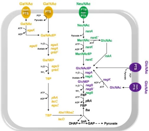
composed of N-acetylglucosamine (GlcNAc), N-acetylgalactosamine (GalNAc), N-acetylneuraminic acid (NeuNAc), galactose (Gal), fucose (Fuc) and sulphated sugars linked to the protein core. Importantly, S. pneumoniae possesses a large set of extracellular glycosidases that act
over glycans and release free sugars that can potentially be used for grow. Therefore, we hypothesised that the pneumococcus depends on one or multiple glycan-derived sugars to grow.
To disclose prevalent pathways during growth on mucin we resorted to a transcriptome analysis comparing transcript levels during growth on the model glycoprotein porcine gastric mucin and glucose (Glc). The gene expression profile revealed Gal, Man and GlcNAc as the most probable glycan-derived sugars to be used as carbon sources by S. pneumoniae
D39. Accordingly, D39 was able to grow on these sugars, but not on other host-derived glycan monosaccharides (Fuc, NeuNAc, GalNAc), as expected from its genomic potential. An in depth characterization of growth profiles on a chemically defined medium supplemented with each carbohydrate using two different sugar concentrations (30 and 10 mM) was performed. S. pneumoniae displayed a preference for GlcNAc,
whereas Gal was the least preferable carbohydrate for growth, but energetically the most favourable. The inefficient Gal catabolism could be partially explained by the absence of a high affinity Gal transporter. Gal caused a remarkable metabolic shift from homolactic to a truly mixed acid fermentation. The in silico predicted pathways for the catabolism of each
sugar were experimentally validated at the biochemical level by determining sugar-specific intracellular metabolites and pathway specific enzyme activities. Furthermore, mutants were generated in each pathway by inactivation of a gene encoding a pathway specific enzyme, and their analysis proved at the genetic level the biochemical analyses. Curiously, inactivation of galK (Leloir pathway) rendered a strain unable to grow on
(tagatose 6-phosphate pathway), suggesting a subtle regulatory link between the two pathways.
Intranasal mouse infection models of pneumococcal colonisation and disease (bronchopneumonia with bacteraemia) showed that mutants in the Gal catabolic genes (∆lacD, ∆galK, ∆lacD∆galK) were attenuated, but
mutants on Man (∆manA) and GlcNAc (∆nagA) pathways were not. Our
data identified Gal as a key nutrient for growth in the respiratory tract. Strengthening this view is the large fraction of genes committed to Gal catabolism induced by mucin (25%) as well as the considerable group of virulence genes (8.2%) displaying altered expression in response to Gal. The response to Gal, GlcNAc and Man (test sugars) was also evaluated at the transcriptional and metabolic levels. The transcriptional response was substantial and sugar-specific. Gal, GlcNAc and Man affected the expression of up to 8.4%, 11.4%, and 14.2% of the genome, respectively, covering multiple cellular processes, including specific sugar pathways, central metabolism and virulence. An overview of the transcriptional response of central carbon metabolism functions (sugar transport and sugar specific pathways, glycolysis and fermentation pathways) to growth on the glycan-derived sugars as compared to Glc is presented. A complete correlation between expression profiles and pneumococcal phenotypic traits was not observed, denoting regulation at other cellular layers, such as post-transcriptional and/or metabolic levels. Hence, the glycolytic dynamics of S. pneumoniae D39 during catabolism
of the glycan-derived sugars were monitored in resting cells by in vivo13
C-NMR. The rate of GlcNAc utilization was similar to that of the fast metabolizable sugar Glc, and likewise the glycolytic metabolite fructose 1,6-bisphosphate (FBP), a metabolic activator of homolactic metabolism, accumulated to considerably high concentrations (circa 30 mM). In line,
FBP was considerably reduced to circa 12 mM, and a mixed-acid
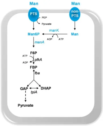
fermentation profile was observed. In addition, specific metabolic intermediates (α-galactose 6-phosphate and mannose 6-phosphate) accumulated, suggesting metabolic bottlenecks in the specific steps converting Gal and Man to glycolytic intermediates. On Man, the metabolic constriction most likely occurs at the level of mannose 6-phosphate isomerase, since growth is improved when this activity is positively modulated by varying the gene expression.
In the host S. pneumoniae has to cope with fluctuating concentrations
of carbon sources. While free sugars, particularly Glc, are scarce in the nasopharynx, in the bloodstream and during infection Glc is comparatively higher. Therefore, we hypothesised that during the transition from colonisation to invasive disease S. pneumoniae adapts to the nutritional
changes through a specific response to a Glc stimulus that result in changes in gene regulation and metabolism.
Thus, cells growing in glycan-derived sugars or in a sugar mixture of Gal, GlcNAc and Man were challenged in exponential phase with a Glc pulse. The response to this stimulus was evaluated at the transcriptional, physiological and metabolic (for Gal-adapted cells) levels. Glc was readily consumed independently of the initial substrate adaptation, strengthening the view that Glc is the preferred carbohydrate for pneumococcal growth. The transcriptional response to a Glc stimulus was large, the most represented COG category being carbohydrate metabolism and transport. Except for Man-adapted cells, Glc exerted mostly negative regulation over the majority of genes encoding central carbon metabolism functions. Additionally, Glc induced shifts to more homolactic profiles and, which on Gal was accompanied by an increase on the FBP concentration following the Glc pulse.
genes. Interestingly, cells adapted to grow on a sugar mixture (Gal, Man and GlcNAc) displayed the smallest transcriptional response to Glc, suggesting improved fitness of S. pneumoniae when exposed to varied
sugars. We suggest that the nasopharynx is the reservoir for the development of niche-specific virulence traits, essential for successful colonisation of the niche. In addition, the majority of these virulence factors are downregulated by a Glc stimulus, and are therefore not required in disease. The link between sugar metabolism and virulence is herein reinforced.
Resumo
Streptococcus pneumoniae é um organismo comensal assintomático
comummente encontrado na nasofaringe humana. Contudo, é mais conhecido no seu papel patogénico ameaçador que causa doenças como a pneumonia, a meningite ou septicémia, bem como outras infeções menos severas mas mais recorrentes (e.g. otite média). Com o aumento
da resistência a antibióticos e a eficácia limitada das vacinas, as infeções pneumocócicas permanecem um importante problema. Assim sendo, há uma grande necessidade em descobrir novos alvos terapêuticos e drogas preventivas. Neste contexto, a atenção tem-se focado em aspetos clássicos da virulência (e.g.,toxinas, cápsula ou componentes da parede
celular). No entanto, o conhecimento da fisiologia in vivo e do
metabolismo de Streptococcus pneumoniae é ainda limitado. Esta
situação é intrigante, considerando que em grande medida, a patogenicidade pneumocócica baseia-se na aquisição eficiente e subsequente metabolismo de nutrientes necessários para o crescimento e a sobrevivência em nichos no hospedeiro. A corroborar esta afirmação, estudos recentes revelam interdependências substanciais entre o metabolismo dos hidratos de carbono e a virulência, e sugerem que o conhecimento sólido da fisiologia pneumocócica básica é mais importante do que antecipado. Contudo, a escassez de dados relativos ao metabolismo de açúcares e a sua regulação em S. pneumoniae limita
a compreensão das ligações entre estes processos.
O objetivo último desta tese é o de melhorar o conhecimento da fisiologia básica de S. pneumoniae e descobrir relações entre o
S. pneumoniae é um microrganismo estritamente fermentativo que
depende do metabolismo glicolítico para obter energia, mas os monossacáridos livres são escassos nas vias respiratórias. Os açúcares mais abundantes presentes no nicho natural de S. pneumoniae são as
glicoproteínas mucinas, compostas geralmente por N-acetylglucosamina (GlcNAc), N-acetylgalactosamina (GalNAc), ácido N-acetilneuramínico (NeuNAc), galactose (Gal), fucose (Fuc) e açúcares sulfatados ligados a um núcleo proteico. De realçar que S. pneumoniae possui um grande
conjunto de glicosidases extracelulares que atuam sobre os glicanos e libertam açúcares que podem potencialmente ser utilizados para crescimento. Por conseguinte, colocámos e testámos a hipótese de que os pneumococos podem depender, para crescimento, de um ou de múltiplos açúcares derivados destes glicanos.
Para revelar as vias predominantes durante o crescimento em mucinas recorremos a uma análise de transcriptómica comparando os níveis de transcrição durante o crescimento na presença de mucina gástrica de suíno, enquanto glicoproteína modelo, e na presença de glucose (Glc). O perfil de expressão génica revelou que a Gal, Man e GlcNAc são os açúcares derivados de glicano mais provavelmente utilizados como fontes de carbono por S. pneumoniae D39. Em
consonância, a estirpe D39 cresce nestes açúcares, mas não em outros monossacáridos derivados de glicanos do hospedeiro (Fuc, NeuNAc, GalNAc), como antecipado do seu potencial genómico. Realizou-se uma caracterização aprofundada dos perfis de crescimento em meio quimicamente definido suplementado com cada hidrato de carbono usando duas concentrações de açúcar (30 e 10 mM). S. pneumoniae
galactose causou um desvio pronunciado da fermentação homolática para ácidos mistos. As vias catabólicas previstas in silico para cada
açúcar foram experimentalmente validadas ao nível bioquímico, pela determinação de metabolitos intracelulares específicos de cada açúcar e atividades enzimáticas específicas de cada via. Adicionalmente, geraram-se mutantes por inativação de um gene que codifica uma enzima específica de cada via, e a sua análise confirmou ao nível genético a análise bioquímica. Curiosamente, a inativação de galK (via
Leloir) originou uma estirpe incapaz de crescer em Gal, apesar de ter uma via alternativa disponível para o processamento deste açúcar (via da tagatose 6-fosfato), sugerindo uma conexão subtil na regulação das duas vias.
Modelos de infeção intranasal de ratinhos de colonização pneumocócica e doença (broncopneumonia com bacteriemia) demonstraram que mutantes em genes catabólicos da Gal (∆lacD, ∆galK,
∆lacD∆galK) são atenuados, mas mutantes nas vias da Man (∆manA) e
GlcNAc (∆nagA), não. Os nossos estudos identificaram a Gal como um
nutriente crucial para o crescimento no trato respiratório. A grande fração de genes do catabolismo da Gal induzida pela mucina (25%) bem como um grupo considerável de genes de virulência (8.2%) com expressão alterada em Gal, reforçam esta conclusão.
comparação com a Glc. Não foi observada uma correlação completa entre os perfis de expressão e as características fenotípicas do pneumococo, demonstrando a existência de outros níveis de regulação, incluindo regulação pós-transcricional e/ou metabólica. Consequentemente, a dinâmica glicolítica de S. pneumoniae D39 durante
o catabolismo de açúcares derivados de glicanos foi monitorizada em células em suspensão por in vivo 13C-NMR. A taxa de consumo de
GlcNAc foi semelhante à do açúcar rapidamente metabolizado Glc e, do mesmo modo o metabolito glicolítico frutose 1,6-bifosfato (FBP), um ativador do metabolismo homolático, acumulou em concentrações elevadas (cerca de 30 mM). Em concordância, este açúcar apresentou um perfil de produtos finais totalmente homolático. Em contraste, as taxas de consumo de Gal e Man foram 2 vezes inferiores, a acumulação de FBP foi consideravelmente reduzida a aproximadamente 12 mM, e o perfil de fermentação de ácidos mistos foi observado. Além disso, foi detectada a acumulação de intermediários metabólicos específicos (α-galactose 6-fosfato e manose 6-fosfato), sugerindo estrangulamentos metabólicos em passos específicos da conversão de Gal e Man a intermediários glicolíticos. Em Man, a constrição metabólica muito provavelmente ocorre ao nível da manose 6-fosfato isomerase, uma vez que o crescimento é melhorado quando esta atividade é positivamente modulada por alteração da expressão do gene.
No hospedeiro, S. pneumoniae tem que lidar com concentrações
variáveis de fontes de carbono. Enquanto os açúcares livres, particularmente Glc, são escassos na nasofaringe, na corrente sanguínea e durante a infeção, a Glc é comparativamente elevada. Deste modo, colocámos a hipótese que durante a transição de colonização para doença invasiva S. pneumoniae adapta-se às variações nutricionais
Deste modo, células a crescerem em açúcares derivados de glicanos ou numa mistura de Gal, GlcNAc e Man foram sujeitas, durante o crescimento exponencial, a um pulso de Glc. A resposta a este estímulo foi avaliada aos níveis transcricional, fisiológico e metabólico (para células adaptadas a Gal). A Glc foi prontamente consumida independentemente da adaptação inicial ao substrato, reforçando que a Glc é o hidrato de carbono preferido para o crescimento dos pneumococos. A resposta transcricional ao estímulo de Glc foi ampla, sendo a categoria COG mais representada a do metabolismo de hidratos de carbono e do transporte. À exceção de células adaptadas a Man, a Glc exerceu principalmente uma regulação negativa sobre a maioria dos genes que codificam funções do metabolismo central de carbono. Adicionalmente, a Glc induziu desvios para perfis mais homoláticos e, em Gal foi acompanhado por um aumento da concentração de FBP após o pulso de Glc.
A expressão de fatores de virulência clássicos foi também dependente do açúcar, mas a Glc exerceu consistentemente um efeito repressivo sobre os genes de virulência. De interesse, notámos que as células adaptadas ao crescimento numa mistura de açúcares (Gal, Man e GlcNAc) originaram uma menor resposta transcricional à Glc, sugerindo uma melhor aptidão de S. pneumoniae quando exposto a uma variedade
de açúcares. Sugerimos que a nasofaringe é o reservatório para o desenvolvimento de traços específicos de virulência, essenciais para a colonização bem-sucedida deste nicho. Adicionalmente, a maioria destes fatores de virulência são reprimidos pelo estímulo de Glc, não sendo por conseguinte necessários na doença. A ligação entre metabolismo de açúcares e virulência é aqui reforçada.
Contents
Thesis outline ... xxi Abbreviations ... xxiii Chapter 1 - General introduction ... 1 Chapter 2 - Host glycan sugar-specific pathways in Streptococcus pneumoniae ... 71 Chapter 3 - Transcriptional and metabolic effects of glucose on S.
Thesis outline
Pneumococcal infections have high socio-economic impact, and are therefore a major burden worldwide. According to the WHO, S. pneumoniae causes over 1 million deaths per year in children under 5
years of age. Moreover, this high mortality is exacerbated by the rate at which the organism acquires resistance to traditional antibiotics and by the re-emergence of non-vaccine type strains. Therefore, the development of new therapeutic and preventive agents demands an increasing understanding of S. pneumoniae physiology and virulence.
The overall goal of this thesis is to gain new insights regarding S. pneumoniae adaptive response to sugar availability and the link to in vivo
fitness and virulence.
Chapter 1 starts with a brief introduction to the main phenotypic traits of S. pneumoniae, its ability to colonise the nasopharynx and cause
disease, and provides an overview of the traits of the bacterium in the perspective of a commensal and a pathogen. An overview of the major virulence factors is given as well as its link to carbohydrate metabolism. The catabolism, of monosaccharides (galactose, mannose, N-acetylglucosamine, N-acetylgalactosamine, N-acetylneuraminic acid and fucose) is reviewed. The relevance of these sugars in the lifestyle of different bacteria is discussed. Lastly, the current knowledge concerning central metabolism in S. pneumoniae is overviewed.
and N-acetylglucosamine is provided. The catabolic pathways of those sugars were, for the first time, experimentally validated at genetic and biochemical levels, confirming the in silico predictions. Finally, the
relevance of sugar-specific catabolic genes to in vivo fitness was
assessed in murine models of colonisation (nasopharyngeal carriage) and pneumococcal disease (bronchopneumonia with bacteraemia).
Chapter 3 describes the transcriptional and metabolic responses in the presence of galactose, mannose and N-acetylglucosamine, by DNA microarrays and in vivo 13C-NMR, respectively. Glucose is used as the
reference sugar.
The effect of adding the fast metabolizable sugar glucose to S. pneumoniae cells actively growing on galactose, mannose,
N-acetylglucosamine or in a mixture thereof is evaluated at the transcriptional, physiological and metabolic levels.
Furthermore, the influence of the carbohydrate source and the glucose pulse in the gene expression of known virulence factors is examined at the transcriptional level by DNA microarrays.
Abbreviations
ABC ATP-binding cassette
BgaA β-galactosidase A
CAP Community-acquired pneumonia
CcpA Carbon catabolite protein A CCR Carbon catabolite repression
CDM Chemically defined medium
COG Clusters of orthologous groups
CRE Catabolite response elements
DHAP Dihydroxyacetone phosphate
DW Dry weight
EI Enzyme I
EMP Embden-Meyerhof-Parnas
Eno Enolase
F6P Fructose 6-phosphate
Fba Fructose-bisphosphate aldolase
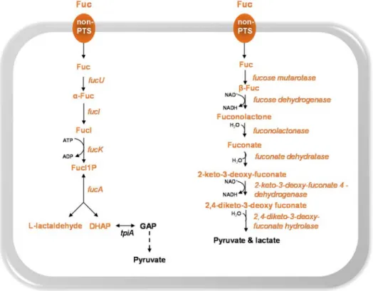
Fuc Fucose
FucA L-fuculose phosphate aldolase
FucI L-fucose isomerase
FucK L-fuculose kinase
FucO L-1,2-propanediol oxidoreductase
FucU Fucose mutarotase
G6P Glucose 6-phosphate
Gal Galactose
Gal6P Galactose 6-phosphate
GalE UDP-glucose 4-epimerase
GalK Galactokinase
GalM Aldolase 1-epimerase (galactose mutarotase)
GalN6P Galactosamine 6-phosphate GalNAc N-acetylgalactosamine
GalNAc6P N-acetylgalactosamine 6-phosphate
GalP Galactose permease
GalT Galactose 1-phosphate uridylyltransferase
Gap Glyceraldehyde 3-phosphate
Glc Glucose
GlcN Glucosamine
GlcN6P Glucosamine 6-phosphate GlcNAc N-acetylglucosamine
GlcNAc6P N-acetylglucosamine 6-phosphate HPLC High performance liquid chromatography Hpr Histidine phosphocarrier protein HPr
IPD Invasive pneumococcal disease
LacAB Galactose 6-phosphate isomerase subunits LacA andLacB LacC Tagatose 6-phosphate kinase
LacD Tagatose 1,6-diphosphate aldolase
Lcto Lactate oxidase
LDH Lactate dehydrogenase
Man Mannose
Man6P Mannose 6-phosphate
ManA Mannose 6-phosphate isomerase ManNAc N-acetylmannosamine
ManNAc6P N-acetylmannosamine 6-phosphate
NagA N-acetylglucosamine 6-phosphate deacetylase NagB Glucosamine 6-phosphate isomerase
NAD+ Nicotinamide adenine dinucleotide
NADH Dihydronicotinamide adenine dinucleotide
NanA Neuraminidase A
NanE N-acetylmannosamine 6-phosphate epimerase
NBD ATP- or nucleotide-binding domains NeuNAc N-acetylneuraminic acid
NMR Nuclear magnetic resonance
ODmax Maximum optical density
PCV Pneumococcal conjugate vaccine
PDHC Pyruvate dehydrogenase complex
PFL Pyruvate formate-lyase
PFL-AE Pyruvate formate-lyase activating enzyme
Pgk Phosphoglycerate kinase
Pgm Phosphoglucomutase
PME Phosphomonoester
PTS Phosphoenolpyruvate-dependent carbohydrate phosphotransferase system
Pyk Pyruvate kinase
SBP Solute-binding proteins
Adh Alcohol dehydrogenase
SpxB Pyruvate oxidase
StrH β-N-acetylglucosaminidase
T6P Tagatose 6-phosphate
TBP Tagatose 1,6-bisphosphate
TMD Transmembrane domains
WHO World Health Organization α-G1P α-glucose 1-phosphate α-Gal1P α-galactose 1-phosphate α-Gal6P α-galactose 6-phosphate μmax Maximum specific growth rate
Chapter 1
General introduction
Chapter 1 - Contents
Streptococcus pneumoniae ... 4
At the core of great discoveries ... 4 General characteristics ... 5
S. pneumoniae, a commensal and a pathogen ... 7
Transition from colonisation to disease ... 11 Virulence factors ... 12 Virulence and carbohydrate metabolism ... 17 Glycans at the interface of bacteria-host interactions ... 20 Pneumococcal carbohydrate metabolism ... 22
Carbohydrate metabolism and its importance for pneumococcal lifestyle ... 22 Sugar transport systems in S. pneumoniae ... 24
Glycan-derived monosaccharides: pathways and functions ... 27 Galactose ... 27 Galactose catabolism ... 28 Mannose ... 31 Mannose catabolism ... 32 Amino sugars: N-acetylneuraminic acid, N-acetylglucosamine and N-acetylgalactosamine ... 34
Streptococcus pneumoniae
At the core of great discoveries
Streptococcus pneumoniae, also known as the pneumococcus, was
discovered in 1881 by Louis Pasteur and George Miller Sternberg in two independent studies [1,2]. Shortly after its isolation S. pneumoniae was
identified as a major causative agent of human lobar pneumonia [1,2]. Due to its morphology (pairs of coccoid bacteria) and the involvement in pneumonia the bacterium was then named Diplococcus pneumoniae [1].
The present nomenclature remains since 1974 [1].
The story of S. pneumoniae is intertwined with the history of different
fields of research that range from microbiology, molecular biology, biochemistry to immunology (reviewed in [1,2]). Indeed, the study of the pneumococcus was at the centre of critical important discoveries. In 1928, Griffith demonstrated that when mice were inoculated with a mixture of dead virulent pneumococcal strains and live avirulent pneumococci, virulent phenotype strains could be recovered from the pneumonia dead animals. This phenomenon was denominated the “transforming principle” [3], and preceded the discovery of DNA as the hereditary material. Indeed, in 1944, Avery, McLeod and McCarthy proved, that the DNA was the transforming principle responsible for the phenotypic changes [4].
molecular processes such as quorum sensing, autolysis and opsonisation, as well as to the development of molecular tools, the Gram’s stain technique and other identification methods (e.g. Quellung reaction
and bile solubility test) still in use today [1,2].
General characteristics
Streptococcus pneumoniae is a low-GC (40%), Gram-positive,
lanceolate cocci (elongated ellipsoid-shape) [11]. These ovoid cells are commonly designated ovococci (Fig. 1.1) [12,13]. It is usually arranged in pairs (diplococci), but can occur as single cells or in short chains. Individual cells can range from 0.5 to 1.25 micrometres in size. S. pneumoniae is non-motile and a non-spore forming bacterium. It is an
aerotolerant anaerobe and a strictly fermentative microorganism that mainly converts carbohydrates to lactic acid (homolactic fermentation) [11].
S. pneumoniae is a fastidious bacterium that requires nutritionally rich
media for growth. Its nutritional requirement for choline is a unique characteristic of this microorganism [14]. It grows optimally at 37ºC in a pH range of 6.5-7.5. When cultured until stationary phase of growth the bacterium displays a tendency to undergo autolysis due to the presence of efficient autolysins, such as autolysin, LytA [15]. The pneumococci are also naturally transformable, taking up DNA from the medium (other bacteria), a phenomenon that contributes to a highly variable genome [16].
The genus Streptococcus belongs to the phylum Firmicutes,
similarity within this group of microorganisms together with frequent events of horizontal gene transfer makes it difficult to differentiate S. pneumoniae from other closely related commensals (S. mitis, S. oralis, S. infantis, S. pseudopneumoniae) [18–20].
Figure 1.1. Streptococcus pneumoniae.
Phase contrast image of S. pneumoniae D39 grown in chemically defined medium
supplemented with 10 mM glucose, until exponential phase of growth.
Traditionally, S. pneumoniae has been identified based on colony
morphology and on phenotypic tests [21]. Two properties generally assessed are the pneumococcal alpha-hemolytic activity when cultured on blood agar plates and its catalase-negative phenotype [11]. Commonly, optochin susceptibility and/or bile solubility are used to distinguish S. pneumoniae from other viridans streptococci [20]. In
addition, the ability to ferment inulin has also been used as a differentiating parameter [11].
Despite the application of different phenotypic and genotypic techniques (e.g. PCR targeting virulence genes, pulsed-field gel
electrophoresis, multilocus sequence typing (MLST), multilocus sequence analysis (MLSA)), and recently proteomic profiling (MALDI-TOF MS), identification and classification of S. pneumoniae has been problematic
and is still controversial [18,20,21,25,26]. Atypical reactions to standard identification tests are documented [18,25]. Part of this struggle stems out from the pneumococcus’ vast pan-genome and its genomic plasticity.
S. pneumoniae
, a commensal and a pathogen
The scientific interest on S. pneumoniae was primarily raised because
the organism has been a leading cause of mortality worldwide. In the beginning of the 20th century, Sir William Osler referred to it as the “captain of the men of death” [27,28], and pneumonia was the “natural end of elderly people” [29]. Nowadays, S. pneumoniae is also recognized as a
commensal microorganism, which resides in the mucosal surface lining the upper respiratory tract, i.e. the nasopharynx of humans [30,31].
Pneumococci are members of the respiratory tract microflora alongside with other prominent opportunistic pathogens such as Haemophilus influenzae, Moraxella catarrhalis and Staphylococcus aureus [32].
of pneumococcal carriage and disease are: ethnicity, crowding (e.g.
attendance to day-care centres, hospitals, schools, contact with older siblings and children), environmental (e.g. smoking) and socioeconomic
(e.g. living circumstances, income) ([33], reviewed in [35,36]).
Colonisation is a dynamic process in terms of duration and colonising serotypes in the lifetime of an individual. Carriage with one or more serotypes can occur simultaneously or sequentially. It is a transient stage and its duration is serotype-dependent and influenced by past carriage episodes [31]. It is longer in young than older children [37,38]. Most often pneumococcal infections occur following a newly-acquired strain [31]. Some serotypes are more commonly involved in carriage while others, a relatively small number, are more related with invasive pneumococcal disease [16,39,40]. This finding suggests that the latter clones have specific genes that facilitate progression to disease [41]. Interestingly, high mortality rates were found within serotypes of lower invasive potential [16,39].
The establishment of a carrier state is a pre-requisite for pneumococcal disease with the colonising homologous strain, which usually occurs when the bacterium spreads to other parts of the human body [30,33]. Indeed, the pneumococcus is an opportunistic bacterium and children, elderly (>65 years old) and immunocompromised people are at increased risk of pneumococcal disease [33,42]. S. pneumoniae is an etiological agent of
mild respiratory mucosal infections such as sinusitis or otitis media (non-invasive diseases), but also less prevalent but more serious (non-invasive pneumococcal diseases (IPD), when the pneumococcus gains access to normally sterile areas of the human body, such as bacteremic pneumonia or pneumonia with empyema, meningitis or sepsis [33].
pneumococcus, the limited efficacy of pneumococcal vaccines, the aging of populations and difficulties in clinical diagnosis of pneumococcal diseases makes S. pneumoniae a re-emergent infectious agent and a
matter of concern. Currently, the impact of pneumococcal disease on society is still large, with the pathogen being responsible for high mortalities and morbidities worldwide. It was estimated, by the WHO (World Health Organization), that the pneumococcus is responsible for approximately 700 000 to 1 000 000 children deaths every year worldwide [43]. Among children, pneumococcus is the primary cause of acute otitis media; this illness is the most common manifestation of pneumococcal infection and of antibiotic prescription in the United States of America (USA). Although complications resultant from otitis media are rare the economic costs are high [44].
The pneumococcus is the most common cause of community-acquired pneumonia (CAP), and the clinical and economic burden of CAP is documented [45]. According to the WHO, pneumonia is the largest infectious cause of death in children worldwide, with high incidence in South Asia and sub-Saharan Africa countries [46].
[29,48]. This indirect effect promoted a positive impact on public health systems and economy [48]. However, immunization with PCV accounted for replacement by non-vaccine serotypes (serotype replacement), contributing to a unaltered colonisation prevalence [48]. Moreover, an increase of drug resistant clones not included in PCV7 has also been reported [29,49]. Approximately nine years after the launch of PCV7, a 10- and 13-valent PCV were licensed, PCV10 (Synflorix) and PCV13 (Prevnar13), respectively, expanding the serotype coverage [47]. Despite the availability and positive impact of PCVs, only few countries with high incidence rates of pneumococcal disease have introduced the PCV in their immunization programmes [47,48]. Today PCV13 is recommended for children, and additionally some studies point towards the beneficial use of this vaccine for adults (reviewed in [45]). The use of PCV13 in adults older than 50 was approved by FDA in 2011 [45]. In a very recent study [50], the use of PCV13 was found to have an added benefit in diminishing carriage of antibiotic-nonsusceptible S. pneumoniae over
PCV7, in infants.
The 23-valent pneumococcal polysaccharide vaccine (PPV), which includes the main serotypes that have developed antibiotic resistance and causative agents of IPD, is commonly used in adults [29]. This vaccine has cross-reactivity with serotypes not included in PPV and therefore potentially protects against more than 23 serotypes [51].
The development of protein-based vaccines has emerged as an alternative to the currently available vaccines (reviewed in [45]). Currently, a number of these protein based vaccines are in clinical trial phase I [45]. These products are based in pneumococcal surface proteins (e.g.
pneumolysin, autolysin) and are expected to be cheaper, elicit protection in all age groups in a serotype-independent way [33].
Despite the intrinsic limitations of vaccines (e.g. number of serotypes
pneumococcal vaccination could prevent more than 7 million deaths by 2030 at a global scale [52].
Transition from colonisation to disease
While the exact circumstances that trigger the transition from colonisation to disease are still largely unknown, it is surely a multifactorial event that results from an imbalance of the equilibrium between the host, the pathogen and the other niche residents. Pneumococcal disease starts with a successful colonisation, which relies on the adherence of the pneumococcus to host structures, the ability to acquire nutrients in fluctuating conditions to replicate, compete with co-colonisers (e.g. resist
to toxic molecules such as bacteriocins produced by other microorganisms) and evasion from the host immune system (innate or acquired immunity) [42,53]. Several experimental animal models of pneumococcal colonisation and disease have been used to study different aspects of host-pathogen interactions (e.g. inflammatory host responses,
bacterial virulence factors) (reviewed in [16,30,54]).
During colonisation the expression of specific pneumococcal genes contributes to its commensal lifestyle and persistence in the airways but, simultaneously, might influence the virulence potential of this microorganism. Indeed, virulence can only be seen in the context of host-pathogen interactions, as it is an outcome of these relations [55]. Since nasopharynx provides a suitable environment for pneumococcal growth and proliferation it is likely that disease is accidental as it can result in a dead end for the microorganism. Therefore, some of the virulence factors promote pneumococcal lifestyle and for that reason are also considered colonisation determinants [34,56].
Virulence factors
The pneumococcal capsule is recognized as a major virulence factor. The polysaccharide capsule produced by S. pneumoniae is protective
against host defences. This structure is covalently linked to the outer surface of the cell wall peptidoglycan and is, in general, negatively charged [30]. At first, capsule prevents the entrapment in the airway mucus to allow subsequence access to epithelial surfaces [57]. However, once in this surface, capsule seems to be disadvantageous because it masks pneumococcal molecules that recognize host receptors. Therefore, the pneumococcus spontaneously undergoes a phase variation phenotype between two forms distinguishable by different colony morphologies: opaque and transparent [58]. In the initial stages of colonisation transparent variants express a thinner capsule promoting adherence to host tissues by expressing higher amounts of surface-exposed proteins. In contrast to opaque variants which display increased amounts of capsule that mask pneumococcal molecules recognizing host receptors. These variants are usually isolated from the blood, indicating that are selected to cross the epithelial barriers [56,59–62]. Part of this peculiarity is due to enhanced opsonophagocytic resistance [63]. Indeed, the capsule is highly anti-phagocytic [64], preventing antibodies (e.g. Fc
of IgG) and complement (e.g. iC3b component) associated with bacterial
cell surfaces, from interacting with the correspondent receptors on the phagocytic cells [65,66]. Moreover, capsule diminishes the spontaneous or antibiotic-induced autolysis contributing to antibiotic tolerance and has been associated with reduction of natural competence [30,67–69].
Capsule is regarded as a sine qua non factor for virulence, as clinical
[71]. Furthermore, the capsular thickness and serotype are associated with different degrees of virulence [64,72]. In summary, the capsule allows
S. pneumoniae to evade the host immune system during inflammation
and “invade” the host [56].
Besides the polysaccharide capsule, S. pneumoniae possesses an
incredible array of surface-exposed proteins that enable adherence in various degrees to host structures (glycoconjugates), either directly or by modulating the functional properties of other pneumococcal proteins, and thus influencing the colonisation and invasive effectiveness of this microorganism (reviewed in [57]).
Three main clusters of surface-exposed proteins were identified in S. pneumoniae: 1) proteins carrying a LPxTG motif covalently linked to the
cell wall peptidoglycan, 2) the lipoproteins, embedded in the phospholipidic bilayer and 3) choline-binding proteins (CBPs), non-covalently linked to phosphorylcholine (ChoP) of the cell wall and to lipid-anchored teichoic acids [14,62,73]. Besides this classification, the cell wall is decorated with other proteins that do not fulfil these features (moonlighting proteins) [62,73] (Fig. 1.2).
The LPxTG-anchored proteins encompass a variety of glycosidases (e.g. neuraminidase (NanA), β-galactosidase (BgaA), β-N
-acetylglucosaminidase (StrH), pullulanase (SpuA), endo-β-N
-acetylglucosaminidase (EndoD), endo-α-N-acetylgalactosaminidase
(Eng)), proteases (e.g. PrtA, ZmpABC), one lyase (SpnHL), among others
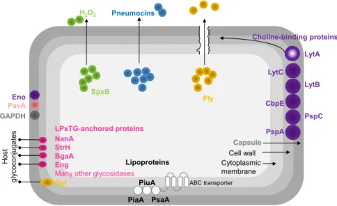
Figure 1.2. Schematic representation of pneumococcal virulence factors.
Abbreviations: NanA, neuraminidase A; StrH, β-N-acetylglucosaminidase; BgaA,
β-galactosidase; Eng, endo-α-N-acetylgalactosaminidase; Hyl, hyaluronate lyase; Eno,
enolase; PavA, pneumococcal adherence and virulence factor A; GAPDH, glyceraldehyde 3-phosphate dehydrogenase; SpxB, pyruvate oxidase; Ply, pneumolysin; LytA, autolysin; LytC, 1,4-β-N-acetylmuramidase; LytB, endo-β-N-acetylglucosamidase; CbpE, choline-binding protein E; PspC, pneumococcal surface protein C; PspA, pneumococcal surface protein A; PiaA, pneumococcal iron acquisition A, PiuA, pneumococcal iron uptake A, PsaA, pneumococcal surface antigen A, H2O2, hydrogen peroxide (adapted from Kadioglu
et al. [30]).
Adherence of the pneumococcus appears to occur through binding to human glycoconjugates [74]. Pneumococcal glycosidases might be implicated in such process [75]. At least 10 extracellular exoglycosidases have already been identified in S. pneumoniae with different specificities
for sugar structures present in human airways. Their activity contribute in different ways to the lifestyle of the pneumococcus and has broad implications in colonisation and the virulence potential of the bacterium (reviewed in King [75]). For example, the sequential cleaving activity of terminal sugars from human glycoconjugates by NanA, BgaA and StrH results in reduction of mucus viscosity, reveals receptors for adherence, releases sugars for growth and promotes resistance to
Capsule Cell wall Cytoplasmic membrane
Pneumocins
H2O2
Ply
Eno PavA
GAPDH
SpxB
LPxTG-anchored proteins NanA
StrH BgaA Eng
Many other glycosidases
Ho
st
glycoco
nj
ug
at
es
Choline-binding proteins
PspA
PspC CbpE
LytC
LytA
PsaA Lipoproteins
ABC transporter
Hyl PiuA PiaA
opsonophagocytosis by neutrophils [75–77]. Recently, BgaA was found to work as an adhesin promoting adherence to epithelial cells [78]. Another glycosidase, hyaluronidase (Hyl), acts over hyaluronic acid of the extracellular matrix, and is believed to assist in dissemination through tissues and colonisation [79,80] (Fig. 1.2).
The exact role of the serine protease, PrtA,in virulence is still unknown, but PrtA-deficient mutants were attenuated in a murine model of disease and all clinical isolates possessed this protein [81].
Among the lipoproteins identified (listed in [14]), PsaA (pneumococcal surface antigen A) is the substrate-binding lipoprotein of an ABC transporter for manganese. Mutations in this protein caused reduced adhesion and virulence and increased sensitivity to oxidative stress [82,83]. Other metal-binding lipoproteins (PiuA and PiaA) were identified to play role in virulence [30].
antibody response that increase complement deposition, conferring protection in diverse models of pneumococcal infection.PspC acts as an adhesin and binds to the complement regulatory protein factor H and to the human secretory IgA receptor, providing resistance to complement. PspC mutants presented attenuated virulence in different models of infection. Both PspA and PspC, are considered good candidates for protein-based vaccines (reviewed by Nieto et al., 2013 [84] and by
Kadioglu et al. [30]).
S. pneumoniae also have other surface proteins, lacking conventional
anchoring motifs or secretory signals (reviewed in [14]). These “non-classical surface proteins” display moonlighting functions. They are usually cytoplasmic with intracellular roles but when they reach the cell surface have other functions. Among these are, the pneumococcal adherence and virulence factor A (PavA) and the glycolytic enzymes enolase (Eno) and glyceraldehyde 3-phosphate dehydrogenase (GAPDH). PavA is an adhesin that binds to fibronectin, a component of the extracellular matrix. PavA mutants were attenuated in diverse models of pneumococcal disease and therefore is considered a crucial virulence factor [86,87]. Eno and GAPDH are plasmin(ogen)-binding proteins that allow transmigration through the basement membrane. Interaction of Eno with plasminogen was found to promote adherence in epithelial and endothelial cells [88–91].
Pneumolysin (Ply) is a potent virulence factor of S. pneumoniae which
development of pneumonia, as mutants lacking Ply were attenuated in models of pneumococcal pneumoniae [30] (Fig. 1.2).
Other aspect influencing the pneumococcal virulence potential is the competitive interactions with other microorganisms occupying the same niche during colonisation. Pneumococci produce bacteriocins (pneumocins), small antimicrobial peptides to target other bacteria from the same or closely species that do not produce the cognate immunity factor. This phenomenon has been hypothesised as the mechanism behind serotype replacement due to PCV administration [95]. Chemical warfare is also thought to contribute to the virulence potential. S. pneumoniae can produce a range of end-products, among them hydrogen
peroxide (H2O2). This metabolite, produced by pyruvate oxidase (SpxB) as a by-product of pyruvate aerobic metabolism, is known to damage host tissues and to kill other organisms in the same niche [96–98]. The high insensitivity of S. pneumoniae to H2O2 is well described [99]. In sum, the
vast diversity of pneumococcal virulence factors endows the bacterium with an “armory” to survive in the host and compete with other bacteria.
Virulence and carbohydrate metabolism
Several studies, using signature target mutagenesis (STM) [100–102], microarrays analysis of gene expression [103–106], and Tn-seq [107] have consistently revealed a link between carbohydrate metabolism and virulence of S. pneumoniae. Importantly these studies have shown the
dynamics and pattern of gene expression in different stages of pneumococcal infection, providing a means to evaluate the role of different genes in colonisation and disease. Carbohydrate uptake systems (e.g. msmK, satABC, SPD_0295-6-7, scrT, susT1T2X) [108–111] and
glycosidases (e.g.nanA, nanB, eng, bgaC) [112–117] were established
as important for in vivo fitness of S. pneumoniae by testing mutants of
and GAPDH, other central metabolic functions such as pyruvate formate-lyase (PFL) [118], lactate dehydrogenase (LDH) [119], pyruvate oxidase (SpxB) [120], have been associated with pathogenicity, as mutant strains were attenuated or avirulent in the ability to colonise or cause disease in different mouse models (Fig. 1.3). The PFL mutant phenotype likely resulted from reduced synthesis of ATP and alterations of membrane lipid composition [118]. A LDH-deficient mutant was avirulent after intravenous inoculation and attenuated post intranasal infection. These features were explained as arising from the different environmental conditions (carbohydrate availability) encountered in the two niches [119]. SpxB was found important in adaptation to different host environments. It enhances colonisation by competing with other host co-colonisers through H2O2 production or biofilm formation. In contrast its role in virulence is still controversial. Some studies showed that downregulation of spxB is
beneficial for survival in the bloodstream, but this behaviour is in contradiction to the study by Spellerberg et al., [120] that attributed the
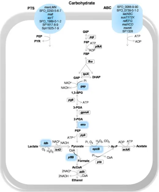
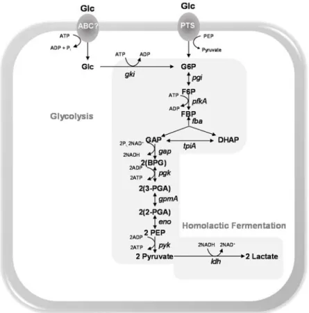
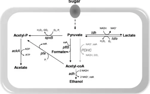
Figure 1.3. Schematic representation of sugar transporters and central carbon metabolism highlighting proteins implied in virulence of S. pneumoniae, as
assessed by in vivo studies (except for Eno and GAPDH).
Reactions are catalysed by the following protein encoding genes: gki, glucokinase; pgi,
glucose 6-phosphate isomerase; pfkA, 6-phosphofructokinase; fba, fructose-biphosphate
aldolase; tpiA, triosephosphate isomerase; gap, glyceraldehyde 3-phosphate
dehydrogenase; pgk, phosphoglycerate kinase; gpmA, phosphoglyceromutase; eno,
enolase; pyk, pyruvate kinase; ldh, L-lactate dehydrogenase; spxB, pyruvate oxidase; lcto,
lactate oxidase; pflB, pyruvate formate-lyase; pta, phosphotransacetylase; ackA, acetate
Intermediates: G6P, glucose 6-phosphate; F6P, fructose 6-phosphate; FBP, fructose 1,6-biphosphate; GAP, glyceraldehyde 3-phosphate; DHAP, dihydroxyacetone phosphate; BPG, 1,3-biphosphoglycerate; 3-PGA, 3-phosphoglycerate; 2-PGA, 2-phosphoglycerate; PEP, phosphoenolpyruvate.
In Gram-positive bacteria, the transcriptional regulator carbon catabolite protein A (CcpA) plays a major role in carbon catabolite control,
i.e., the hierarchical and highly regulated utilization of carbon sources
[122]. CcpA has been reported to contribute to pneumococcal colonisation and virulence [123,124]. The involvement of CcpA has been rationalized as a consequence of regulation of enzymes of central metabolism, impaired capsule production and/or attachment to the cell envelope, and influencing the binding of cell wall components [123–125].
Glycans at the interface of bacteria-host interactions
Glycoconjugates are widespread in nature and encompass a wide variety of molecules consisting of carbohydrates covalently linked to a non-sugar backbone. The principal categories are glycoproteins (N- or O-linked to a protein core) and glycopeptides (O-linked to an oligopeptide), peptidoglycans (bound to amino acids), glycolipids and lipopolysaccharides (linked to a lipid moiety) [126]. Despite the vast diversity of host glyconjugates, their glycan portions are often composed of the monosaccharides acetylglucosamine (GlcNAc), N-acetylgalactosamine (GalNAc), N-acetylneuraminic acid (NeuNAc), galactose (Gal) and fucose (Fuc), mannose (Man), glucose (Glc) and fructose. While the first five sugars are widespread in both N- and O-glycans (e.g. mucins), the other three are generally restricted to
These molecules contribute to important biological functions in different kingdoms of life (reviewed in [127,128]). The bacterial glycome (e.g.
capsule, exopolysaccharides, lipolysaccharides, peptidoglycan, teichoic acids and lipoteichoic acids) is far more diverse than that of animals and is usually located at the cell surface (exception polysaccharides used as energy reserves). As many bacteria (commensal or pathogens) reside in mucosal surfaces, the glycoconjugates on the cell envelope play important roles in bacteria-host interactions [128]. From the bacteria perspective these molecules are highly important for in vivo fitness. On
the other hand, in animals, and particularly in humans, a glycoconjugate barrier often prevents direct contact between bacteria and host cells. Among these, mucins are highly important in the mucosal surfaces (e.g.
Mucus and mucins have a dualistic role. On one hand represent a first defence barrier (innate defensive barrier) by trapping the microbes and protecting the underlying host surfaces from interaction with bacteria, facilitating their removal through mucociliary transport. On the other hand, the carbohydrate content of mucins provide adhesion sites and nutrients for bacteria enabling their survival and colonisation at these surfaces [130,131].
Interactions between mucin and microbes are broad in nature. For example, expression of mucin can be induced by probiotic bacteria, likely limiting the infection by pathogens [133]; the lipopolysaccharides of
Helicobacter pylori, decrease mucin synthesis in gastric epithelial cells in
vitro, modulating the mucus barrier [134]; Bacteroides thetaiotaomicron
induces fucosylation of mucin oligosaccharides, subsequently using fucose as nutrient [135]. Also, microbes have developed strategies to overcome this barrier: adhesins to bind oligosaccharides (H. pylori,
possesses four adhesins with different specificities) [136]; motility (flagella) or production of mucin degrading enzymes (proteases and glycosidases) that destabilize mucus and release carbohydrates that can be used for growth [75,137,138].
Pneumococcal carbohydrate metabolism
Carbohydrate metabolism and its importance for
pneumococcal lifestyle
The incidence and impact of pneumococcal infections have prompt intense research on the identification of factors that contribute to S. pneumoniae pathogenesis. In particular, factors that directly impinge on
adhesins and capsule [30]. Even though the relevance of carbohydrate metabolism has been recognized for in vivo fitness, only recently the
underlying mechanisms have been systematically addressed [109– 111,118,125]. With the availability of the pneumococcal genome the relevance of carbohydrates for pneumococcal fitness became apparent [139]. S. pneumoniae is a strictly fermentative bacterium that relies on
glycolytic metabolism to obtain energy [139]. It lacks a complete set of respiratory genes and is, for that reason, unable to generate energy by respiration [139,140]. Among the bacteria sharing the same niche, S. pneumoniae possesses the highest number of transport systems, and of
those more than 30% were predicted to be involved in the uptake of sugars [140]. A recent functional genomics approach validated the majority of the homology based predictions, and identified 32 carbohydrates that can be used by pneumococci [141]. It has been postulated that the vast diversity in terms of sugar utilization is driven by adaptations to host niches as a means to proliferate [142–144]. Additionally, it has been reported that sugar transporters contribute to S. pneumoniae colonisation and disease [103,109–111]. Furthermore,
numerous studies have revealed a consistent link between genes involved in sugar catabolism and virulence [100–102,105–107,145].
In the human airway free carbohydrates are scarce, being the Glc concentration below 1 mM, in contrast to its content in blood (~4-6 mM) [145,146]. Hexoses, and in particular Glc, are generally the preferred carbon sources for several bacteria and the same seems to be true for the pneumococcus [125,141]. Therefore, in vivo growth in the
nasopharynx requires alternative carbon sources. The host glycoproteins (O- and N-linked glycans, and glycosaminoglycans), secreted or lining the epithelial surfaces appear as good candidates to serve as carbon and energy sources for pneumococcal growth. S. pneumoniae can grow on
Furthermore, it is able to grow on mucin as the sole carbon source [147]. In addition to mucins and other glycans, carbohydrates might also be provided by the host diet and other microbial residents of the same niche [143].
S. pneumoniae is equipped with at least 10 extracellular (exo- or endo)
glycosidases with a broad range of specificities (reviewed by King [75]). These enzymes can break down O-linked glycans (e.g. BgaC, Eng) [114–
116], N-linked glycans (e.g. NanA, StrH, BgaA, NanB) [77,148], and
glycosaminoglycans (hyaluronic acid) (e.g. Hyl) [110], providing free
sugars that can potentially be used by the pneumococcus to grow [110,148].
Sugar transport systems in
S. pneumoniae
Sugar transport across the membrane is the first step in catabolism. In
Streptococcaceae there are three types of transport systems: the
secondary carriers, the primary active transporters ATP-binding cassette superfamily (ABC transporters) and the phosphoenolpyruvate-dependent
carbohydrate phosphotransferase systems (PTS systems) (Fig. 1.4). Secondary carriers couple the translocation of the carbohydrate to an electrochemical gradient and comprise symporters (a carbohydrate is imported together with a solute), antiporters (carbohydrate and solute are transported in opposite directions) and uniporters (catalyse the unidirectional translocation of the sugar across the cytoplasmic membrane) [149].
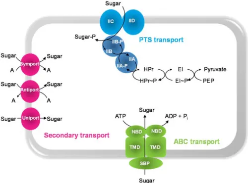
Figure 1.4. Schematic representation of sugar transport systems in S. pneumoniae.
Abbreviations: A, general solute; SBP, solute-binding protein; NBD, nucleotide-binding domains; TMD, transmembrane domains; ABC, ATP-Binding Cassette transporter; PEP, phosphoenolpyruvate; PTS, phosphoenolpyruvate-dependent-carbohydrate
phosphotransferase systems; EI, enzyme I and HPr, histidine phosphocarrier protein are general components of the PTS system; IIAB cytoplasmic proteins and IICD, transmembrane domains of the PTS system. The occurrence of IID component is typical of mannose type-PTS transporter. P~ corresponds to the phosphorylated form of each component.
In S. pneumoniae there are 1 CUT2 family ABC transporter and 6-7
CUT1 ABC transporters, differing in the nature of the substrate and structure. Each locus of the CUT1 family ABC transporter lacks the predicted ATPase [141]. Recently, a single ATPase (MsmK), was found to energize multiple carbohydrate ABC transporters, that are involved in the uptake of sialic acid, raffinose and maltotetraose [108].
The PTS system involves uptake and concomitant phosphorylation of the incoming sugar via a phosphorylation cascade that transfers a phosphate group from PEP. It consists of a series of cytoplasmic and membrane-bound proteins [152]. The PTS permeases (EII complexes), generally comprise 3 or 4 domains (the cytoplasmic subunits IIA, IIB and membrane-bound subunits IIC, IID) arranged in different ways, which allow the transport and phosphorylation of the substrate. While IIAB domains participate in the phosphorylation of the substrate, IICD correspond to the membrane permeases. The PTS is also composed by two PTS energy-coupling proteins, Enzyme I (EI) and the heat stable histidine phosphocarrier protein HPr, encoded by ptsI and ptsH,
respectively. While the EII subunits are usually substrate-specific, the EI and HPr are common to all PTS carbohydrates and therefore denominated general proteins [153,154]. PTS systems are energetically more favourable since transport and phosphorylation occurs in one step spending a PEP molecule instead of ATP. PEP is energetically equivalent to one molecule of ATP as one ATP is formed from PEP to pyruvate in glycolysis. In non-PTS systems more ATP is expended in both processes. This likely explains the high number of these systems in anaerobic bacteria [154].
In S. pneumoniae, the functional genomics approach conducted by
Bidossi et al. [141], identified twenty-one PTS systems, six to seven CUT1
carbohydrate uptake. The total number of transporters varies with the strain [141]. At the time, only a few transporters (beta-glucosides, sucrose, sialic acid, maltose and raffinose) had been partially characterized in S. pneumoniae [141]. Thirty-two carbohydrates can be
metabolized by the pneumococcus. Some of these were internalized by more than one transporter and the same transporter could take up different sugars [141]. Even though the complement of transporters is well covered in Bidossi et al. [141], the specificity for substrates is only fully
elucidated for a reduced number of transport systems. More in-depth studies combining biochemical and genetic approaches are needed to establish the specificity range of pneumococcal transport systems.
Glycan-derived monosaccharides: pathways and functions
In this section, catabolic pathways for the assimilation of selected carbohydrates, i.e. monosaccharides present in host glycans, will be
described. The focus will be on S. pneumoniae and closely related
organisms, but alternative pathways in other bacteria are also presented. In S. pneumoniae the pathways for the catabolism of monosaccharides
can be deduced from genome annotations, but their functionality remains largely to be proven. In this work we focused on the six carbohydrates constituents of the highly available glycoprotein, mucin.
Galactose
Galactose is abundant in the airway glycoconjugates (e.g. mucins)
Galactose catabolism
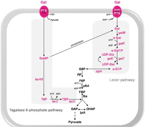
In several Streptococcaceae, metabolism by the Leloir pathway is
preceded by the entry of Gal in the cell through the secondary carrier galactose permease (GalP) [158–160]. However, homologues of this protein were not found in the pneumococcus. Recently, Bidossi et al.
[141], implicated the CUT1-family ABC transporter SPD_0088-9-0 in the uptake of Gal, but the evidence presented was relatively weak, since inactivation of the transporter resulted in a very mild reduction in Gal utilization. Possible explanations can be that the inactivation of the ABC transporter is masked by the activity of other transporters or Gal is exclusively taken up via PTS-type transporters.
Metabolism of Gal via tagatose 6-phosphate (T6P) pathway is preceded by transport via a PTS system. In other Streptococcaceae it
occurs through a lactose-specific PTS (PTSlac), encoded by lacFE genes, albeit with a low efficiency [161–164]. In S. pneumoniae, duplication of lacFE genes appears to have occurred, but the involvement of these
transporters is not yet disclosed. Nevertheless, our team showed that
lacFE genes were induced by Gal, suggesting a potential contribution of
the lactose-PTS to the uptake of the sugar [125]. Transport of Gal by a PTS complex other than PTSlac was proposed for Lactococcus lactis,
Streptococcus mutans, Streptococcus gordonii and Streptococcus oligofermentans (e.g. galactose-specific PTS and/or PTSMan)
[156,157,163,165,166].
In the pneumococcus Gal uptake was attributed to the galactitol-PTS SPD_0559-1 [141,167]. This seems to be the major Gal uptake system [141]. Two additional PTS systems were then allocated to Gal, the mannose-family PTS (ManMNL) and probably a mannose-family PTS (SPD_0066-7-8-9) [141]. The latter, was assigned based on the presence of a beta-galactosidase (bgaC) with specificity for Galβ1-3GlcNAc