online | memorias.ioc.fiocruz.br
Congenital toxoplasmosis: evaluation of serological methods for the
detection of anti-Toxoplasma gondii IgM and IgA antibodies
IMX Rodrigues, AM Castro, MBF Gomes, WN Amaral, MM Avelino/+
Hospital das Clínicas, Universidade Federal de Goiás, 1ª Avenida s/n, Setor Universitário, 74605-050 Goiânia, GO, Brasil
A study was carried out to evaluate the presence of serological markers for the immunodiagnosis of the vertical transmission of toxoplasmosis. We tested the sensitivity, specificity and predictive values (positive and negative) of different serological methods for the early diagnosis of congenital toxoplasmosis. In a prospective longitudinal study, 50 infants with suspected congenital toxoplasmosis were followed up in the ambulatory care centre of Congenital Infections at University Hospital in Goiânia, Goiás, Brazil, from 1 January 2004-30 September 2005. Microparticle Enzyme Immunoassay (MEIA), Enzyme-Linked Fluorescent Assay (ELFA) and Immune-Fluorescent Antibody Tech-nique (IFAT) were used to detect specific IgM anti-Toxoplasma gondii antibodies and a capture ELISA was used to detect specific IgA antibodies. The results showed that 28/50 infants were infected. During the neonatal period, IgM was detected in 39.3% (11/28) of those infected infants and IgA was detected in 21.4% (6/28). The sensitivity, specific-ity and predictive values (positive and negative) of each assay were, respectively: MEIA and ELFA: 60.9%, 100%, 100%, 55.0%; IFAT: 59.6%, 91.7%, 93.3%, 53.7%; IgA capture ELISA: 57.1%, 100%, 100%, 51.2%. The presence of specific IgM and IgA antibodies during the neonatal period was not frequent, although it was correlated with the most severe cases of congenital transmission. The results indicate that the absence of congenital disease markers (IgM and IgA) in newborns, even after confirming the absence with several techniques, does not constitute an exclu-sion criterion for toxoplasmosis.
Key words:congenital toxoplasmosis - specific IgM identification - specific IgA identification - diagnosis
Maternal primary infection by Toxoplasma gondii
acquired during pregnancy is of great importance since it also poses a risk of infection to the foetus and a number of serious sequelae in the infant. Approximately 40% of infected pregnant women transmit toxoplasmosis to their foetuses and this percentage is higher when the infection occurs in the third trimester. The incidence of congenital toxoplasmosis is variable, ranging from one in 1,000-one in 12,000 births (Guerina et al.1994) in various countries and in Goiânia, Goiás (GO), Brazil, Rodrigues (2006) found it to be six in 1,000 live newborns.
Both precocious diagnosis of toxoplasmosis and suit-able anti-parasitic treatment of pregnant women have shown to be effective in reducing transmission severity, although they do not greatly alter the possibility of foetal infection (Foulon et al. 1999). The clinical spectrum of congenital infection by T. gondii ranges from apparent developmental alterations at birth, with high perinatal morbidity and mortality (microcephaly, intrauterine growth retardation and hydrocephaly), to subclinical in-fection, with a risk of developing retinochoroiditis and/ or later complications (Roizen et al. 1995).
Aiming to reduce the sequelae caused by this pro-tozoan, definite diagnosis of congenital infection is mandatory and must be promptly carried out.
Serologi-Financial support: Secretaria Municipal de Saúde de Goiânia +Corresponding author: mariza.avelino@gmail.com Received 5 March 2008
Accepted 3 March 2009
cal tests are the fastest way to diagnose this infection since they can indicate the presence of two classes of immunoglobulins that do not cross placental barrier in the newborn or nursing infant serum: anti-T. gondii IgM and IgA (Remington et al. 2001).
Demonstration of specific IgA anti-Toxoplasma an-tibodies have been the recommended antibody isotype for diagnosing neonatal infection because they can be as sensitive as, or more sensitive than, the demonstration of IgM antibodies (Stepick- Biek et al. 1990). By contrast, the presence of specific IgG antibodies may only repre-sent transmission of maternal antibodies, which disap-pear in the first year of life.
There are many difficulties in diagnosing congeni-tal toxoplasmosis; for example, approximately 80% of infected newborns are asymptomatic at birth and may not present any manifestation of this infection for years. However, this does not prevent the late appearance of lesions, mainly in the eyes and brain, which can lead to blindness or mental retardation (Caiaffa et al. 1993). Thus, the need for sensitive and specific serological methods for the precocious diagnosis of congenital toxo-plasmosis prompted this study.
MATERIAL AND METHODS
investi-gation signed a Free and Informed Consent form. The maternity ward of Hospital das Clínicas is a reference centre for the diagnosis and treatment of newborns with suspected congenital toxoplasmosis and receives pregnant women diagnosed with acute toxoplasmo-sis during pregnancy from all of the Health Units of Goiânia and other cities in GO.
The Pregnant Protection Program sorted the women with suspected acute infection by identifying specific IgM anti-T. gondii antibodies in blood samples taken from the digital pulp (filter-paper ELISA test) at the first prenatal check-up. The presence of these antibodies was confirmed by an ELISA test carried out on peripheral blood samples, followed by IgG avidity test. A low level of IgG avidity (< 25%) indicated the acute phase of T. gondii infection. Moreover, since other serological reac-tions were performed, the probability of cross-reactivity and/or misdiagnosis of other infections, such as syphilis, rubella, hepatitis B, hepatitis C, HIV, HTLV-1, Chagas disease, herpes virus and cytomegalovirus, is remote.
Inclusion criteria were: (i) mother’s acceptance to participate in this research and (ii) final conclusion whether vertical transmission was present. Exclusion criteria were: (i) no return to the service and (ii) incon-clusive toxoplasmosis diagnosis.
The selected women were sent to the Reference Ser-vice of the hospital, where they underwent exams after 20 weeks of pregnancy to diagnose infections in samples col-lected by cordocentesis and amniocentesis. At the time of delivery, the women were directed to the maternity ward of the same hospital and samples were taken from the umbilical cord of all newborns with suspected congenital toxoplasmosis. These samples were tested for the detec-tion of specific IgM and IgG anti-T. gondii antibodies.
In this study, 50 infants born to mothers with acute T. gondii infection were followed until the congenital tox-oplasmosis diagnostic was concluded and one of them was a possible case of T. gondii reinfection during preg-nancy. Blood samples of these individuals were tested using a Microparticle Enzyme Immunoassay (MEIA), an Enzyme-Linked Fluorescent Assay (ELFA) and an Immune-Fluorescent Antibody Technique (IFAT) to detect specific IgM anti-T. gondii antibodies. Then they were stored at -20ºC until detection of specific IgA by a double-sandwich ELISA (capture). The MEIA was eval-uated with automated equipment (Axsym, Abbott) to de-tect anti-T. gondii IgG and IgM antibodies in the blood samples of the newborns. Reference values for IgG were: > 3 UI/mL: reactive; between 2-3 UI/mL: indeterminate; and < 2 UI/mL: nonreactive. Reference values for IgM were: > 0.600 UI/mL: reactive; between 0.500-0.600 UI/ mL: indeterminate; and < 0.500 UI/mL: nonreactive. Blood samples were sent to a private laboratory to de-tect anti-T. gondii IgM antibodies using ELFA (VIDAS, Bio-Mérieux) and IFAT (Camargo & Leser 1976). Also, double-sandwich ELISA (capture) was used to detect specific IgA anti-T. gondii. Reference values for IgM us-ing ELFA were: < 0.55 UI/mL: nonreactive; > 0.55 and < 0.65 UI/mL: indeterminate; and > 0.65 UI/mL: reac-tive (Bio-Mérieux Instruction Manual Toxo-M).
Refer-ence values for IFAT were: < 1/10: nonreactive; and ≥
1/10: reactive. Reference values for specific IgA were: < 4.5 UA/mL: nonreactive; between 4.5-5 UA/mL: in-determinate; and > 5 UA/mL: reactive. The ELFA was performed because it captures IgM antibodies, avoids false-positive (due to the presence of rheumatoid fac-tor) and false-negative (due to excess IgG) reactions, and presents sensitivity and specificity comparable to the immunosorbent agglutination assay (ISAGA) of 93.5% and 99.3%, respectively.
In order to prevent possible cross-reactions of IgM anti-T. gondii antibodies with anti-cytomegalovirus, rubella, Epstein-Barr virus antibodies and anti-syphilis antibodies, serology for these viruses was carried out in the reactive IgM samples using MEIA for the first two, indirect agglutination for the third and Venereal Disease Research Laboratory test for the last group.
Congenital toxoplasmosis diagnosis was achieved using the following service protocol: identification of specific IgM and/or IgA anti-T. gondii antibodies in the umbilical cord of newborns with suspected congenital toxoplasmosis, approximately two weeks after birth, confirmed in peripheral blood; presence of characteris-tic clinical signs (retinochoroiditis or intracranial calci-fication); identification of protozoan through culture and inoculation in mice followed by histopathological exam of the brains of inoculated animals; identification of spe-cific anti-T. gondii antibodies in the cephalo-rachidian liquid (CRL) of newborns with suspected congenital infection; IgG persisting for more than six months in infants treated since birth; detection of specific IgM during laboratory control or detection of antibodies after ceasing treatment of infants who received sulfadiazine in association with pyrimethamine and folinic acid since birth (Berger et al. 1992). The recognised gold standard diagnostic method for congenital toxoplasmosis has been the isolation of the parasite. However, specific IgM and IgA anti-T. gondii antibodies, which are not trans-mitted through the placenta, have also been considered serological markers of vertical transmission. Further-more, characteristic symptoms, such as retinochoroidi-tis, diffuse intracranial calcifications, microcephaly and hydrocephaly in the newborn (in the absence of sero-logical positivity for syphilis, rubella, cytomegalovirus, HIV, hepatitis B and C, herpes and Chagas disease in the mother) have also been considered diagnostic, since these symptoms indicate the neuro-ophthalmic form of toxoplasmosis. The presence of specific antibodies in CRL, followed or not by the growth of cells or proteins and without cranial volume changes, was considered a possible acute meningitic form of congenital toxoplas-mosis and the infants were considered infected until confirmation of toxoplasmosis through other results was provided, such as: persistence of antibodies six months after birth; positivity of antibodies after ceasing treat-ment in the case of past negative exams; increase of IgG antibodies during the follow-up; or detection of retino- choroiditis during the follow-up.
IgM antibodies were determined using MEIA, ELFA and IFAT and of IgA antibodies using a capture ELISA to diagnose congenital toxoplasmosis. Calculations were made using Thomas Bayes’s mathematical formu-las (Kawamura 2002).
Data were analysed using Excel version 2007 (Micro-soft, Brazil) and EPI-INFO (version 6.04, CDC, USA). The statistical significance level was set at p < 0.05 with a 95% confidence interval.
RESULTS
Among the 50 newborns, 28 were infected. In the 22 non-infected patients, IgG antibodies disappeared from peripheral blood between the fourth and 10th months of age (negative results were confirmed after ceasing treat-ment). The liquor exam was normal and presented negative serology, both the cranial ultrasonography and the ophthal-moscopy were normal, and the infants presented normal neuropsychomotor development at one year of age.
Serology for cytomegalovirus, rubella, mononucleosis and syphilis was carried out for the samples presenting reactive specific IgM anti-T. gondii antibodies, and all were nonreactive, demonstrating that there was no cross-reaction with the antibodies against these diseases.
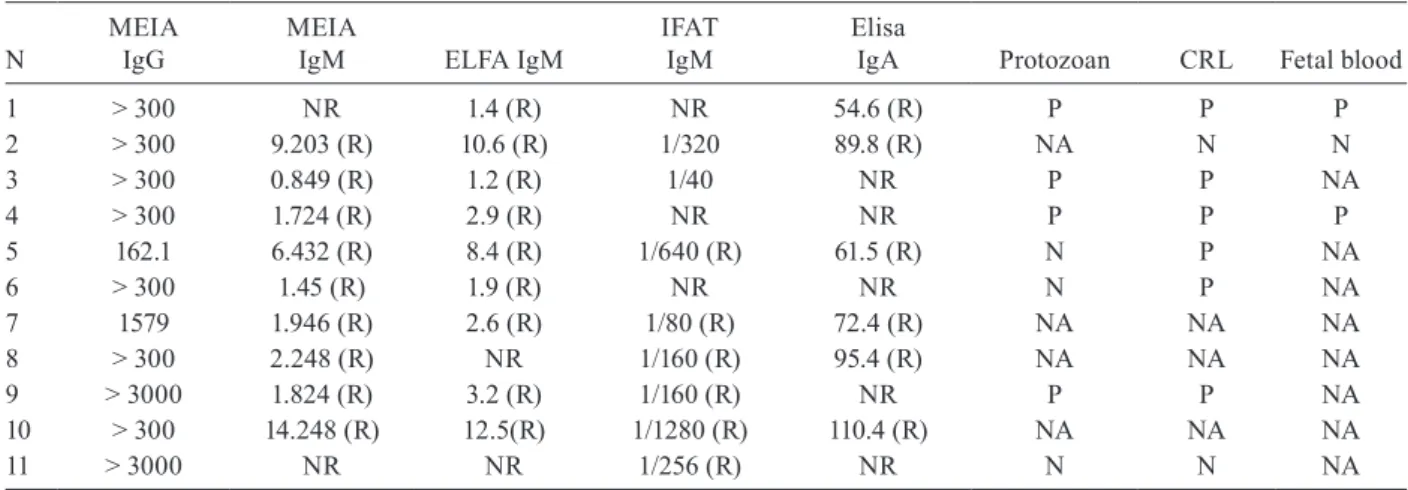
During the neonatal period, congenital toxoplasmo-sis was diagnosed through laboratory exams (serology) in 11 out of the 28 infected newborns (39.3%). This was confirmed in the first 15 days after birth based on the presence of specific IgM anti-T. gondii antibodies in the umbilical cord blood serum and also confirmed in the peripheral blood (Table I). Specific IgA was found in 21.4% (6/28) of the infants and the highest results for IgM coincided with IgA positivity, except for case 1.
Among the 11 newborns with serological markers of congenital infection, 90.9% (10/11) presented very high IgG titres and 72.7% (8/11) did not undergo complemen-tary exams for the diagnosis of foetal toxoplasmosis (Table I). One asymptomatic newborn, who had nonre-active IgM and IgA 27 days after birth, was positive for both parameters using the four techniques described two months after birth. The liquor exam was abnormal (posi-tive serology), the opthalmoscopy was abnormal, the protozoan parasite was positive and the patient received a late serological diagnosis of congenital toxoplasmo-sis. Among the newborns who were infected and symp-tomatic at birth (10/28 - 35.7%), 25% (7/28) presented serological markers of vertical transmission and severe forms of congenital toxoplasmosis.
Table II shows the clinical alterations presented by the infants who had serological markers of congenital
T. gondii infection during the neonatal period. The most severe form of congenital toxoplasmosis (neuro-oph-talmic) was present in 25% of the cases (7/28) and the presence of serological markers in their peripheral blood showed positive correlation with severity of infection.
Among the mothers of the 11 newborns with con-genital toxoplasmosis markers, five were seronegative (45.4%) in the prenatal screening (Table II) and there-fore did not receive medication. Only two newborns were asymptomatic and persisted in this condition after ceasing treatment. It was also verified that the clinical
manifestations were more severe in one individual who presented the highest concentrations of IgM and whose mother had not been treated previously (case 10).
In this study, we observed that there were alterations in CRL in 57.1% of the cases (16/28) and 31.2% of these (5/16) already presented sequelae of neurotoxoplasmosis due to precocious intrauterine infection. The remaining 68.8% (11/16) were in the acute phase of meningitis; six of them did not present any sequelae after ceasing spe-cific treatment, two presented hearing deficit and two presented cortico-subcortical dysfunction (Tables I, II).
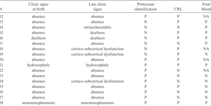
Table III displays the criteria used for the diagno-sis of congenital infection in the newborns who did not present serological markers of vertical transmission. We observed that the best method for this diagnosis in our study was the identification of specific antibodies in CRL in 57.1% of the cases (16/28), followed by the identification of protozoan in 50% of the cases (14/28). Foetal IgM was diagnosed in 32.1% of the cases (9/28), although this was tested in only 16 out of the 28 in-fected newborns (Tables I, III). Furthermore, IgM and/ or IgA were identified only two months after birth in one infant who, neither at birth nor 27 days after birth, presented serological markers of specific IgM and/or IgA. On the other hand, lack of diagnosis and specific treatment of the pregnant women seemed to have had positive correlation with the most severe clinical forms of the congenital disease at birth. Also, it is worth men-tioning that only three pregnant women underwent complementary exams for the diagnosis of foetal toxo-plasmosis and used spyramicin from the diagnosis up to the time of their infant’s birth.
Table IV presents the different criteria for the diag-nosis of congenital toxoplasmosis as well as the impor-tance of each criterion in the precocious diagnosis of this infection.
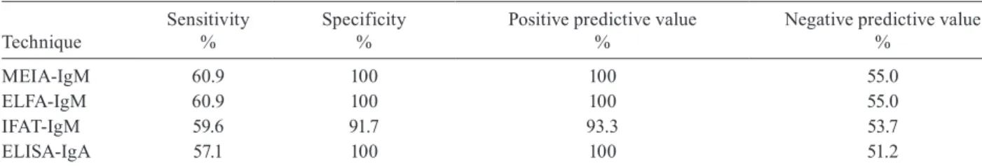
Table V displays the sensitivity, specificity and pre-dictive values (positive and negative) for the tests used. Specificity of MEIA and ELFA was 100%, since none of the newborns not affected by the infection presented specific IgM anti-T. gondii antibodies; their sensitivities were each 60.9%, as 10 out of the 28 infected patients presented IgM antibodies using both of these techniques. Specificity of IFAT was 91.7%, since two out of the 22 non-infected newborns presented false-positive results for IgM; the sensitivity of this technique was 59.6%, since nine out of the 28 infected individuals presented reactive IgM antibodies.
According to the literature, MEIA had not been tested in newborns before this study and even though it is not a capture test, it presented specificity and sensitivity com-parable to ELFA for the detection of anti-T. gondii IgM antibodies, which makes it the most indicated technique for this purpose due to its lower cost and similar efficacy.
DISCUSSION
anti-bodies do not cross the placental barrier and when they do so at birth, they have a lifespan of only five days, which allows them to be used as serological markers of vertical transmission.
Specific IgM anti-T. gondii antibodies were not de-tected in 57.1% (16/28) of the patients presenting con-genital toxoplasmosis, which confirms the low sensitiv-ity of this technique for newborns, as is widely seen in the literature. This might have happened due to techni-cal difficulties in reading the laboratory results because of: (i) maternal IgG binding in high titres to the binding
site of IgM; (ii) foetal inhibition of antibody produc-tion because of high titres of maternal antibodies in the foetus’s blood; or (iii) the decrease of parasitaemia as a consequence of the mother’s treatment (Pinon et al. 2001). This finding shows that the pregnant woman’s treatment modifies the foetus and newborn immune response as a result of the significant reduction of T. gondii circulating antigens.
The low possibility of diagnosis using serological tech-niques demonstrates the need for using several criteria for the precocious detection of congenital toxoplasmosis. In TABLE I
Immunologic and parasitologic profile of 11 newborns presenting serological markers of congenital toxoplasmosis vertical trans-mission during the neonatal period using different techniques
N
MEIA IgG
MEIA
IgM ELFA IgM
IFAT IgM
Elisa
IgA Protozoan CRL Fetal blood
1 > 300 NR 1.4 (R) NR 54.6 (R) P P P
2 > 300 9.203 (R) 10.6 (R) 1/320 89.8 (R) NA N N
3 > 300 0.849 (R) 1.2 (R) 1/40 NR P P NA
4 > 300 1.724 (R) 2.9 (R) NR NR P P P
5 162.1 6.432 (R) 8.4 (R) 1/640 (R) 61.5 (R) N P NA
6 > 300 1.45 (R) 1.9 (R) NR NR N P NA
7 1579 1.946 (R) 2.6 (R) 1/80 (R) 72.4 (R) NA NA NA
8 > 300 2.248 (R) NR 1/160 (R) 95.4 (R) NA NA NA
9 > 3000 1.824 (R) 3.2 (R) 1/160 (R) NR P P NA
10 > 300 14.248 (R) 12.5(R) 1/1280 (R) 110.4 (R) NA NA NA
11 > 3000 NR NR 1/256 (R) NR N N NA
CRL: cephalo-rachidian liquid; ELFA: Enzyme-Linked Fluorescent Assay; IFAT: Immune-Fluorescent Antibody Technique; MEIA: Microparticle Enzyme Immunoassay; N: negative; NA: not applicable; NR: nonreactive; P: positive; R: reactive. Serology for cytomegalovirus (MEIA), rubella (MEIA), Epstein-Barr virus (indirect agglutination) and syphilis was negative.
TABLE II
Correlation between the diagnosis of toxoplasmosis of the pregnant women and the presence of clinic alterations in the newborns who presented serological markers of congenital infection during the neonatal period
N
Pregnant women Newborns
Prenatal diagnostics Fetal diagnosis Treatment Clinic signs at birth Beginning treatment Late clinic signs
1 previous pegnancy R S absence at birth absence
2 IgM + NR S absence at birth seizures
3 seronegative NA none retinochoroiditis 3 months after birth no recurrence
4 IgM + NA none neurotoxoplasmosis no follow-up no follow-up
5 seronegative NA none absence 17 days after birth absence
6 IgM + R none neurotoxoplasmosis 2 months after birth neurotoxoplasmosis
7 seronegative NA none absence no follow-up no follow-up
8 seronegative NA none neurotoxoplasmosis 11 months neurotoxoplasmosis
9 IgM + NA none neurotoxoplasmosis at birth neurotoxoplasmosis
10 seronegative NA none fatal systemic infection deceased deceased
11 IgM + NA S retinochoroiditis at birth no recurrence
TABLE III
Distribution of diagnostic criteria for one newborn presenting late serological markers and 16 newborns that did not present serological markers of congenital toxoplasmosis during the neonatal period
N
Clinic signs at birth
Late clinic signs
Protozoan
identification CRL
Fetal blood
12 absence absence P P NA
13 absence absence N P P
14 absence retinochoroiditis N N P
15 absence deafness N P P
16 deafness deafness N P P
17 absence absence N N P
18 absence cortico-subcortical dysfunction N P NA
19 absence cortico-subcortical dysfunction N P N
20 absence absence P P NA
21 hydrocephaly hydrocephaly P P P
22 absence absence P N NA
23 absence absence P N N
24 absence cortico-subcortical dysfunction P N N
25 absence absence P P N
26 absence absence P N N
27 absence absence P N N
28 neurotoxoplasmosis neurotoxoplasmosis P P P
CRL: cephalo-rachidian liquid; ELFA: Enzyme-Linked Fluorescent Assay; IFAT: Immune-Fluorescent Antibody Technique; MEIA: Microparticle Enzyme Immunoassay; N: negative; NA: not applicable; P: positive. Serology for cytomegalovirus (MEIA), rubella (MEIA), Epstein-Barr virus (indirect agglutination) and syphilis was negative. 12-MEIA IgM = 1.824 (reative); ELFA = 2.4 (reative); IFAT = 1/80 two months after birth.
our study, the variety of criteria employed allowed the di-agnosis of 28 newborns, 19 (67.8%) of whom started the specific treatment at birth. Precocious therapy is necessary because the development of late sequelae (in the brain and eyes) is a common occurrence. The present study showed that 57.1% (16/28) of the infected newborns presented al-terations in CRL; 31.2% of them (5/16) presented seque-lae of neurotoxoplasmosis due to intrauterine infection; the remaining 68.8% (11/16) were in the acute phase of meningitis; six of them did not present any sequelae after ceasing specific treatment; two presented hearing deficit; and two presented cortico-subcortical dysfunction. These two last sequelae are considered mild since the evolution of non-treated meningitis is hydrocephaly and the intrac-ranial calcifications can be responsible for seizures and mental retardation.
This study showed that the exam that best detected congenital toxoplasmosis was the one carried out in CRL because we found specific anti-T. gondii antibodies in 57.2% of the cases, followed by parasite isolation (50%), and identification of serological markers (42.8%), as pre-sented in Table III. These exams permitted a confirma-tory diagnosis of congenital toxoplasmosis in all 28 cases (Tables I-III). Nonetheless, one isolated criterion, such as the identification of IgM in neonatal screening, should not be used in place of prenatal serological tests for sero-conversion surveillance since it presented a good result in only 39.3% of the infected newborns.
On the other hand, the presence of serological mark-ers of vertical transmission was positively correlated with a higher severity of the congenital infection 63.6% (7/11) and one newborn died as a consequence of system-ic toxoplasmosis. Retinochoroiditis identified at birth was observed in 18.2% (2/11) of the infected newborns who presented serological markers of congenital infec-tion and in 27.3% (6/22) among the other infected new-borns, for a total of 28.6% (8/28), as shown in Table IV.
Among the newborns presenting congenital toxoplas-mosis, 39.3% (11/28) showed symptoms of the infection and IgM antibodies were not detected in two of those (18.2%) who presented the neurologic form. There is a consensus regarding congenital syphilis (Borobio et al. 1980, Serra et al. 1982, Srinivasan et al. 1983) that IgM levels are high in symptomatic newborns and can be nor-mal or low in asymptomatic individuals (Horwitz 1980). It seems that the same remains true for toxoplasmosis, since in the present study, 70% (7/10) of the symptomatic newborns presented anti-T. gondii IgM antibodies.
The sensitivity of identifying anti-T. gondii antibodies by using three serological techniques (MEIA, ELFA and IFAT) was just 42.8% (12/28), a fact of great concern, as in many paediatric services the detection of this antibody with the newborn screening test is the only exam carried out in order to diagnose congenital toxoplasmosis.
sensitiv-ity (57.1%) than MEIA (60.9%), ELFA (60.9%) and IFAT (59.6%). This low sensitivity might result from the tech-nique used (double-sandwich ELISA) and/or the treat-ment of pregnant women and newborns with suspected congenital infection (Pinon et al. 1996).
It should be emphasised that the suspected congeni-tal toxoplasmosis cannot be dismissed simply by taking into consideration the absence of IgM and IgA antibod-ies or the negative result obtained for IgG antibodantibod-ies, mainly for newborns who had been treated during
in-trauterine and/or post-natal phases. The follow-up of the newborns with suspected congenital toxoplasmosis must be carried out in specialised centres, using all possible diagnostic resources available, so that no child suffers the consequences of misdiagnosed toxoplasmosis based solely on the absence of IgM or IgA antibodies.
ACKNOWLEDGEMENTS
To Maria Heloísa Mesquita, who made this scientific research possible.
TABLE V
Sensitivity, specificity and predictive values (positive and negative) of serological reactions used for the diagnosis of congenital toxoplasmosis
Technique
Sensitivity %
Specificity %
Positive predictive value %
Negative predictive value %
MEIA-IgM 60.9 100 100 55.0
ELFA-IgM 60.9 100 100 55.0
IFAT-IgM 59.6 91.7 93.3 53.7
ELISA-IgA 57.1 100 100 51.2
ELFA: Enzyme-Linked Fluorescent Assay; IFAT: Immune-Fluorescent Antibody Technique; MEIA: Microparticle Enzyme Im-munoassay. CI: MEIA/ELFA-IgM = 44.15-77.64; ELISA-IgA = 41.42-72.78; IFAT-IgM = 43.22-75.98.
TABLE IV
Evaluation of criteria for the diagnosis of congenital toxoplasmosis in 50 newborns followed-up in a longitudinal study
Diagnostic criteria
Toxoplasmosis
(%) NA
No toxoplasmosis
(%) NA Total
IgM fetal (+)a 32.1 (9/28) 0 (0/22) 18.0 (9/50)
IgM fetal (-) 42.8 (7/28) 50.0 (11/22) 36.0 (18/50)
Total 57.1 (16/28) 42.9 (12/28) 50.0 (11/22) 50.0 (11/22) 54.0 (27/50)
IgM (+)b 42.8 (12/28) 0 (0/22) 24.0 (12/50)
IgM (-) 57.2 (16/28) 100.0 (22/22) 76.0 (38/50)
Total 100.0 (28/28) 0 (0/28) 100.0 (22/22) 0 (0/22) 100.0 (50/50)
retinochoroiditis (+)c 28.6 (8/28) 0 (0/22) 16.0 (8/50)
retinochoroiditis (-) 67.8 (19/28) 100.0 (22/22) 84.0 (42/50)
Total 96.4 (27/28) 3.6 (1/28) 100.0 (22/22) 0 (0/22) 100.0 (50/50)
hidrocephaly (+)d 21.4 (6/28) 0/22 12.0 (6/50)
hidrocephaly (-) 78.6 (22/28) 100.0 (22/22) 88.0 (44/50)
Total 100.0 (28/28) 0 (0/28) 0 (0/22) 0 (0/22) 100.0 (50/50)
protozoan (+)e 50.0 (14/28) 0 (0/22) 28.0 (14/50)
protozoan (-) 35.7 (10/28) 72.7 (16/22) 52.0 (26/50)
Total 85.7 (24/28) 14.3 (4/28) 72.7 (16/22) 27.3 (6/22) 80.0 (40/50)
CRL (+)f 57.2 (16/28) 0 (0/22) 32.0 (16/50)
CRL (-) 32.1 (9/28) 81.8 (18/22) 54.0 (27/50)
Total 89.3 (25/28) 10.7 (3/28) 81.8 (18/22) 18.2 (4/22) 86.0 (43/50)
REFERENCES
Berger R, Sturchler D, Rudin C 1992. Cord blood screening for congen-ital toxoplasmosis: detection and treatment of asymptomatic new-borns in Basel, Switzerland. Scan J Infect Dis Suppl 84: 46-50.
Borobio MV Nogales MC, Palomares SC 1980. Value of serological diagnosis in congenital syphilis. Report of nine cases. Br J Vener Dis 56: 377-380.
Caiaffa WT, Chiari CA, Figueiredo AR, Orefice F, Antunes CM 1993. Toxoplasmosis and mental retardation. Report of a case-control study. Mem Inst Oswaldo Cruz 88: 253-261.
Camargo ME, Leser PG 1976. Diagnostic information from serologi-cal tests in human toxoplasmosis. II Evolutive study of antibodies and serological patterns in acquired toxoplasmosis, as detected by hemagglutination, complement fixation, IgG and IgM-immu-nofluorescence tests. Rev Inst Med Trop São Paulo 18: 227-238.
Foulon W, Villena I, Stray-Pedersen B, Decoster A, Lappalainen M, Pinon JM, Jenum PA, Hedman K, Naessens A 1999. Treatment of toxoplasmosis during pregnancy: a multicenter study of impact on fetal transmission and children’s sequelae at age 1 year. Am J Obstet Gynecol 180: 410-415.
Guerina NG, Hsu HW, Meissner HC, Maguire JH, Lynfield R, Stech-enberg B, Abroms I, Pasternack MS, Hoff R, Eaton RB, Grady GF 1994. Neonatal serologic screening and early treatment for con-genital Toxoplasma gondii infection. The New England Regional Toxoplasma Working Group. N Engl J Med 330: 1858-1863.
Horwitz CA 1980. Laboratory investigation of syphilis. Postgrad Med 68: 71-76, 78-79, 81.
Kawamura T 2002. Interpretação de um teste sob a visão epidemi-ológica. Eficiência de um teste. Arq Bras Cardiol 79: 437-444.
Pinon JM, Chemla C, Villena I, Foudrinier F, Aubert D, Puygauthier-Toubas D, Leroux B, Dupouy D, Quereux C, Talmud M, Trenque T, Potron G, Pluot M, Remy G, Bonhomme A 1996. Early
neona-tal diagnosis of congenineona-tal toxoplasmosis: value of comparative enzyme-linked immunofiltration assay immunological profiles and anti-Toxoplasma gondii immunoglobulin M (IgM) or IgA immunocapture and implications for postnatal therapeutic strate-gies. J Clin Microbiol 34: 579-583.
Pinon JM, Dumon H, Chemla C, Franck J, Petersen E, Lebech M, Zufferey J, Bessieres MH, Marty P, Holliman R, Johnson J, Luyasu V, Lecolier B, Guy E, Joynson DH, Decoster A, Enders G, Pelloux H, Candolfi E 2001. Strategy for diagnosis of congenital toxoplas-mosis: evaluation of methods comparing mothers and newborns and standard methods for postnatal detection of immunoglobulin G, M, and A antibodies. J Clin Microbiol 39: 2267-2271.
Remington JS, Mcleod R, Thulliez P, Desmonts G 2001. Toxoplasmo-sis. In JS Remington, JO Klein, Infectious diseases of the fetus and newborn infant, 5th ed., WB Saunders, Philadelphia, p. 205-346.
Rodrigues IMX 2006. Diagnóstico pós-natal da toxoplasmose con-gênita através da detecção de anticorpos das classes IgG, IgM e IgA anti-Toxoplasma gondii, MSc thesis, Universidade Federal de Goiás, Goiânia, 115 pp.
Roizen N, Swisher CN, Stein MA, Hopkins J, Boyer KM, Holfels E, Mets MB, Stein L, Patel D, Meier P, Withers S, Remington J, Mack D, Heydemann P, Patton D, McLeod R 1995. Neurologic and developmental outcome in treated congenital toxoplasmosis. Pediatrics 95: 11-20.
Serra G, Bruschettini PL, Bonacci W, Magliano P, Mastromatteo L, Cacciabue E 1982. La lue connatale: criteri diagnostici e condotta terapeutica. Minerva Pediatr 34: 749-756.
Srinivasan G, Ramamurthy RS, Bharathi A, Voora S, Pildes RS 1983. Congenital syphilis: a diagnostic and therapeutic dilemma. Pe-diatr Infect Dis 2: 436-441.