Polymorphism of the rDNA and tDNA loci in clinical isolates of
Pseudomonas
aeruginosa
: A perspective for molecular epidemiology surveillance
Isabel Cristina Guerra Spacov
1,2, Suzileyde Alberto Marques da Silva
2, Marcos Antônio de Morais Júnior
1,2and Márcia Maria Camargo de Morais
2,31
Departamento de Genética, Universidade Federal de Pernambuco, Recife, PE, Brazil.
2
Laboratório de Imunopatologia Keizo Asami, Universidade Federal de Pernambuco, Recife, PE, Brazil.
3
Departamento de Patologia, Instituto de Ciências Biológicas, Universidade de Pernambuco, Recife, PE,
Brazil.
Abstract
The Gram-negative bacteriumPseudomonas aeruginosa has a wide environmental and ecological distribution. It is an opportunistic pathogen that acquires resistance to multiple antimicrobial agents and can infect plants, animals and humans. We used rDNA and tDNA PCR markers to characterize the bacterial diversity ofP. aeruginosa strains isolated at a Brazilian teaching hospital (Oswaldo Cruz University Hospital, Recife, Brazil) between March 2003 and February 2004. Clonal groups ofP. aeruginosa clinical isolates were identified from different patients in different hos-pital units using either rDNA or tDNA markers, or a combination of both in a duplex PCR. These PCR-typing methods together with drug-resistance profiles were used to trace the distribution of antibiotic resistantP. aeruginosa clones and to identify cross-infection of the same patient with a different bacterial clone after being moved to a different hos-pital unit. The data presented here demonstrates a rapid, reliable and useful method for epidemiological surveillance that can contribute to the control ofP aeruginosa infections in hospital environments.
Key words:antibiotic resistance, molecular typing, nosocomial infection,P. aeruginosa,rDNA-PCR, tDNA-PCR.
Received: July 4, 2005; Accepted: April 4, 2006.
Introduction
The routine identification of hospital isolates of Pseu-domonas aeruginosais based on phenotypic analysis using
characteristic biochemical reactions. However, horizontal dissemination cannot be traced properly using phenotypic methods based only on genus and specie identification. On the other hand, methods based on the polymerase chain re-action (PCR) have been widely used for the analysis of the genetic diversity of many microorganisms (Agodi et al.,
2000; Lopeset al., 2005). Depending on the primers and
amplification conditions employed, the results allow the discrimination of organisms at not only the genus and spe-cies level but also at the level of individual strains. By using molecular typing methods it is possible to identify genetic similarities between both phenotypically unrelated and phenotypically analogous strains. These molecular ap-proaches provide the precise information necessary for the monitoring and control of hospital infections. Moreover,
epidemiological relationships can be detected and/or con-firmed and clonal groups can be delimited (Severinoet al.,
1999).
The fact that the completeP. aeruginosagenome
se-quence is known allows the identification of polymorphic regions present in different bacterial strains and the design of methods to discriminate isolates based on such poly-morphisms (see The Pseudomonas Genome Project at www.pseudomonas.com).
The P. aeruginosa rRNA cluster
(5S-23S-(ISR)-tRNAALA-(ISR)-tRNAILE-(ISR)-16S) is distributed four ti-mes throughout the genome of the sequencedP. aeruginosa
strain Pa01. The intergenic spacer regions (ISRs) are sub-ject to lower evolutionary pressure, and therefore show wider genetic variation (Gürtler and Stanisich, 1996) that is dependent on the number and type of both tRNA genes and
boxAsequence between 23S and 16S sequences and by the
presence of enzyme-recognition sites at the ISRs, besides the length of the ISRs themselves (García-Martínezet al.,
1999). Two PCR-based methods have been proposed for identifying this cluster. Intergenic Transcribed Spacer PCR ribotyping (PCR-ribotyping) uses specific primers that am-plifies sequences between the 16S and 23S gene and has www.sbg.org.br
Send correspondence to Márcia Maria Camargo de Morais. Depar-tamento de Patologia, Instituto de Ciências Biológicas, Univer-sidade de Pernambuco, Rua Arnóbio Marques 310, 50100-130 Recife, PE, Brazil. E-mail: camargo@icb.upe.br.
been applied for molecular identification of bacteria at the species level (Jensenet al., 1993; Daffonchioet al., 1998;
Agodi et al., 2000) and the discrimination of bacterial
strains (Kostman et al., 1992; Clementino et al., 2001;
Pereiraet al., 2002). This method detects both the number
of tRNA genes and the spacer length within the cluster. An-other highly promising method for bacterial identification is based on the PCR length polymorphisms of the inter-genic spacers between tRNA genes (tDNA-PCR) spread along the bacterial genome. The tRNA genes are highly conserved among eubacteria and occur in multiple copies throughout the bacterial genome, within and outside rDNA gene clusters. These genes are generally clustered and are separated by spacers whose length and sequences are sub-jected to a higher degree of variations (Welsh and McClelland, 1992). The tDNA-PCR method uses consen-sus primers complementary to the highly conserved edges of the flanking tRNA genes that are directed outwards (Welsh and McClelland, 1991). This method is regarded as producing species-specific banding patterns and has been applied to the differentiation of various bacterial species (Vaneechoutteet al., 1998; De Gheldreet al., 1999;
Cle-mentinoet al., 2001).
A significant increase in the number of multi-drug re-sistant (MDR)P. aeruginosainfections has been detected by our group at the Oswaldo Cruz University Hospital, Recife, Brazil (unpublished results). However, biochemi-cal routine identification was not able to distinguish clonal groups of multi-drug resistant strains. We tested rDNA-PCR and tDNA-rDNA-PCR for their capacity to type P. aeruginosahospital isolates, our aim being to contribute to the epidemiological control of P. aeruginosainfections. Our results indicate that rDNA/tDNA duplex PCR might be useful forP. aeruginosatyping of isolates from different hospital units. Moreover, the sequence of acquisition and/or expression of antimicrobial resistance by cells of distinct clonal groups could be identified.
Material and Methods
Bacterial strains and microbiological methods
The Pseudomonas Genome Project (Boston, USA) kindly providedPseudomonas aeruginosastrain PA01 and
The Oswaldo Cruz Foundation Microbial Collection (INCQS, Rio de Janeiro, Brazil) provided the Pseudomo-nasreference strainsP. aeruginosaATCC 9027,P. putida
ATCC 15175 andP. fluorescensATCC 13525.
We collected 34 P. aeruginosa isolates from
nosocomial infections of patients hospitalized in different units of the Oswaldo Cruz University Hospital (Recife, Brazil) between March 2003 and February 2004, the iso-lates being obtained from a variety of clinical samples (Ta-ble 1). The isolates were identified by colony pigmentation, grape-like odor, motility and biochemical tests (carbohy-drate fermentation (-), citrate assimilation (+), lysine
decar-boxylase (-), indol (-), oxidase (+), beta-hemolysis on blood-agar (+), DNAse (-). Identifications were confirmed using the ID32 Mini-Api automatic system (BioMerieux, France). Antibiogram data were registered by disk diffu-sion method according to the criteria ofNational Commit-tee for Clinical Laboratory Standards (NCCLS M100).
Strains were maintained on nutrient agar (pH 7.4) contain-ing (gL-1) beef extract, 10; tryptone, 5; NaCl, 5; bacterio-logical agar 12 g or by freezing in 15% (v/v) glycerol at -80 °C. All reagents were of at least analytical purity, and beef extract, tryptone and bacteriological agar were sup-plied by Oxoid (Oxoid Brasil Ltda, São Paulo, Brazil).
Of the 34P. aeruginosahospital isolates collected, 33
were confirmed by automated identification except for iso-late 22 (Table 1) which gave an unacceptable profile forP. aeruginosa. All the isolates, including isolate 22, were
sub-mitted to antibiogram assays with nine antibacterial agents suitable forPseudomonas(Pellegrinoet al., 2002). Isolates
6, 22 and 26 were susceptible to all drugs tested while the other isolates displayed different individual susceptibility profiles, with some showing multiple drug resistance (MDR) to up to eight different antibacterial agents. No cor-relation was observed between bacterial antibacterial sus-ceptibility profiles and site of infection or hospital unit (Table 1). Genomic DNA from each isolate was extracted for further molecular analysis.
Genotyping
Genomic DNA was extracted according to Ausubelet al(1989), separated on 0.8% (w/v) agarose gel, stained
us-ing ethidium bromide and visualized under UV light. We carried out rDNA-PCR by amplification of ISRs regions between the rRNA16S-23S genes with the 03 pri-mer (5’-TTGTACACACCGCCCGTCA-3’) complemen-tary to the rRNA 23S conserved region and the 04 primer (5’-GGTACCTTAGATTGTTTCAGTTC-3’), comple-mentary to the rRNA 16S conserved region according to Kostmanet al. (1992). The PCR reactions were prepared
containing 50 ng of genomic DNA, 20 pmol of each primer, 4 pmols of dNTP, 37.5 pmols of MgCl2, 1U ofTaqDNA polymerase and 1x reaction buffer (Invitrogen) in a final volume of 25mL and the amplifications carried out in a
Hybaid Touchdown PCR thermocycler (Thermo Electron Corp., Waltham, MA) using an initial denaturing step of 2 min at 94 °C, followed by 30 cycles of 1 min at 94 °C, 1 min at 55 °C and 1 min at 72 °C with a final 7 min exten-sion at 72 °C. Amplification products were separated on 1.6% (w/v) agarose gel in 0.5x TBE buffer at 10 V.cm-1and stained with ethidium bromide.
Ampli-fication conditions consisted of denaturation for 2 min at 94 °C followed by 30 cycles of 30 s at 94 °C, 30 s at 55 °C and 2 min at 72 °C with a final 10 min extension at 72 °C. Amplification products were separated and visualized as described above.
Duplex PCR used both sets of primer pairs, rDNA and tDNA markers, and the reaction was conducted accord-ing to the rDNA-PCR protocol described above, with minor modifications (2 min for the cyclic extensions at 72 °C and 10 min final extension). Amplification products were
sepa-Table 1- Phenotypic and molecular characterization ofPseudomonas aeruginosaclinical isolates.
Clinical isolate1 Isolate
code2 Antibacterial resistance profile
2 PCR pattern
cp gn am pt cf cm im az pl rRNA-PCR tRNA-PCR
GICU-6Mar03-Sk 1 r r r s r r r s s R1 R1T1
CU-16Apr03-Rt 2 r r r r r s r r s R1 R1T2
CU-23Apr03-Sk 3 s s s s r r s s s R1 R1T1
CU-30Apr03-Bl 4 - r r s r r r s s R1 R1T1
ASU-27Jun03-Be 5 r r r s r r r s s R1 R1T1
IDU-23Jul03-Ur 6 s s s s s s s s s R1 R1T1
IDU-11Sep03-Bl 7 r r r r r r r r s R1 R1T1
IDU-18Sep03-Ur 8 s r s s s s s s s R1 R1T1
GICU-6Oct03-Rt 9 r r r s r r r r s R2 R2T5
GICU-7Oct03-Rt 10 r r r s r r r s s R1 R1T1
CU-10Oct03-Bl 11 s r s s r s s s s R1 R1T1
AU-23Oct03-Ur 12 r r s r r r s r s R1 R1T1
CU-31Oct03-Sk 13 s r s s r r s s s R1 R1T1
IDU-31Oct03-Rt 14 r r r s r r r s s R1 R1T1
IDU-4Nov03-Bl 15 s r s r r r r r s R4 R4T7
CEU-10Nov03-Sk 16 r r r r r r r s s R1 R1T1
IDU-11Nov03-Ur 17 r r r r r r r s s R1 R1T1
GPU-18Nov03-Ur 18 r r r r r r r s s R1 R1T1
GPU-26Nov03-Ur 19 r r r s r r r s s R1 R1T1
CU-28Nov03-Sk 20 r r r r r r r s s R1 R1T1
PU-28Nov03-Rt 21 r r r s r r r s s R1 R1T4
CU-2Dec03-Bl* 22 s s s s s s s s s R1 R1T1
CEU-3Dec03-Ur 23 r r r s r r r s s R1 R1T1
GICU-3Dec03-Sk 24 s s s s r r s s s R5 R5T8
PU-4Dec03-Rt 25 r r r s r r s s s R1 R1T1
IDU-5Dec03-Bl 26 s s s s s s s s s R1 R1T1
PU-13Dec03-Rt 27 r r r s r r s s s R1 R1T1
OU-18Dec03-Sk 28 s r s s r s s s s R1 R1T1
IDU-31Dec03-Ur 29 s r s s r s s s s R1 R1T1
GICU-9Jan04-Bl 30 - r r r r - r r s R3 R3T6
ASU-16Jan04-Rt 31 s s s s r s s s s R1 R1T1
OU-19Jan04-Bl 32 s r s s r s r s s R1 R1T3
GPU-22Jan04-Ur 33 s r r s r r r s s R1 R1T1
IDU-27Jan04-Sk 34 r r r r r r r s s R1 R1T1
1The initial letters give the hospital unit from which the clinical isolate was obtained (GICU- = General Intensive Care Unit; IDU = Infectious Diseases
Unit; CU = Cardiology Unit; ASU = Abdominal Surgery Unit; AU = Ambulatory Unit; CEU = Cardiology Emergency Unit; CU = Coronary Unit; GPU = General Practice Unit; IDU = Infectious Diseases Unit; OU = Oncology Unit; and PU = Pneumology Unit) followed by the isolation date and the clinical sample (Be = Bile; Bl = Blood; Sk = Skin, Rt = Respiratory tract; and Ur = Urine).
2This simplified code was used in the phylogenetic analysis and the text.
3Antibacterial agent code: Amicacin = am; Aztreonam = az; Cefepime = cm; Cefotaxime = cf; Ciprofloxacin = cp; Gentamicin = gn, Imipinem = im;
rated on 1% (w/v) agarose gel and stained as described above.
Computational analysis
The bacterial isolates were analyzed using the Net-Work program version 4.1.0.8 (Fluxus Technology Ltd, www.fluxus-engineering.com) designed for phylogeny analysis and showing the geographic distribution of haplo-types. Binary data, grouped according median analysis, consisted of susceptibility (0) and resistance (1) to the anti-biotics tested.
Results
Genetic typing
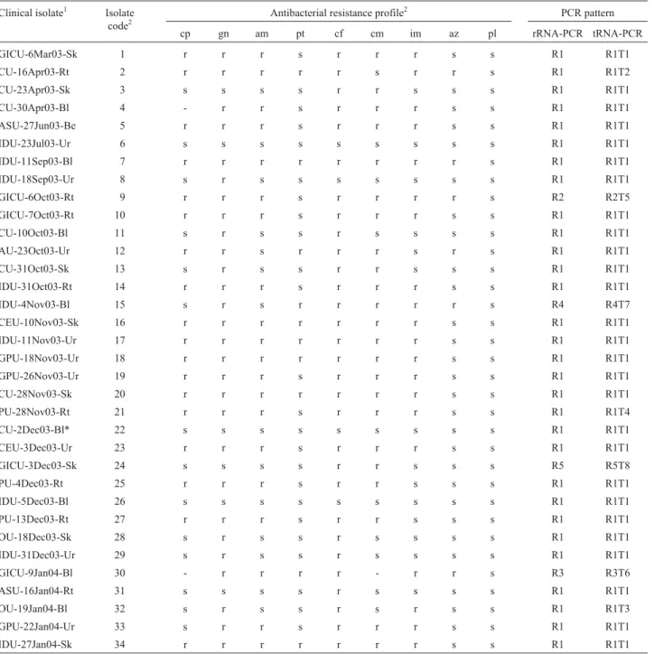
For the rDNA marker the reference strains P. aeruginosaPA01 and ATCC 9027 showed the same ampli-fication profile (banding pattern) and a characteristic 800 base pair (bp) band, which was not shown byP. putida
ATCC 15175 andP. fluorescensATCC 13525 (Figure 1A). We includedP. putidabecause it had been reported in cases of bacteremia in intensive care units for newborn babies (Bouallegue,et al., 2004) whileP. fluorescenswas selected due to reports of its presence in oncology units (Hsueh,et al., 1998). We found that isolate 22 showed an rDNA marker banding pattern similar to the twoP. aeruginosa
reference strains (Figure 1B), suggesting that was indeed a
P. aeruginosaisolate. The rDNA marker banding patterns for the remaining 33 isolates were classified in five distinct rDNA-PCR banding patterns (profiles R1 to R5, Figure 1B) of which the R1 pattern was observed in 88% of the isolates (34/30) and the twoP. aeruginosareference strains. The other four patterns were observed for only one isolate each.
The rDNA marker showed 15% of genetic variability among the isolates (5/34).
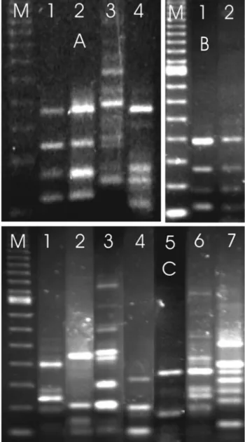
For the tDNA marker the twoP. aeruginosareference
strains again presented the same banding pattern, including four common bands (365, 250, 170 and 125 bp). As was seen for the rDNA marker banding patterns, the tDNA marker banding patterns for the P. putida and P. fluorescensreference strains also differed from those
ob-served for theP. aeruginosareference strains (Figure 2A).
The tDNA marker banding pattern of isolate 22 was similar to the reference strainP. aeruginosaATCC 9027 (Figure
2B), as also observed for the rDNA-PCR marker. Analysis
Figure 1- Amplification products generated by rDNA-PCR. A. lane 1 =P. aeruginosaPA01, lane 2 =P. aeruginosaATCC 9027, lane 3 =P. putidaATCC 15175 and lane 4 =P. fluorescensATCC 13525. B. Refer-ence strains: lane 1 =P. aeruginosaPA01, lane 2 = isolate 22 rDNA pat-tern 1, lanes 3 to 6 one representative of the other four rDNA-PCR patterns produced by automated identifiedP. aeruginosaisolates. Lane M = 100 bp ladder.
Figure 2- Amplification products generated by tDNA-PCR. A. lane 1 =P. aeruginosaPA01, lane 2 =P. aeruginosaATCC 9027; lane 3 =P. putida
of the other 33 isolates revealed eight different tDNA-PCR banding patterns (profiles T1 to T8, Figure 2C) of which the T1 pattern was seen in 79% of our bacterial isolates (27/34) as well as the two P. aeruginosa reference strains. The
tDNA marker showed 23% of genetic variability among the isolates (8/34).
Using the R1 rDNA profile and four of the tDNA profiles together resulted in four additional tDNA pro-files (propro-files R1T1, R1T2, R1T3 and R1T4), with R1T1 representing the most common pattern found among the isolates. Although the polymorphism occurring for both primers cannot be considered species-specific for P. aeruginosait can, however, be concluded that any
bacte-rial isolate presenting the R1T1 profile can be identified as belonging to this species, even, as the case of isolate 22, with ambiguous identification by the biochemical tests.
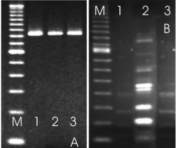
The results showed the discrimination power of both markers, which can be used as an epidemiological tool for detection of strain dispersion. In this sense, the results of PCR typing and the resistance profiles showed that bacte-rial isolates 16, 17, 118, 20 and 34 may correspond to a uniqueP. aeruginosastrain which was isolated from four
different hospital units and infected at least five different patients during the period of study (Table 1). Moreover, in one case two genotypically different isolates were collected from the same patient. Isolate 21 ( genotype R1T4) was iso-lated first but five days after the patient had been trans-ferred to another hospital unit isolate 23 (genotype R1T1) was recovered from the same patient (Figure 3), suggesting that isolate 23 may represent a second nosocomial infection episode.
Duplex-PCR typing
The results above suggested the possibility of com-bining both markers in a duplex PCR for use in typing and screening ofP. aeruginosaclinical isolates. Duplex PCR of
the twoP. aeruginosareference strains and theP. putida
andP. fluorescensreference strains generated the expected
profiles resulted from the combination of the both markers (data not shown). When duplex PCR was used for typing the 34 isolates, the profiles observed corresponded to the eight profiles detected, showing the same percentage of ge-netic variability observed for the tDNA marker (data not shown). Duplex rDNA/tDNA-PCR may thus be used as a reliable approach to follow the spread of different bacterial clonal lineages across different hospital units.
Evolution of the resistance to antibiotics
The molecular typing results suggested that some strains were disseminated to different units in the hospital. In addition, such strains may suffer distinct selective pres-sures at the different units, resulting in the acquisition and/or expression of differential resistance to antibiotics. In such a context, the isolates of the clonal lineage R1T1 were submitted to computational analysis that revealed a haplo-typic distribution among its isolates and the emergence of antibiotic resistance (Figure 4). In this analysis, isolates 6, 22 and 26 (clonal group 6) were susceptible to all the antibi-otics tested (Table 1) and could be the basal isolates from which isolate 8 (gentamicin resistant) and 31 (cefotaxime resistant) evolved by distinct events of acquiring or ex-pressing resistance. The expression of resistance to cefo-taxime by isolate 8 or to gentamicin by isolate 31 could
Figure 3- Amplification products of two bacterial isolates from the same patient. A. rDNA-PCR ofP. aeruginosaATCC 9027 (lane 1), bacterial isolate 21 (lane 2) and bacterial isolate 23 (lane 3). B. tDNA-PCR ofP. aeruginosaATCC 9027 (lane 1), bacterial isolate 21 (lane 2) and bacterial isolate 23 (lane 3). The molecular marker 100 bp ladder is shown in lane M.
have generated clonal group 11 (isolates 11, 28 and 29) and, likewise, expression of resistance to cefepime could have generated isolate 3. The expression of resistance to cefe-pime by group 11 isolates or to gentamicin by isolate 3 could have generated the isolate 13 and so on. Our analysis suggests that isolate 7 emerged from group 6 after sequen-tial expression of resistance to eight different antibiotics. Moreover, thisin silicoanalysis suggests that at least one
representative of the R1T1 genotype has not been isolated among our bacterial samples (Figure 4). This clone, show-ing resistance to four antibiotics, could have been the basal strain for clonal groups 4 and 25 and may be present, but occult, in one of the hospital units. Obviously, this analysis is an attempt to establish the relatedness among the ob-served clonal lineages, which does not excluded the possi-bility that new unrelated R1T1 clones could enter the hospital.
Discussion
At 6.3 Mb theP. aeruginosagenome is the largest bacterial genome so far sequenced, and this size, coupled with the high number of regulatory genes and subsequent complex metabolism, is consistent with its versatility in adapting to different environments (Stover et al., 2000). Based on this genomic versatility we used two PCR mark-ers that were sufficiently polymorphic to discriminate bac-terial isolates from different clonal origins, our results being reproducible and allowed the establishment of com-mon amplification patterns detected for the type strains and for the majority of hospital isolates. However, because of the polymorphism presented by both markers, we cannot suggest their use for routine bacterial identification, except, as in the case of the isolate 22, when bacterial isolates pres-ent the common pattern generated by type-strains.
Many techniques have been described for typingP. aeruginosa, such as arbitrarily primed PCR (Kersulyte et
al., 1995; Bennekovet al., 1996), pulsed field gel
electro-phoresis (Kersulyte et al., 1995; Agodiet al., 2000;
Naka-muraet al., 2001), traditional ribotyping (Bennekovet al.,
1996; Agodiet al., 2000) and more recently multi-locus
se-quence typing (MLST; Curran et al, 2004) and
entero-bacterial repetitive intergenic consensus-based PCR (ERIC-PCR; Yang et al., 2005). One of these methods, rDNA-PCR typing, has been reported as a rapid and accu-rate method for typing P. aeruginosa (Liu et al, 1996;
Agodi et al., 2000) and Burkholderia (Pseudomonas) cepacia(Dasenet al. 1994; Kostmanat al., 1992). In our
present work the rDNA marker revealed four distinct clonal groups among ourP. aeruginosaclinical isolates.Our
tech-nique was based on the amplification of ITS loci at rDNA gene clusters and any polymorphism detected may have been due to significant genetic variation such as nucleotide deletions or insertions of tRNA genes or ISRs. Therefore, two bacterial isolates presenting the same amplification pattern could be considered as originating from the same
clonal origin. The use of ITS locus restriction analysis and RFLP-ribotyping has been proposed for strain discrimina-tion and clustering by evaluating genetic differences caused by point mutations that create or remove restriction sites (Gruneret al., 1993).
Several authors have used tDNA-PCR typing to iden-tify bacterial genera and species,e.g. Lactobacillus(Baele et al., 2002) and other bacteria (Welsh and McClelland,
1991) but our results suggest that this approach is more ap-propriated for intraspecific discrimination and we have found similar results for clinical isolates of Klebsiella pneumoniae(manuscript in preparation). Our work is the
first report of the application of tDNA toP. aeruginosa
typ-ing, which increased the discriminatory power of the rDNA marker and the combination of both markers in a duplex PCR proved to be a reliable tool with high discriminatory power for typingP. aeruginosaisolates. The combination
of both markers generated amplification patterns that var-ied from three to ten bands, which may be enough for strain discrimination.
Strain discrimination and clustering can also be ob-tained using RAPD analysis (Mataret al., 2005), which
generates more bands than tDNA-PCR typing. However, RAPD analysis has been criticized because of its low repro-ducibility and reliability and the fact that amplification can be influenced by factors other than the genetic variation be-tween bacterial isolates. Therefore, it can be difficult to re-produce RAPD results between different laboratories, which is a great disadvantage for epidemiological surveil-lance studies. On the other hand, the specific primer anneal-ing which occurs with both rDNA-PCR and tDNA-PCR ensures reproducibility wherever these techniques are used. Gencer et al. (2002) have pointed out that P. aeruginosa strains are the third most prevalent pathogen
isolated from cases of hospital infections, while Pellegrino
et al. (2002) reported that in some hospitals it is the main
bacterial species isolated. The Intensive Care Unit (ICU) at the hospital where our isolates were collected has shown an increase in the number of cases ofP. aeruginosainfections
over the last five years as well as in the antibiotic resistance (data not shown) and diversity of the clonal groups of theP. aeruginosa isolates recovered (Table 1). Similar genetic
variability in ICUP. aeruginosaclinical isolates has been
reported by Sarwariet al. (2004) and Stoveret al. (2000)
have noted that the genome complexity ofP. aeruginosa
seems to be responsible for the versatility, environmental adaptation and intrinsic drug resistance exhibited by this organism.
Nosocomial infections related toP. aeruginosa are
frequently caused by multi-drug resistant (MDR) strains (Sarwariet al., 2004), which we also detected in our study
(Table 1). Nevertheless, data from susceptibility tests and amplification profiles could be used to follow the spread of clonal groups through the hospital units. In addition, such data could be useful for analysis of the evolution of the phenotype of resistance to antibiotics, which may rep-resent the expression of intrinsic mechanisms or the ac-quisition of resistance mechanisms by horizontal genetic transmission (Poole and Srikumar, 2001). Analysis of the distribution of antibiotic resistance could help in develop-ing an effective control of nosocomial infections by iden-tifying the origin of contamination. By assuming that all the isolates in the R1T1 group were genetically similar we were able to follow the spread of members of this group (and of the other seven groups) to the various hospital units and infection sites. This was the case for clonal group 16 (isolates 16, 17, 18, 20 and 34), members of which were detected in five different patients in distinct units of the hospital, indicating that members of this group were dispersed in different points within the hospital. On the other hand, some isolates seemed to be confined to a particular unit (Table 1). Our results support those of Galeset al. (2001) and suggest that informative molecular
markers can be a powerful method for following the dis-persion of clonal groups and to detect the occurrence of cross-infections (Blanc et al., 1997; Ruiz et al., 2004).
Another example of informative data produced by our analysis is the identification of the two clones which were collected from the same patient at different times and in different hospital units, these clones showing the same an-tibiotic resistance phenotype but distinct tDNA profiles. These results indicate that phenotypic convergence for an-tibiotics resistance can occur in different bacterial strains submitted to similar selective pressure.
In conclusion, our results indicate that the use of rDNA and tDNA markers forP. aeruginosatyping was suf-ficiently discriminatory to generate very informative data regarding the clonal dispersion of nosocomial hospital strains of P. aeruginosa. Moreover, results from the
du-plex-PCR associated to antibiotic susceptibility analysis may be used as an epidemiological approach to preventP. aeruginosaoutbreaks.
Acknowledgments
The authors thank the following: Oswaldo Cruz Uni-versity Hospital; UniUni-versity of Pernambuco Bacteriology Laboratory; and the Keizo Asami Immunopathology Lab-oratory of the Federal University of Pernambuco, Recife, Pernambuco, Brazil. We also thank INCQS/FIOCRUZ for the reference strains and thePseudomonas Genome Pro-jectfor the sequenced strain and the public information
about the bacterial genome. This work was supported by grants from the Brazilian agencies CAPES, CNPq and FACEPE.
References
Agodi A, Sciacca A, Campanile F, Messina C, Barchitta M, Sciacca S and Stefani S (2000). Molecular epidemiology of
Pseudomonas aeruginosafrom cystic fibrosis in Sicily: Ge-nome macrorestriction analysis and rapid PCR-ribotyping. New Microbiol 23:319-327.
Ausubel FM, Brent R, Kingston RE, Moore DD and Smith JA (1989) Current Protocols in Molecular Biology. v 1 and 2. Jonh Willey & Sons, Inc. New York, 650 pp.
Baele M, Vaneechoutte M, Verhelst R, Vancanneyt M, Devriese LA and Haesebrouck F (2002) Identification of
Lactobacillus species using tRNA-PCR. J Microbiol Methods 50:263-271.
Benekov T, Colding H, Ojeniyi B, Bentzon MW and Hoiby N (1996) Comparison of ribotyping and genome fingerprinting ofPseudomonas aeruginosaisolates from cystic fibrosis
pa-tients. J Clin Microbiol 34:202-204.
Blanc DS, Parret T, Janin B, Raselli P and Francioli P (1997) Nosocomial infections and pseudoinfections from contami-nated bronchoscopes: Two-year follow up using molecular markers. Infect Control Hosp Epidemiol 18:134-136. Bouallegue O, Mzoughi R, Weill FX, Mahdhaoui N, Ben Salem
Y, Sboui H, Grimont F and Grimont PA (2004) Outbreak of
Pseudomonas putida bacteraemia in a neonatal intensive care unit. J Hosp Infect 57:88-91.
Clementino MM, Filippis I, Nascimento CR, Branquinho R, Ro-cha CL and Martins O (2001) PCR analysis of tRNA intergenic spacer, 16S-23S internal transcribed spacer, and randomly amplified polymorphic DNA reveal inter- and intraspecific relationships ofEnterobacter cloacaestrains. J
Clin Microbiol 39:3865-3870.
Curran B, Jonas D, Grundmann H, Pitt T and Dowson CG (2004) Development of a multilocus sequence typing scheme for the opportunistic pathogenPseudomonas aeruginosa. J Clin Microbiol 42:5644-5649.
Daffonchio D, Borin S, Giuseppe F, Manachini PL and Sorlini C (1998) PCR fingerprinting of whole genome: The spacers between the 16S and 23S rRNA genes and of intergenic tRNA gene regions reveal a different intraspecific genomic variability ofBacillus cereusandBacillus licheniformis. Int
J Syst Bacteriol 48:107-116.
Dasen SE, LiPuma JJ, Kostman JR and Stull TL (1994) Character-ization of PCR-ribotyping forBurkholderia(Pseudomonas)
cepacia. J Clin Microbiol 32:2422-2424.
De Gheldre Y, Vandamme P, Gooses H and Struelens MJ (1999) Identification of clinically relevant viridans streptococci by analysis of transfer DNA intergenic spacer length polymor-phism. Int J Syst Bacteriol 49:1591-1598.
Gales AC, Jones RN, Turnidge J, Rennie R and Ramphal R. (2001) Characterization of Pseudomonas aeruginosa
iso-lates: Occurrence rates, antimicrobial susceptibility patterns, and molecular typing in the global SENTRY anti-microbial surveillance program, 1997-1999. Clin Infect Dis 15:146-155.
García-Martínez J, Acinas SG, Antón AI and Rodríguez-Valera F (1999). Use of the 16S-23S ribosomal genes spacer region in studies of prokaryotic diversity. J Microbiol Meth 36:55-64. Gencer S, Ak O, Benzonana N, Batirel A and Ozer S (2002)
Sus-ceptibility patterns and cross resistances of antibiotics againstPseudomonas aeruginosain a teaching hospital of
Gürtler V and Stanisich A (1996) New approaches to typing and identification of bacteria using the 16S-23S rRNA spacer re-gion. Microbiology 142:3-6.
Gruner E, Kropec A, Huebner J, Altwegg M and Daschner F (1993) Ribotyping ofPseudomonas aeruginosastrains
iso-lated from surgical intensive care patients. J Infect Dis 167:1216-20.
Hsueh PR, Teng LJ, Pan HJ, Chen YC, Sun CC, Ho SW and Luh KT (1998) Outbreak ofPseudomonas fluorescens bactere-mia among oncology patients. J Clin Microbiol 36:2914-2917.
Jensen MA, Webster JA and Straus N (1993) Rapid identification of bacteria on the basis of polymerase chain reaction-amplified ribosomal DNA spacer polymorphisms. Appl En-viron Microbiol 59:945-952.
Kersulyte D, Struelens MJ, Deplano A and Berg DE (1995) Com-parison of arbitrarily primed PCR and macrorestriction (pulsed field gel electrophoresis) typing ofPseudomonas aeruginosa strains from cystic fibrosis patients. J Clin Microbiol 33:2216-2219.
Kostman JR, Edlind TD, Lipuma JJ and Stull TL (1992) Molecu-lar epidemiology ofPseudomonas cepaciadetermined by polymerase chain reation. J Clin Microbiol 30:2084-2087. Liu Y, Davin-Regli A, Bosi C, Charrel RN and Bollet C (1996)
Epidemiological investigation ofPseudomonas aeruginosa
nosocomial bacteraemia isolates by PCR-based DNA fin-gerprinting analysis. J Med Microbiol 45:359-365. Lopes ACS, Rodrigues JF and Morais Jr MA (2005) Molecular
typing ofKlebsiella pneumoniaeisolates from public hospi-tals in Recife, Brazil. Microbiol Res 160:37-46.
Matar GM, Chaar MH, Araj GF, Srour Z, Jamaleddine G and Hadi U (2005) Detection of a highly prevalent and potentially vir-ulent strain ofPseudomonas aeruginosafrom nosocomial infections in a medical center. BMC Microbiol 20:29. Nakamura ALC, Novales MGM, Miranda BL, Gómez LP,
Cha-vez AH, Rendón JÁ and Benavides AS (2001) Epidemiologic study ofPseudomonas aeruginosain critical patients and reservoirs. Arch Med Res 32:238-242. Pellegrino FL, Teixeira LM, Carvalho M da G, Aranha NS, Pinto
OM, Mello SJL, D’Avila FA, Ferreira AL, Amorim EL, Riley LW and Moreira BM (2002) Occurrence of a multidrug-resistantPseudomonas aeruginosaclone in dif-ferent hospitals in Rio de Janeiro, Brazil. J Clin Microbiol 40:2420-4.
Pereira MSV, Leal NC, Leal TCA, Sobreira M, Almeida AMP, Siqueira-Júnior JP and Campos-Takaki GM (2002) Typing of human and bovine Staphylococcus aureus by RAPD-PCR and ribotyping-PCR. Lett Appl Microbiol 35:32-36. Poole K and Srikumar R (2001) Multidrug efflux inPseudomonas
aeruginosa: Components, mechanisms and clinical
signifi-cance. Curr Top Med Chem 1:59-71.
Ruiz L, Dominguez MA, Ruiz N and Vinas M (2004) Relation-ship between clinical and environmental isolates of Pseudo-monas aeruginosa in a hospital setting. Arch Med Res
35:251-257.
Sarwari A, Hasan R, Lim CB, Ng Y, Ng C and Zaman S (2004) PCR identification and automated ribotyping of Pseudomo-nas aeruginosaclinical isolates from intensive care patients. Scand J Infect Dis 36:342-349.
Severino P, Darini AL and Magalhães VD (1999) The discrimina-tory power of ribo-PCR compared to conventional riboty-ping for epidemiological purposes. APMIS 107:107910-84. Stover KC, Pham XQ, Erwin AL, Mizoguchi SD, Warrener P,
Hickey MJ, Brinkman FSL, Hufnagle WO, Kowalik DJ, Lagrou M, Garber RL, Goltry L, Tolentino E, Westbrock-Wadman S, Yuan Y, Brody LL, Coulter SN, Folger KR, Kas A, Larbig K, Lim R, Smith K, Spencer D, Wong GK-S, Wu Z, Paulsen I, Reizer J, Saier MH, Hancock REW, Lory S and Olson MV (2000). Complete genome sequence of Pseudo-monas aeruginosaPAO1: An opportunistic pathogen.
Na-ture 406:959-964.
Vaneechoutte M, Boerlin P, Tichy HV, Bannerman E, Jäger B and Bille J (1998) Comparison of PCR-based DNA fingerprint-ing techniques for the identification of Listeria species and their use for atypical Listeria isolates. Int J Syst Bacteriol 48:127-139.
Yang W, Shi L, Jia WX, Yin X, Su JY, Kou Y, Yi X, Shinoda S and Miyoshi S (2005) Evaluation of the biofilm-forming ability and genetic typing for clinical isolates of Pseudomo-nas aeruginosaby enterobacterial repetitive intergenic
con-sensus-based PCR. Microbiol Immunol 49:1057-1061. Welsh J and McClelland M (1991) Genomic fingerprints
pro-duced by PCR with consensus tRNA gene primers. Nucleic Acids Res 19:861-866.
Welsh J and McClelland M (1992) PCR-amplified length poly-morphisms in tRNA intergenic spacers for categorizing staphylococci. Mol Microbiol 6:1673-168.