online | memorias.ioc.fiocruz.br
Revisiting steroid treatment for septic shock: molecular actions
and clinical effects - A review
André M Japiassú1,2,4, Jorge IF Salluh1,3, Patrícia T Bozza1, Fernando A Bozza2,
Hugo C Castro-Faria-Neto1/+
1Laboratório de Imunofarmacologia, Instituto Oswaldo Cruz 2Unidade de Terapia Intensiva, Instituto de Pesquisa Clínica Evandro Chagas-Fiocruz, Av. Brasil 4365, 21045-900 Rio de Janeiro, RJ, Brasil 3Unidade de Terapia Intensiva, Instituto Nacional de Câncer,
Rio de Janeiro, RJ, Brasil 4Unidade de Terapia Intensiva, Casa de Saúde São José, Rio de Janeiro, RJ, Brasil
Corticosteroids are widely used to treat a diversity of pathological conditions including allergic, autoimmune and some infectious diseases. These drugs have complex mechanisms of action involving both genomic and non-genomic mechanisms and interfere with different signal transduction pathways in the cell. The use of corticosteroids to treat critically ill patients with acute respiratory distress syndrome and severe infections, such as sepsis and pneumonia, is still a matter of intense debate in the scientific and medical community with evidence both for and against its use in these patients. Here, we review the basic molecular mechanisms important for corticosteroid ac-tion as well as current evidence for their use, or not, in septic patients. We also present an analysis of the reasons why this is still such a controversial point in the literature.
Key words: sepsis - septic shock - glucocorticoids - therapeutics - bacterial infections
Glucocorticoids are used in the treatment of a wide range of inflammatory, allergic, autoimmune and infec-tious conditions. They have been applied successfully in the clinical setting for more than half a century. Despite their unquestionable utility in the treatment of several diseases, glucocorticoids have serious unwanted effects and their use is still a matter of intense debate in sev-eral pathological conditions. The use of glucocorticoids for the treatment of sepsis is probably one of the most striking examples of this contentiousness in the medical community. Several questions remain regarding the use of steroids for the treatment of sepsis. Should they be used at all? What dose of corticosteroid is appropriate? When should treatment begin? How should the steroids be used? Which patients will benefit from the treat-ment? What are the important molecular mechanisms involved? These and many other questions remain un-answered in the debate about the use of corticosteroids in septic patients. In the present review, we will sum-marise the current knowledge of the molecular mecha-nisms and clinical aspects of corticosteroid use in sepsis. In a recent meta-analysis, Annane et al. (2004) found that treatment with full doses of corticosteroids did not significantly affect mortality, but the use of long courses of low dose corticosteroids decreased mortality at 28 days. However, a subsequent multicenter, randomised, double-blind placebo-controlled trial could not confirm this finding and showed that the corticosteroid
hydrocor-Financial support: PAPES, FAPERJ, CNPq + Corresponding author: [email protected] Received 15 April 2009
Accepted 30 June 2009
tisone did not improve survival or cause the reversal of shock in patients with septic shock (Sprung et al. 2008). Our aim in this review is not to answer the questions stated above, but rather to make them clear in order to reinforce the need to answer these questions and to spur the undertaking of additional studies that can point us in the right direction.
History
Several years later, additional studies demonstrated that corticosteroid replacement improved survival in adrenalectomised dogs challenged with lethal doses of bacteria and treated with standard antibiotics (Hinshaw et al. 1979, 1980).
The first clinical evidence for the use of therapeu-tic cortherapeu-ticosteroids in severe generalised infections ap-peared in 1951 (Hahn et al. 1951). A study that enrolled paediatric and adult subjects with severe infection was published in 1954 and the authors noted that “There is no question that the administration of ACTH or cortisone in sufficient amounts to patients with severe infections will result in rapid and striking clinical improvement.” (Jahn et al. 1954). After those initial observations, multiple small clinical investigations were conducted, but it was only in 1963 that Bennett et al. published the first pro-spective, randomised placebo-controlled trial of hydro-cortisone in sepsis (Bennett et al. 1962). Based primarily on results of impressive animal experiments (described previously) and initial clinical studies, corticosteroids were routinely used for adjunctive treatment of sepsis in 1960s. However, in the past 48 years we have witnessed a growing debate about if and how corticosteroids are useful in sepsis and waves of pro and con evidence have turned the issue of corticosteroid use for treating septic
patients into an almost religious dispute. We will next summarise the basic molecular mechanisms of action and current evidence for the use of corticosteroids in pa-tients with severe infections and sepsis.
Molecular mechanisms of corticosteroid action
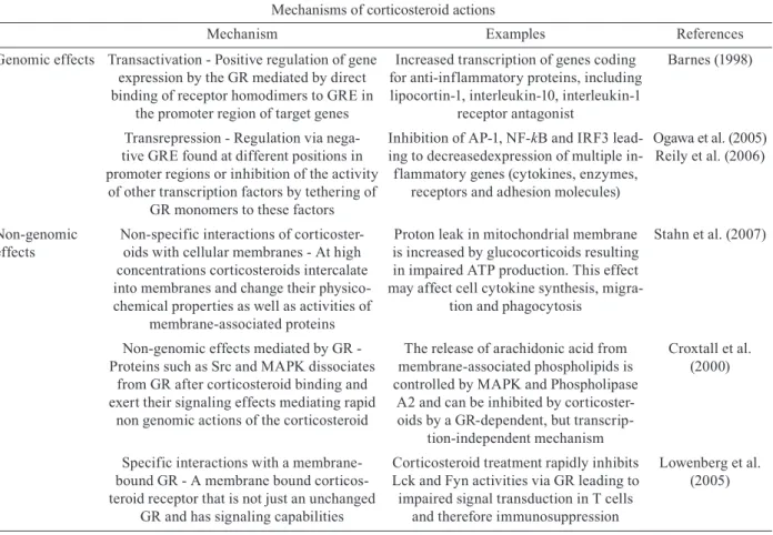
Different studies have shown that corticosteroids ac-tivities can be divided into the classical genomic effects, mediated by the cytosolic glucocorticoid receptor (GR) and the more recently identified non-genomic effects (Pratt & Dittmar 1998, Almawi & Melemedjian 2002, Buttgereit & Scheffold 2002, Adcock & Lane 2003). These non-genomic corticosteroid activities can be fur-ther divided in three modes of action: GR-mediated non-genomic effects, non-specific non-non-genomic effects and effects that are considered to be mediated by membrane-bound GRs (mGCR) (Table I) (Buttgereit & Scheffold 2002, Spies et al. 2006).
GR mediates the classical genomic actions of corticosteroids
The GR is classically described as a 94-kD protein member of the steroid-hormone-receptor family and is expressed ubiquitously, although several isoforms gen-erated by differential splicing or translation have been
TABLE I
Mechanisms of corticosteroid actions
Mechanism Examples References
Genomic effects Transactivation - Positive regulation of gene expression by the GR mediated by direct binding of receptor homodimers to GRE in
the promoter region of target genes
Increased transcription of genes coding for anti-inflammatory proteins, including lipocortin-1, interleukin-10, interleukin-1
receptor antagonist
Barnes (1998)
Transrepression - Regulation via nega-tive GRE found at different positions in promoter regions or inhibition of the activity of other transcription factors by tethering of
GR monomers to these factors
Inhibition of AP-1, NF-kB and IRF3 lead-ing to decreasedexpression of multiple in-flammatory genes (cytokines, enzymes,
receptors and adhesion molecules)
Ogawa et al. (2005) Reily et al. (2006)
Non-genomic effects
Non-specific interactions of corticoster-oids with cellular membranes - At high concentrations corticosteroids intercalate into membranes and change their physico-chemical properties as well as activities of
membrane-associated proteins
Proton leak in mitochondrial membrane is increased by glucocorticoids resulting in impaired ATP production. This effect may affect cell cytokine synthesis,
migra-tion and phagocytosis
Stahn et al. (2007)
Non-genomic effects mediated by GR - Proteins such as Src and MAPK dissociates
from GR after corticosteroid binding and exert their signaling effects mediating rapid
non genomic actions of the corticosteroid
The release of arachidonic acid from membrane-associated phospholipids is controlled by MAPK and Phospholipase
A2 and can be inhibited by corticoster-oids by a GR-dependent, but
transcrip-tion-independent mechanism
Croxtall et al. (2000)
Specific interactions with a membrane-bound GR - A membrane membrane-bound corticos-teroid receptor that is not just an unchanged
GR and has signaling capabilities
Corticosteroid treatment rapidly inhibits Lck and Fyn activities via GR leading to impaired signal transduction in T cells
and therefore immunosuppression
Lowenberg et al. (2005)
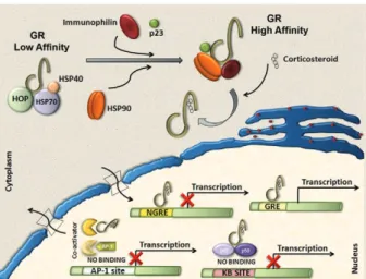
identified (Pratt & Dittmar 1998, Almawi & Melemed-jian 2002, Wikstrom 2003). The GR consists of three domains with different functions: an N-terminal domain containing transactivation functions, a DNA-binding domain with a zinc-finger motif that is common to DNA interaction proteins and a ligand-binding domain con-sisting of 12 alpha-helices, which is involved in the for-mation of the hydrophobic ligand-binding pocket (Wik-strom 2003). Corticosteroids can easily pass through the plasma membrane due to their highly lipophilic charac-teristics. In its inactive form, the receptor is located in the cytoplasm of cells associated with molecular chap-erones and immunophilins and is able to bind different corticosteroids with high affinity. After the binding of ligand to the GR, the receptor dissociates from this com-plex and is transported into the nucleus to modulate the expression of specific genes either positively (transacti-vation) or negatively (transrepression) (Pratt 1998, Al-mawi & Melemedjian 2002, Wikstrom 2003).
Molecular chaperones shape responses to corticosteroids
Chaperones facilitate the initial folding, maturation and, in some cases, regulate function of various proteins referred to as clients. Two main chaperone systems, the Hsp70 and Hsp90, influence GR assembly and activity. Hsp70 and Hsp90 are abundant, mainly cytosolic pro-teins. Hsp70 binds short, hydrophobic peptides and as-sists in the folding of nascent chains, while Hsp90 binds prefolded or even completely folded proteins and helps them to achieve or to maintain their active states (Grad & Picard 2007). Efficient maturation of GR to a conforma-tion capable of high-affinity hormone binding requires Hsp70, Hsp40, Hsp90, Hop and p23 (Pratt & Dittmar 1998). The first molecular chaperone system that rec-ognises de novo synthesised GR is the Hsp70 complex (Smith & Toft 1993). Hsp70 binds nascent proteins by recognizing hydrophobic segments of unfolded polypep-tides in an ATP-regulated process. Hsp40 has also been shown to be a crucial component of the GR “foldosome” complex in reticulocyte extracts (Dittmar et al. 1998). It accelerates ATP hydrolysis by Hsp70, which results in tight binding of the substrate (GR) to Hsp70, which facilitates the binding of the next component of the com-plex, the tetratricopeptide repeat (TPR)-containing the cochaperone, Hop. Hop contains three TPR domains, two of which allow the simultaneous interaction with Hsp70 and Hsp90 and therefore allow a transfer of the substrate from the Hsp70 to the Hsp90 system (Grad & Picard 2007). Hsp90 regulates the final maturation of GR, facilitating a hormone-activatable state, which is dependent on ATP binding to Hsp90 (Grenert et al. 1999). GR has a 100-fold lower affinity for corticoids without Hsp90, as was shown in cell-free steroid bind-ing assays (Nemoto et al. 1990). The affinity of Hsp90 for Hop is lower in the presence of ATP. Hop can then exit the complex making the TPR binding domain of Hsp90 available for the TPR-containing immunophilins, FKB52, FKBP51 and cyclophilin 40 and other
cochap-erones modulating GR-Hsp90 activity. Once bound to corticoids, GR has to move to the nucleus to function as a transcription factor. This step is also controlled by the Hsp90 machinery, specifically by the recruitment of immunophilin FKBP52 to the GR–Hsp90 complex. However, the details of the nuclear transport of GR re-main controversial and incompletely understood (Grad & Picard 2007) (Fig. 1).
Transactivation
Positive regulation of gene expression by the GR has been shown to be mainly mediated by the direct binding of receptor homodimers to specific sequences [gluco-corticoid responsive elements (GREs)] in the promoter regions of target genes. Three different types of GREs, simple, composite or tethering have been described (Lef-stin & Yamamoto 1998), sugge(Lef-sting different molecular mechanisms for GR interaction with GREs. Activation of gene transcription by the GR via simple and composite GREs is dependent on the direct binding of the activated GR homodimer to DNA, although dimerization may not be required in composite GREs. On the contrary, GR binds to other DNA-bound transcription factors such as AP1, Stat3 and NFkB to increase transcription activity at tethering GREs. Regulation of gene transcription by GR requires the recruitment of coregulators. Among these coregulators, the p160 steroid receptor coactivator (Src) gene family contains three homologous members (Src-1, Src-2 and Src-3) that are crucial in facilitating chromatin remodelling, assembly of general transcrip-tion factors and transcriptranscrip-tion of target genes by the re-cruitment of histone acetyltransferases and methyltrans-ferases to specific enhancer/promoter regions (Xu & Li 2003, Chinenov & Rogatsky 2007).
Transrepression
Different mechanisms account for negative regula- tion by the GR (Fig. 1). One of the best described is regulation via negative GREs that differ in structure and function from positive GREs (Dostert & Heinzel 2004). Such elements can be found at different positions in pro-moter regions and interfere either with the binding sites of other transcription factors or with the binding site of the basal transcription initiation complex (Schacke et al. 2007). However, only a few genes are known to be regula- ted via these negative GREs and the contribution of this kind of negative regulation to the overall activities of corticosteroids is probably of minor importance. The major mechanism of negative regulation by the GR is the inhibition of the activity of other transcription fac-tors by being tethered to these facfac-tors. Interestingly, this mechanism is thought to occur through GR monomers (Heck et al. 1994, Reichardt et al. 1998). The transcrip-tion factors to which the GR binds include activator pro-tein 1 (AP-1), nuclear factor-kB (NF-kB) and interferon regulatory factor 3 (IRF3) (Ogawa et al. 2005, Reily et al. 2006). Adding complexity to the system, Bladh et al. (2005) demonstrated that different domains of the GR seem to be responsible for either NF-kB or AP-1 inter-actions. A GR (R488Q) mutant unable to repress
NF-kB activity retained the ability to repress AP-1 activity while transactivation activities were unaffected. Because all of these transcription factors regulate the expression of pro-inflammatory genes, their negative regulation by the GR has been considered the hallmark for the anti-inflammatory and immune suppressive action of corti-costeroids. Other mechanisms identified that may help explain the transrepressive action of the GR is its bind-ing to Jun N-terminal kinase (JNK) leadbind-ing to suppres-sion of JNK activity and, subsequently, to the inhibition of AP-1 (Caelles et al. 1997, Bruna et al. 2003)
Cofactors
It has been known for quite some time that GR does not function alone as a transcription factor. To achieve highly coordinated regulation of gene transcription, co-factors that modify and remodel chromatin structures are needed. Cofactors can be categorised as positive reg-ulators of transcription (coactivators) or negative regula- tors of transcription (corepressors) depending on their function (Rogatsky & Ivashkiv 2006, Feige & Auwerx 2007). These cofactors do not bind directly to DNA, but are recruited through protein-protein interactions be-tween transcription activation domains of the GR and regulatory sequences where they exert enzymatic activi-ties such as histone acetylases (HATs) or deacetylases. Among cofactors that function as coactivators are CBP/ p300, HATs and p160 proteins, including TIF2/GRIP1/ NCoA3, pCIP/RAC3/ACTR/AIB1/NCoA3 and Src1/ NCoA1 (Xu & O´Malley 2002). On the other hand, TIF2/ GRIP1 is also able to function as a corepressor of the GR at the AP-1 and NF-kB tethering sites (Rogatsky et al. 2001). Despite the fact that the repression of many pro-inflammatory molecules seems to be a major part of the anti-inflammatory effects of corticosteroids, detailed
knowledge about the molecular mechanisms involved is still scarce. For instance, additional corepressors, such as NCoR and SMRT, have also been described as being recruited to GR, although their role in the negative regula- tion of pro-inflammatory genes is not well recognised (Schacke et al. 2007)
Rapid onset effects of corticosteroids are mediated by non-genomic mechanisms
Mounting evidence suggests that corticosteroid effects cannot be entirely accounted for by the genomic mecha-nism of action explained above. Some effects are produced in a very short time, vanish rapidly and do not involve protein synthesis. Interestingly, some evidence suggests that these mechanisms are more ancient than the genomic ones, despite the fact that the latter were discovered earlier (Dallman 2005). There are three proposed non-genomic based mechanisms to explain the rapid anti-inflammatory and immunosuppressive effects of corticosteroids that are not compatible with the genomic mechanism of ac-tion (Croxtall et al. 2000, Buttgereit & Scheffold 2002, Hafezi-Moghadam et al. 2002): (i) non-specific interac-tions of corticosteroids with cellular membranes; (ii) non-genomic effects which are mediated by the cytosolic GR and (iii) specific interactions with a mGCR.
Non-specific interactions of corticosteroids with cel-lular membranes - Plasma and mitochondrial membranes are targets for corticosteroids. At high concentrations corticosteroids intercalate into membranes and change their physicochemical properties as well as the activities of membrane-associated proteins (Buttgereit & Schef-fold 2002, Buttgereit et al. 2004). As a consequence, cal-cium and sodium cycling across plasma membranes of immune cells is reduced and might contribute to rapid immunosuppression and decreased inflammation (But-tgereit & Scheffold 2002). In addition, proton leakage in the mitochondrial membrane was shown to be increased by glucocorticoids, resulting in impaired ATP produc-tion. This effect may contribute to clinically relevant outcomes in immune cells affecting cytokine synthesis, migration and phagocytosis, in conditions such as sepsis (Stahn et al. 2007) and may explain why high doses of corticoids can be harmful to septic patients.
Specific interactions with a mGCR - The existence of a membrane bound corticosteroid receptor was first shown in lymphoma and leukemia cells (Gametchu et al. 1999). This receptor was later identified on human pe-ripheral blood mononuclear cells through high-sensitiv-ity immunofluorescent staining. Overexpression of GR did not cause increased mGCR expression on the cell sur-face. This indicates that mGCR is not just an unchanged GR that has been transported to the cell membrane. One possibility is that the mGCR is a variant of GR produced by differential splicing or promoter switching or by post-translational editing (Bartholome et al. 2004), but its ori-gin still remains unexplained. It has been reported that stimulation with lipopolysaccharide (LPS) increases the percentage of mGCR-positive monocytes, suggesting that immunostimulation is responsible for up-regulation and transcellular transport of mGCR (Bartholome et al. 2004, Stahn et al. 2007). In addition, patients with rheu-matoid arthritis, ankylosing spondylitis and systemic lupus erythematosus were shown to have high/increased numbers of mGCR-positive monocytes (Buttgereit et al. 2005, Spies et al. 2006, Tryc et al. 2006). The correlation between immune stimulation and increased expression of mGCR may have a clear impact on the importance of non-genomic effects for the corticosteroid actions in patients at an early phase of sepsis. This possibility, however, remains to be explored in future studies. More recent work has shed light on the molecular mechanism of the non-genomic mGCR-mediated immunosuppres-sive effects of corticosteroids in T cells. p56lck (Lck) and p59fyn (Fyn) kinases, members of the Src family of tyrosine kinases, were identified as cellular targets for non-genomic corticosteroid activities (Lowenberg et al. 2005). Lck and Fyn are expressed in T cells and they are involved in T cell receptor (TCR)-mediated signal transduction. Their association with the TCR complex is essential for efficient TCR signalling (Palacios & Weiss 2004). Lowenberg et al. (2005) have recently demon-strated that corticosteroid treatment rapidly inhibits Lck and Fyn activities in vitro and in vivo, via a mGCR-dependent pathway. These observations add complexity to the molecular mechanism of immune suppression by corticosteroids and indicate that a specific non-genomic mechanism may be of central relevance to corticoster-oid-impaired TCR signalling.
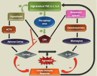
Corticosteroid effects on innate immune responses
Innate immune responses play a central role in the pathophysiology of sepsis and are markedly influenced by corticosteroids and other endocrine and neural sys-tems (Fig. 2). Mammalian cells sense the presence of pathogens through a family of transmembrane and cy-toplasmic receptors, which detect conserved microbial components [lypopolysaccharide (LPS), peptidoglycans, single-stranded and double-stranded DNA and RNA and others) collectively referred to as pathogen-associated molecular patterns (PAMPs). This family of receptors is known as a group of pattern recognition receptors. Toll-like receptors (TLR) are one of the best characterised members of this family. To date, there are 10 members of the TLR subfamily that have been identified in
hu-mans. TLRs recognise a wide variety of PAMPs and upon activation, initiate a cascade of signalling events through several adaptor proteins and protein kinases that converge on the transcriptional regulators, NFkB, AP1 and IRFs, which, in turn, induce transcription of di-verse cytokines and chemokines (Chinenov & Rogatsky 2007). The TLR signalling network is a target for GR-mediated actions and different mechanisms are involved in this effect. One of the most characterised mechanisms is the induction of endogenous inhibitors of the TLR sig-nalling pathway. The induction of IkB by GR is a clas-sic example of this mechanism (Scheinman et al. 1995), but other relevant targets were later revealed. JNK and p38 stress kinase pathways are activated by all known TLRs. Negative regulation of these pathways involves dephosphorylation of JNK, p38 and their upstream ki-nases, MEK3,4,6, by MAPK phosphatases (Abraham & Clark 2006). Interestingly, glucocorticoids inhibit JNK and p38 without altering their levels, suggesting that GR does not repress transcription of those kinases (Caelles et al. 1997, Hirasawa et al. 1998). One possi-ble explanation is that GR induces transcription of an inhibitory phosphatase, MKP1, as already described in mast cells and endothelial cells (Kassel et al. 2001, Furst et al. 2007). In fact, in MKP1-/- mouse macrophages, glucocorticoid treatment failed to inhibit LPS-induced JNK and p38 activation and the production of inflamma-tory mediators (Abraham & Clark 2006). In addition, the MKP-1-/- mouse is extremely sensitive to LPS challenge and partially insensitive to the anti-inflammatory effects of corticosteroids (Chi et al. 2006, Wang et al. 2008). The precise mode of GR action on MKP1 gene transcription and to what extend MKP1 induction contributes to the anti-inflammatory actions of corticosteroids in in vivo models and in the clinical setting is not yet determined.
Cytokines signal through the JAK/STAT pathways, which are sensitive to inhibition by the suppressors of cytokine signalling (SOCS) (Yoshida et al. 2004). In addition, SOCS1 also directly inhibit TLR 2 and 4 sig-nalling by inducing rapid degradation of their TIR do-main-containing adaptor TIRAP (Mansell et al. 2006). Corticosteroids induce SOCS1 mRNA in hematopoietic cell lines and in acute lymphoblastic leukemia patients. The precise mechanism of this up-regulation is pres-ently unknown, but several potential GREs are scat-tered throughout the SOCS1 regulatory region and may play a role in corticosteroid-induced SOCS1 expression (Chinenov & Rogatsky 2007).
Yet another potential mechanism for corticosteroid-induced immune modulatory effects involves glucocor-ticoid-inducible leucine zipper (GILZ), a small protein with a wide spectrum of anti-inflammatory activities, including the inhibition of LPS-induced expression of TLR2 in macrophages and of TLR 2 and 3-mediated NFkB activation in airway epithelial cells (D’Adamio et al. 1997, Ayroldi et al. 2001, Eddleston et al. 2007). GILZ also seems to be involved in enhancement of spontaneous thymocyte apoptosis, likely through down-regulation of the anti-apoptotic factor, Bcl-X (Delfino et al. 2004). GILZ is markedly induced by corticoster-oids in a wide range of cell types by a mechanism that requires GR dimerization and DNA binding (Tonko et al. 2001, Berrebi et al. 2003, Rogatsky et al. 2003), sug-gesting that GILZ is a direct transcriptional target for the receptor. Indeed, several GREs were identified in the GILZ 5’-regulatory region (Wang et al. 2004, Chen et al. 2006). GILZ knockdown by siRNA prevents glucocorti-coid inhibition of IL-1b-induced chemokine production in airway epithelial cells, suggesting that at least some of GR’s anti-inflammatory actions require GILZ (Eddles-ton et al. 2007). Importantly, GILZ was also reported to physically interact with several effectors of TLR sig-nalling, most notably Fos, Jun and p65/RelA (Ayroldi et al. 2001, Mittelstadt & Ashwell 2001). Experiments with GILZ knockout mice, presently unavailable, might provide some additional information on the role of this protein in the regulation of inflammation by GR.
As described throughout this section, corticosteroids have many different mechanisms by which they inter-fere with innate immune responses. The intricate nature of corticosteroid effects can be easily illustrated by the apparent paradox involving TLR2 overexpression. Hy-peractivation of TLRs, including TLR2, is thought to play an important role in excessive proinflammatory cytokine production and the pathogenesis of sepsis. Ex-pression of TLR2 in response to TLR agonists is activat-ed by NFkB (Liu et al. 2000, 2001) and could, therefore, be expected to be corticosteroid-sensitive. Surprisingly, several reports show corticosteroid induction of TLR4 and TLR2 mRNAs in multiple cell types (Shuto et al. 2002, Homma et al. 2004, Rozkova et al. 2006) through an incompletely known mechanism (Chinenov & Ro-gatsky 2007). While this seems like a paradoxical ef-fect in light of corticosteroid anti-inflammatory efef-fects, recent discovery of TLR2 and TLR4 expression in the adrenals (Bornstein et al. 2004a, b) may offer a
poten-tial explanation, since LPS and lipoteichoic acid directly stimulate cortisol release in adrenal cells (Vakharia & Hinson 2005). Furthermore, cortisol release upon TLR stimulation is impaired in TLR2 and TLR4-deficient mice (Zacharowski et al. 2006). Conceivably, a positive feedback loop is activated upon exposure to TLR li- gands with the net output being the increased release of corticosteroids into the bloodstream, which would trig-ger classic mechanisms of inflammation reversal. As a net clinically observed effect, steroid infusion is able to modulate the inflammatory response in septic shock, as demonstrated by decreased levels of IL-6, IL-8, E-selec-tin and monocyte HLA-DR levels in a crossover study (Keh et al. 2003). Hydrocortisone was given early or late in different groups of septic shock patients and the func-tion of monocytes and these cytokines were significantly influenced by steroid administration.
Corticosteroid effects on vascular reactivity
Clinical trials evaluating corticosteroid administration in sepsis and acute respiratory distress syndrome (ARDS)
Corticosteroids have been given to patients with se-vere infections for more than 90 years, as described by Friderichsen (1918) in hemorrhagic necrosis of adrenal glands in children with purpura fulminans (Waterhouse-Friderichsen syndrome). An association with meningo-coccemia was done by the same author after almost 40 years. Since then, corticosteroids have been used in the treatment of a myriad of severe infections: Haemophi-lus influenzae and Streptococcus pneumoniae meningi-tis, herpes zoster, tuberculosis (meningimeningi-tis, ophthalmic or pericardial commitment), typhoid fever, infectious mononucleosis, Pneumocystis pneumonia, tetanus, hepa- titis B, pertussis and acute bronchiolitis, just to name a few (McGee & Hirschmann 2008).
Many patients with severe infections also have clini-cal shock and the reduction in arterial pressure and tis-sue perfusion is associated with microbial load and severity of illness. Severe bacterial infections cause systemic inflammatory response defined as sepsis in earlier consensus conference (Bone 1992). Because of our understanding that sepsis is an exaggerated host re-sponse to infections, steroids were quickly proposed for its treatment. Table II summarises the most important results and notes about clinical trials of steroid therapy for severe sepsis and septic shock.
Steroids became a standard treatment for severe infections between the 1960s and 70s. Some small tri-als showed consistent findings that steroid treatment caused faster reversal of shock and lower mortality rates. Schumer (1976) published a trial with more than 300 pa-tients with an almost 60% reduction in mortality rate among those treated with dexamethasone or methylpred-nisolone. However, like this study, most of the studies only included a small population, were poorly controlled or were retrospective trials.
In the early 80s, randomised clinical trials analysed the administration of high doses of methylprednisolone (30 mg/kg/day). It was preconized that corticosteroid therapy should begin early, usually on the first day of septic shock and it was rarely sustained beyond this time. Three studies were representative of this strategy. Sprung et al. (1984) enrolled 59 patients with no more than 24h of septic shock and treated them with meth-ylprednisolone, dexamethasone or placebo. Although the reversal of shock in 24 h was more evident in both steroid groups, mortality was not significantly different. Bone et al. (1987b) used methylprednisolone in 382 pa-tients with severe sepsis (most of them presented septic shock) and found that there was no benefit to the steroid treatment. In fact, more harm than good was observed in the renal insufficiency subgroup. Finally, Luce et al. (1988) used methylprednisolone or placebo in acute lung injury patients; the majority of patients had concomi-tant septic shock. The main goal was the prevention of ARDS, but there was no benefit in the degree of paren-chymal lung injury or mortality. By the negative results of these major trials, corticosteroid treatment in sepsis
was restricted to rare selected cases for more than a dec-ade. Criticism was attributed to these studies by the fact that adrenal dysfunction could last more than 24-48 h, as originally described and large doses were administered, which could have contributed to the higher incidence of hospital-acquired infections as a consequence of pro-found immunosuppression.
After almost 10 years, new evidence for the ben-eficial effects of steroid treatment was shown in small randomised trials. Most authors aimed to analyse the ef-fects of low doses of hydrocortisone on the reversal of septic shock. Corticosteroids were also used for a pro-longed time (usually 7 days) and tapered over several days. Bollaert et al. (1998) studied 41 patients with late refractory septic shock (more than 48 h of evolution) and randomised them to receive 100 mg of hydrocortisone or placebo for five days. The steroid group showed a sig-nificant improvement in shock reversal, although they showed no difference in regard to the response to corti-cotropin test. There was also a trend toward reduced 28-day mortality. Briegel et al. (1999) enrolled 40 patients within 24-28 h of septic shock to either receive or not receive an intravenous infusion of hydrocortisone. The group receiving the steroid infusion had reduced time to the cessation of vasopressor therapy (median 2 vs. 7 days, p < 0.01). In this trial steroid therapy was rapidly tapered after catecholamine withdrawal. Two other tri-als found similar results in reversal of shock and a trend toward reduced early mortality in patients with steroids (Chawla et al. 2000, Yildiz et al. 2002). These small ran-domised trials brought hope again to the usefulness of corticosteroids in critically ill septic patients and mul-ticenter clinical trials with larger population were then planned to confirm this newer successful evidence.
When adding the French study to other studies that have been conducted since 1998, steroids became the standard of care in early septic shock, as recommended by the worldwide task force to reduce sepsis mortality, the Surviving Sepsis Campaign, in 2004 (Dellinger et al. 2004). Hydrocortisone was given to septic shock patients, usually in the first 24 h after presentation, in doses of 200 to 300 mg per day for 7-10 days; fludrocor-tisone was not routinely recommended.
In order to confirm the benefits of corticosteroid, a second large trial (CORTICUS) was conducted in which researchers in 11 European countries designed a
randomised, placebo-controlled trial with septic shock patients (Sprung et al. 2008). Again, the main goal was 28-day mortality in non-responders to the corticotropin test. Hydrocortisone was administered as a 50 mg dose every 6 h for 11 days and tapering was done by day 5-11, in order to avoid rebound hypotension. Enrolment in-cluded 499 patients, of whom 47% were non-responders to the corticotropin test. Mortality on day 28 or in the hospital was not different between the two groups and the corticosteroid group had a higher incidence of super-infections (infection superimposed upon previous ones) and higher rates of new septic shock episodes.
Hyper-TABLE II
Scheduled treatment and 28-day mortality in septic shock patients in selected placebo-controlled, randomized clinical trials
Author n Treatment 28-day mortality Notes
Schumer (1976) 172 Methylprednisolone or dexameta-sone/placebo; initiation immediately
after diagnosis; up to two doses in 24 h
10 x 38% Not blinded; two groups with different steroids
Sprung et al. (1984) 59 Methylprednisolone or dexameta-sone/placebo; initiation immediately
after diagnosis; up to two doses in 24 h
76 x 69% Reversal of shock oc-curred earlier in steroid group; no difference in
hospital mortality
Bone et al. (1987b) 382 Methylprednisolone/placebo; initia-tion up to 2 h after diagnosis
34 x 25% Higher mortality in renal insufficiency group
VASSCSG (1987) 223 Methylprednisolone/placebo; ad-ministration within 2 h; initiation up
to 2 h after diagnosis
20 x 21% Treatment was initiated within 2 h of shock; par-ticipation of 10 centres
Luce et al. (1988) 87 Methylprednisolone/placebo 58 x 54% Most patients had septic shock; ARDS evolution 15
x 16%
Bollaert et al. (1998) 41 Hydrocortisone/placebo; initiation after 48 h of septic shock
32 x 63% Significant reversal of shock at day 7
Briegel et al. (1999) 40 Hydrocortisone/placebo; adminis-tration within 72 h from shock onset
15 x 20% Significant reversal of shock at day 7
Chawla et al. (2000) 44 Hydrocortisone/placebo; adminis-tration within 72 h from shock onset
26 x 48% Significant reversal of shock at day 7
Yildiz et al. 2002) 40 Prednisolone/placebo 40 x 60% Treatment duration of 10 days
Annane et al. (2002) 299 Hydrocortisone + fludrocortisone/ placebo; inclusion till 8 h after
shock onset
61 x 55%; 63 x 53% in
non-responders to ACTH test
Both steroids given for seven days; responders to ACTH test can offset positive results; significant
reversal of shock at day 7
Sprung et al. (2008) 499 Hydrocortisone/placebo; inclusion till 72 h after shock onset
34 x 31% Steroids given for 11 days; significantly shorter time to reversal of shock in steroid group; higher
inci-dence of superinfections and hyperglycemia
Total 1,886 Various 36 x 37% No difference in 28-day
mortality
glycemia was also more frequent in the corticosteroid group. One possible explanation for these contrasting observations is that the mode of hydrocortisone admin-istration could have an impact on glycemic control, as shown with bolus injections and a higher fluctuation in blood glucose levels would impact the outcome (Loisa et al. 2007, Weber-Carstens et al. 2007).
These two randomised double-blinded clinical trials had good methodological quality, but their results were opposite. In both studies, there were problems in study design, as for example, low recruitment during enrol-ment, possibly because of the strict deadline by which to give the first dose of hydrocortisone. When analyz-ing their sample sizes, CORTICUS had less patients than previously calculated for 80% statistical power and a 10% absolute decrease in mortality (n = 800). In the French study (Annane et al. 2002), sample size was cal-culated for 270 patients (they actually included 299) and non-responders to the corticotropin test reached a sig-nificant 20% relative reduction in mortality. Some other differences are noteworthy: in the French trial, patients received steroids in the first 8 h of septic shock presenta-tion, while in CORTICUS there was a looser deadline of 72 h for inclusion. Maybe for this reason, reversal of shock occurred more frequently in the former study. The severity of illness was calculated with a prognostic score (Simplified Acute Physiology Score II - SAPS II), which predicts hospital mortality based on demographic and physiologic data collected in the first 24 h after pa-tient admission to intensive care unit (ICU). The score was higher in the former study by Annane, with a great difference between SAPS II scores: 58 vs. 48 points in CORTICUS. This difference denotes the different se-verity of these two studies populations. More severely ill patients could possibly benefit more from immune therapy, as already shown in the case of other therapies (Bernard et al. 2001). Also, in the French study statisti-cal analyses were done for 1-sided formulation, which means that the authors did not consider the adverse ef-fects of steroids in the treatment of septic shock. An-other possible reason was the difference in the site of infections: pneumonia was the major source of sepsis in the French study, while abdominal/peritoneal infections were the majority in CORTICUS. There is evidence sup-porting benefits to the use of corticosteroids in pneumo-nia and cortisol being a biomarker of prognosis in this infection (Confalonieri et al. 2005, Salluh et al. 2008a), while abdominal infections are usually dependent on maintenance of the macrophage population in the peri-toneum, antibiotic penetration in necrosis and surgical intervention (Hausmann et al. 2000, Sotto et al. 2002). In addition to the differences in the sites of infection, appropriate antimicrobial therapy was more prevalent in the CORTICUS placebo group (78.8% vs. 72.8%), which can obviously lessen the rate of success in corticoster-oid therapy. Patients received corticostercorticoster-oids for longer periods in CORTICUS (11 days), even if they recovered from shock and hyperglycemia and new infections were far more frequent than in previous studies. Annane et al. (2004) performed a meta-analysis of steroid use in septic shock in 2004 and he identified that, even in older
studies (high-dose and short duration), incidence of su-perinfections and serum glucose levels were not altered by intervention. CORTICUS was unique in showing that longer therapy is associated with adverse events, which could offset eventual benefits of the anti-inflammatory effects of corticosteroids.
An international task force was created to develop a consensus statement for the diagnosis and manage-ment of corticosteroid insufficiency in critically ill pa-tients and a meta-analysis demonstrated that the addition of CORTICUS data to the earlier studies confirmed a greater rate of shock reversal up to seven days of sep-tic shock, although there was no effect on the 28-day mortality (Marik et al. 2008). Early reversal of shock was achieved in almost 65% of all patients in the low-dose steroid studies, while only 47.5% presented the same response in the placebo group (relative risk: 1.39, 95% confidence interval: 1.24-1.55). Mortality after one month was not lowered by corticosteroid administration (relative risk: 0.92, 95% confidence interval: 0.79-1.06). The task force recommended corticosteroid treatment in septic shock to patients with poor response to fluids and vasopressor, irrespectively of any diagnostic test of criti-cal illness-related corticosteroid insufficiency (basal to-tal or free serum cortisol or rising cortisol levels after synthetic ACTH administration).
In the view of these studies, corticosteroids cannot be widely recommended for critically ill patients with ARDS. In recent years, the use of corticosteroids has been suggested to be effective in selected patients with community acquired pneumonia (CAP) (Confalonieri et al. 2005, Annane & Meduri 2008, Sibila et al. 2008). Nevertheless, recent reviews concluded that available studies cannot support recommendation for corticoster-oids as standard of care for patients severe CAP (Salluh et al. 2008b, Sibila et al. 2008). In a recent study, we have studied 191 patients with CAP and concluded that in pa-tients with severe CAP, adjunctive therapy with corti-costeroids may facilitate the weaning of vasopressors but has no influence on mortality, on the clinical course assessed by the SOFA score or on the C reactive protein course. However, if necessary, corticosteroid prescrip-tion seems to be safe in these patients (JI Salluh et al., unpublished observations).
Critical illness-related corticosteroid insufficiency (CIRCI)
Any critical illness is able to blunt adrenal synthesis of cortisol or its action (increased resistance). Low levels of cortisol were demonstrated in sepsis, pancreatitis, se-vere burns and liver failure (Murphy et al. 1993, Marik et al. 2005, Widmer et al. 2005, Annane et al. 2006a, Ho et al. 2006). Decreased production of cortisol or ACTH is common in severe sepsis or septic shock, particularly in patients with risk factors, such as female gender, posi-tive blood cultures or Gram-negaposi-tive infections (Annane et al. 2006a, Salgado et al. 2008). Based on experimen-tal data, severe inflammation-associated corticosteroid resistance is thought to primarily exist due to decreased corticosteroid receptor nuclear translocation after endo-toxin or proinflammatory cytokine challenges (Liu et al. 1993, Pariante et al. 1999). Some interesting evidence came from a clinical study that demonstrated the same phenomenon in patients with ARDS; patients had severe gas exchange compromise and low baseline cytoplasmic and serum cortisol levels, indicating impairment of corti-costeroid action (Meduri et al. 2005).
CIRCI is considered the most correct term for in-adequate relative adrenal response to a severe critical disease, mainly sepsis (Marik et al. 2008) and was for-merly known as relative adrenal insufficiency or failure. Diagnosis of CIRCI in critically ill patients is based on two methods: random total serum cortisol or change in cortisol levels after synthetic ACTH administration (Ta-ble III). High levels of random serum cortisol (collected anytime from the beginning of sepsis) have been recog-nised as a sign of severe infection for many years. In a cohort of 20 patients with shock due to infection, it was observed that non-survivors had significantly higher se-rum cortisol than survivors (Melby & Spink 1958). Ma-rik and Zaloga (2003) determined that any critically ill septic patient with a random serum cortisol of 25 mcg/ dL or less has high probability of adrenal insufficiency. Admitting severe infections in the emergency room can also reveal a subset of early CIRCI patients. Their pres-entation is somewhat different from other severe sepsis patients, with lower levels of glucose and sodium and baseline ACTH and cortisol levels (Manglik et al. 2003).
The synthetic ACTH stimulation test is advocated for a more specific diagnosis of CIRCI. It can be done with a full dose (250 mcg) or a low dose (1 mcg). Annane et al. (2000) conducted this test in a large cohort of early sep-tic shock patients and could identify a subset of higher death probability group with a poor response to synthetic ACTH and higher levels of basal serum cortisol. Better prognosis was shown in patients with basal cortisol less than 34 mcg/dL and delta cortisol after ACTH test high-er than 9 mcg/dL (26% mortality). The othhigh-er response patterns were associated with significantly higher mor-tality: basal cortisol higher than 34 mcg/dL and/or delta cortisol less than 9 mcg/dL patients had only 18-33% survival. Bollaert et al. (2003) also obtained a similar re-sult with late (around 4 days) septic shock patients: basal cortisol > 20 mcg/dL and delta cortisol < 9 mcg/dL were independent predictors of mortality.
CIRCI can also be a dynamic process. Adrenal ex-haustion is thought to be a different form of CIRCI and septic shock patients can exhibit normal or high basal cortisol levels at hospital admission, but adrenal response can become severely altered after a few days. After re-testing a cohort of septic shock patients with long vaso-pressor dependency, Guzman and Guzman (2007) con-firmed that these patients presented very low levels of random serum cortisol levels after six days with normal or high cortisol levels at admission (from initial mean ~42 mcg/dL to repeated 10 mcg/dL levels). Hydrocorti-sone administration aided in vasopressor weaning in all of these patients (Guzman & Guzman 2007). Cortisol changes among septic shock patients is very dynamic and can persist for up to 28 days and there is wide difference in random cortisol and delta cortisol (synthetic ACTH test) between survivors and non-survivors (Goodman et al. 2005). These important data suggest that accompany-ing cortisol levels in vasopressor-dependent patients can be useful and sometimes dictate the need for a steroid course during septic patients’ ICU stay.
Even when diagnosing CIRCI, corticosteroid treat-ment is not straightforward for improving septic shock management. Elsouri et al. (2006). analysed 92 pa-tients with septic shock and CIRCI was present in al-most half of them, based on a 250 mcg synthetic ACTH test. CIRCI patients needed rescue vasopressor therapy more frequently, but maximal doses of conventional catecholamines were similar to non-CIRCI patients. Besides, hydrocortisone therapy was given to only half CIRCI patients and did not affect mortality. Morel et al. (2006) made an overview of vasopressor-dependent septic patients and studied parameters associated with hemodynamic improvement after corticosteroid initia-tion (reducinitia-tion of more than 50% in vasopressor dose up
to 3 days of steroid treatment). They compared differ-ent diagnostic tests for CIRCI (random total cortisol or delta cortisol to 1 and 250 mcg synthetic ACTH test) and verified that hemodynamic improvement was not asso-ciated with diagnosis of CIRCI. Another retrospective study conversely stated that CIRCI diagnosed patients have a higher mortality if not treated with glucocorti-coids, although this was a very small subgroup of pa-tients (n = 7) (de Jong et al. 2007). Interestingly, possible CIRCI patients had similar survival when compared to non CIRCI patients who were also treated with corticos-teroids. Based on these small retrospective data, mak-ing the diagnosis of CIRCI does not alter indications for steroid treatment in septic shock.
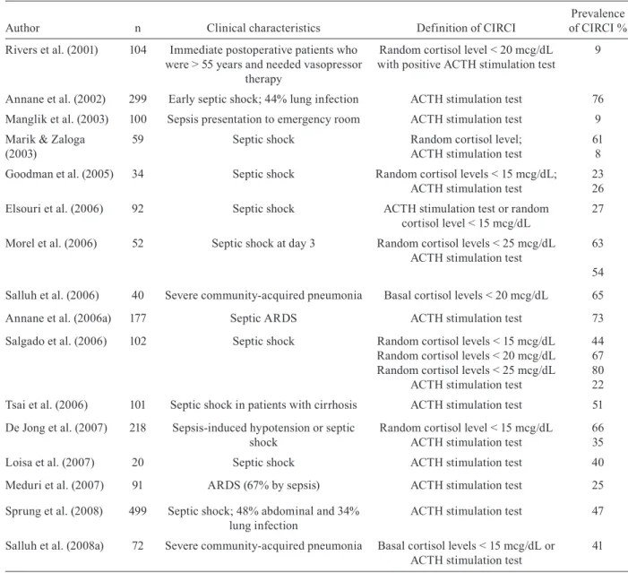
TABLE III
Diagnostic criteria for critical illness related corticosteroid insufficiency (CIRCI) for septic patients in studies since 2001
Author n Clinical characteristics Definition of CIRCI
Prevalence of CIRCI %
Rivers et al. (2001) 104 Immediate postoperative patients who were > 55 years and needed vasopressor
therapy
Random cortisol level < 20 mcg/dL with positive ACTH stimulation test
9
Annane et al. (2002) 299 Early septic shock; 44% lung infection ACTH stimulation test 76
Manglik et al. (2003) 100 Sepsis presentation to emergency room ACTH stimulation test 9
Marik & Zaloga (2003)
59 Septic shock Random cortisol level;
ACTH stimulation test
61 8
Goodman et al. (2005) 34 Septic shock Random cortisol levels < 15 mcg/dL; ACTH stimulation test
23 26
Elsouri et al. (2006) 92 Septic shock ACTH stimulation test or random cortisol level < 15 mcg/dL
27
Morel et al. (2006) 52 Septic shock at day 3 Random cortisol levels < 25 mcg/dL ACTH stimulation test
63
54
Salluh et al. (2006) 40 Severe community-acquired pneumonia Basal cortisol levels < 20 mcg/dL 65
Annane et al. (2006a) 177 Septic ARDS ACTH stimulation test 73
Salgado et al. (2006) 102 Septic shock Random cortisol levels < 15 mcg/dL Random cortisol levels < 20 mcg/dL Random cortisol levels < 25 mcg/dL
ACTH stimulation test
44 67 80 22
Tsai et al. (2006) 101 Septic shock in patients with cirrhosis ACTH stimulation test 51
De Jong et al. (2007) 218 Sepsis-induced hypotension or septic shock
Random cortisol level < 15 mcg/dL ACTH stimulation test
66 35
Loisa et al. (2007) 20 Septic shock ACTH stimulation test 40
Meduri et al. (2007) 91 ARDS (67% by sepsis) ACTH stimulation test 25
Sprung et al. (2008) 499 Septic shock; 48% abdominal and 34% lung infection
ACTH stimulation test 47
Salluh et al. (2008a) 72 Severe community-acquired pneumonia Basal cortisol levels < 15 mcg/dL or ACTH stimulation test
41
When and how to use steroids in septic shock patients?
Common sites of infection in critically ill patients are lungs, abdomen, catheter-related and the urinary tract. The severity of each of these sites is different and is thought to be higher in abdominal and pulmonary in-fections. Incidence of organ dysfunction and response to specific therapies are also different, as recently shown with recombinant activated protein C (Abraham et al. 2005, Sevransky et al. 2008). Another example is severe CAP, which was successfully treated with low doses of hydrocortisone in a small clinical trial, although cer- cer-tainty about its indication remained in low to moder-ate level of recommendation (Confalonieri et al. 2005, Salluh et al. 2008b). Conversely, severe abdominal in-fections present with a poor prognosis, since antibiotic tissue penetration and drainage of abscesses are often harder to guarantee. Urinary tract and venous catheters are also other common sites of infections in the critically ill and their treatment encompasses early antibiotic ad-ministration and device removal, with a generally rapid and good response. Therefore, different infections show diverse patterns of presentation and evolution, making it difficult to predict what is the best treatment for each situation. This reason may be explain why most anti-inflammatory therapies tried in septic patients such as corticosteroids, recombinant activated protein C, anti-thrombin, tifacogin, anti-TNF antibodies and polyclonal immunoglobulins just to name few, did not work as ex-pected based on the initial pre-clinical studies.
There are interesting data regarding the use of corti-costeroids as an adjunctive treatment for different infec-tions. One early example is the use of corticosteroids in hypoxemic Pneumocystis jiroveci pneumonia; a meta-analysis revealed that there is a reduction in need for mechanical ventilation and in 1 and 3-month mortality (Briel et al. 2005). There are only six clinical trials on this topic, with great heterogeneity and a diverse number of patients. Oxygenation entry criteria were also diverse (from PaO2 of 51-75 mmHg or oxygen saturation less than 85-90%). Intravenous methylprednisolone and oral prednisone were the tested routes of administration. The gap between the beginning of antibiotic treatment to first dose of corticosteroids ranged from 24 h to un-limited and there was no mention to tapering schedules. Nevertheless, steroids showed a clear benefit to these pa-tients, lowering need for mechanical ventilation by more than 60% and mortality by 33%. This result means that in the treatment of at least nine P. jiroveci pneumonia patients, adjunctive steroid therapy will save one indi-vidual. Besides all of the critiques to the results of these trials, there is no disagreement in adding corticosteroids to hypoxemic P. jiroveci pneumonia patients.
A recent systematic review concluded that, while corticosteroids cannot be recommended as a standard of care for patients with severe community-acquired pneu-monia, studies that evaluated the use of moderate doses for 7-10 days demonstrated clinical benefits (Salluh et al. 2008b). It is also interesting to observe that signifi-cant derangements in the adrenal response have been
described previously in patients with severe community-acquired pneumonia and is associated with worse out-comes (Salluh & Fuks 2006, Christ-Crain et al. 2007, Gotoh et al. 2008, Salluh et al. 2008a).
Another excellent example is use of corticosteroids as adjunctive treatment in bacterial and tuberculous men-ingitis. Several clinical trials showed conflicting results and efficacious treatment depends on bacterial species, mainly with H. influenzae,Mycobacterium tuberculosis
and S. pneumoniae (Lebel et al. 1988, de Gans & van de Beek 2002). Patients with meningococcal infections saw no benefit upon adding corticosteroids to routine treat-ment in these two large trials, although it is usually more severe in early clinical presentation than other forms of meningitis (van de Beek et al. 2007). Controversies con-tinue in the literature, but corticosteroids are often given to comatose patients with meningitis, mainly with low glucose and high protein in cerebrospinal fluid, until bacterial identification is done.
Severe cases of typhoid fever are classical indications for the administration of corticosteroids (Hoffman et al. 1984). Stratification of steroid need for these patients was done many years ago. Torpor and shock preclude severe presentations, which usually respond better to chloramphenicol and corticosteroid administration than to antibiotic alone, although mild and moderate cases do not benefit from the same strategy.
On the other hand, corticosteroid treatment has no place in other several infections, such as cerebral malaria, acute bronchiolitis by respiratory syncytial virus or viral hepatitis. Treatment with corticosteroids can worsen their prognosis (Gregory et al. 1976, Warrell et al. 1982).
When analyzing incidence and mortality from infec-tious diseases around the globe, we must take into ac-count the very higher number of cases in undeveloped and in-developing countries (Cheng et al. 2008). Pneu-monia and meningitis are the main causes of infectious disease-related morbidity and mortality in lower income locations, which are theoretically very different from higher income ones. Therefore, as higher quality clinical trials of steroid use in severe sepsis or septic shock came from Europe and the United States of America, data regarding this treatment can dramatically differ in the rest of the world. For confirming or denying this theory, clinical studies regarding corticosteroid administration in severe infections (and sepsis) must be applied to dif-ferent parts of the globe.
50-100 mg doses every 6-8 h; (iii) it seems reasonable to insist on giving fluids and low-dose vasopressors in the first 24 h of septic shock before initiating steroids, be-cause many patients recover from this condition in a few hours or days (Rivers et al. 2001). Steroids may be useful in longer and/or high-dose vasopressor-dependent shock; (iv) any diagnostic test for CIRCI (total serum cortisol or ACTH test) may be useful for prognosis, but cannot be indicated for guiding steroid therapy, although very low levels of serum cortisol (below 10 mcg/dL) are indica-tive of adrenal failure; (v) serum levels of cortisol can be influenced by serum protein (particularly albumin) levels (Hamrahian et al. 2004); (vi) a positive response to steroid treatment generally corresponds to fast tapering of vasopressors (up to 3 days) and (vii) based on recent evidence, steroid therapy longer than one week can cause adverse effects, such as hyperglycemia and superinfec-tions (Sprung et al. 2008). If there is a positive response, steroid can be tapered over few days; on the contrary, a negative response must not be indicated for longer ad-ministration and can be suspended up to 5-7 days.
Concluding remarks
Systemic corticosteroids have been used as adjunc-tive therapy for severe infections for more than 60 years; however, their prescription is still a source of intense controversy. Corticosteroid use in sepsis and acute lung injury was initially proposed to counteract the intense systemic and pulmonary inflammation. Different cor-ticosteroid molecules (natural versus synthetic), dosage and therapy duration is a wide field of heterogeneity among many studies, opening possibilities for future researches. Perhaps one of the crucial aspects for test-ing pharmacologic interventions is patient selection. Acknowledging that point, severe sepsis encompasses a wide range of infections of distinct sources and etiologi-cal agents in heterogeneous groups of patients; therefore, it is difficult to generalise the results of any successful in-tervention as valid for all patients with severe infections. Besides, the inflammatory response to infection is not static, but is dynamic with marked changes in biomarkers and mediators over time. The assessment of the immune status (either hyper or hyporeactive), although crucial, re-mains unattainable with the currently available methods (Marshall et al. 2003, Bozza et al. 2005, 2007, Salluh et al. 2008b). More effective stratification of patients, along with more precise evaluation of the immune status of the patients, seem, to be critical to the success of corticoster-oid therapy in the critically ill and should be pursued in future studies aimed to confirm or rebut the usefulness of corticosteroid in patients with severe infection.
ACKNOWLEGDMENTS
To Kelly Grace Magalhães, for her skilled help with prepa-rations of figures, and to Adriana Vieira de Abreu Broxado, in organizing the references cited in this article.
REFERENCES
Abraham E, Laterre PF, Garg R, Levy H, Talwar D, Trzaskoma BL, François B, Guy JS, Brückmann M, Rea-Neto A, Rossaint R, Per-rotin D, Sablotzki A, Arkins N, Utterback BG, Macias WL 2005. Drotrecogin alfa (activated) for adults with severe sepsis and a low risk of death. N Engl J Med 353: 1332-1341.
Abraham SM, Clark AR 2006. Dual-specificity phosphatase 1: a critical regulator of innate immune responses. Biochem Soc
Trans 34: 1018-1023.
Adcock IM, Lane SJ 2003. Corticosteroid-insensitive asthma: mo-lecular mechanisms. J Endocrinol 178: 347-355.
Almawi WY, Melemedjian OK 2002. Molecular mechanisms of gluco-corticoid antiproliferative effects: antagonism of transcription fac-tor activity by glucocorticoid recepfac-tor. J Leukoc Biol 71: 9-15.
Annane D, Bellissant E, Bollaert PE, Briegel J, Keh D, Kupfer Y 2004. Corticosteroids for severe sepsis and septic shock: a sys-Corticosteroids for severe sepsis and septic shock: a sys-tematic review and meta-analysis. BMJ 329: 480.
Annane D, Maxime V, Ibrahim F, Alvarez JC, Abe E, Boudou P 2006a. Diagnosis of adrenal insufficiency in severe sepsis and septic shock. Am J Respir Crit Care Med 174: 1319-1326.
Annane D, Meduri GU 2008. Corticosteroids for community-acquired pneumonia: time to act! Crit Care 12: 166.
Annane D, Sebille V, Bellissant E 2006b. Effect of low doses of cor-Effect of low doses of cor-ticosteroids in septic shock patients with or without early acute respiratory distress syndrome. Crit Care Med 34: 22-30.
Annane D, Sebille V, Charpentier C, Bollaert PE, Francois B, Korach JM, Capellier G, Cohen Y, Azoulay E, Troché G, Chaumet-Rif-faud P, Bellissant E 2002. Effect of treatment with low doses of hydrocortisone and fludrocortisone on mortality in patients with septic shock. JAMA 288: 862-871.
Annane D, Sébille V, Troché G, Raphaël JC, Gajdos P, Bellissant E 2000. A 3-level prognostic classification in septic shock based on cortisol levels and cortisol response to corticotropin. JAMA
283: 1038-1045.
Ayroldi E, Migliorati G, Bruscoli S, Marchetti C, Zollo O, Cannarile L, D’Adamio F, Riccardi C 2001. Modulation of T-cell activation by the glucocorticoid-induced leucine zipper factor via inhibition of nuclear factor kappaB. Blood 98: 743-753.
Barnes PJ 1998. Anti-inflammatory actions of glucocorticoids: mo-lecular mechanisms. Clin Sci 94: 557-572.
Bartholome B, Spies CM, Gaber T, Schuchmann S, Berki T, Kunkel D, Bienert M, Radbruch A, Burmester GR, Lauster R, Schef-fold A, Buttgereit F 2004. Membrane glucocorticoid receptors (mGCR) are expressed in normal human peripheral blood mono-nuclear cells and up-regulated after in vitro stimulation and in patients with rheumatoid arthritis. FASEB J 18: 70-80.
Bellissant E, Annane D 2000. Effect of hydrocortisone on phenyleph-rine--mean arterial pressure dose-response relationship in septic shock. Clin Pharmacol Ther 68: 293-303.
Bennett IL Jr, Finland M, Hamburger M, Kass EH, Lepper M, Wais-bren BA 1962. A double-blind study of the effectiveness of cor-A double-blind study of the effectiveness of cor-tisol in the management of severe infections. Trans Assoc Am
Physicians 75: 198-207.
Bernard GR, Luce JM, Sprung CL, Rinaldo JE, Tate RM, Sibbald WJ, Kariman K, Higgins S, Bradley R, Metz CA 1987. High-dose corticosteroids in patients with the adult respiratory distress syn-drome. N Engl J Med 317: 1565-1570.
Bernard GR, Vincent JL, Laterre PF, LaRosa SP, Dhainaut JF, Lopez-Rodriguez A, Steingrub JS, Garber GE, Helterbrand JD, Ely EW, Fisher CJ Jr 2001. Efficacy and safety of recombinant human acti-vated protein C for severe sepsis. N Engl J Med 344: 699-709.
Bladh LG, Liden J, Dahlman-Wright K, Reimers M, Nilsson S, Okret S 2005. Identification of endogenous glucocorticoid repressed genes differentially regulated by a glucocorticoid receptor mu-tant able to separate between nuclear factor-kappaB and activator protein-1 repression. Mol Pharmacol 67: 815-826.
Bollaert PE, Charpentier C, Levy B, Debouverie M, Audibert G, Lar-can A 1998. Reversal of late septic shock with supraphysiologic doses of hydrocortisone. Crit Care Med 26: 645-650.
Bollaert PE, Fieux F, Charpentier C, Levy B 2003. Baseline cortisol levels, cortisol response to corticotropin and prognosis in late septic shock. Shock 19: 13-15.
Bone RC 1992. Sepsis and coagulation. An important link. Chest 101: 594-596.
Bone RC, Fisher CJ Jr, Clemmer TP, Slotman GJ, Metz CA 1987a. Early methylprednisolone treatment for septic syndrome and the adult respiratory distress syndrome. Chest 92: 1032-1036.
Bone RC, Fisher CJ Jr, Clemmer TP, Slotman GJ, Metz CA, Balk RA 1987b. A controlled clinical trial of high-dose methylpredniso-lone in the treatment of severe sepsis and septic shock. N Engl J
Med 317: 653-658.
Bornstein SR, Schumann RR, Rettori V, McCann SM, Zacharowski K 2004a. Toll-like receptor 2 and Toll-like receptor 4 expression in human adrenals. Horm Metab Res 36: 470-473.
Bornstein SR, Zacharowski P, Schumann RR, Barthel A, Tran N, Papewalis C, Rettori V, McCann SM, Schulze-Osthoff K, Scherbaum WA, Tarnow J, Zacharowski K 2004b. Impaired ad-renal stress response in Toll-like receptor 2-deficient mice. Proc
Natl Acad Sci USA 101: 16695-16700.
Bozza FA, Bozza PT, Castro Faria Neto HC 2005. Beyond sepsis pathophysiology with cytokines: what is their value as biomark-ers for disease severity? Mem Inst Oswaldo Cruz 100 (Suppl. I): 217-221.
Bozza FA, Salluh JI, Japiassu AM, Soares M, Assis EF, Gomes RN, Bozza MT, Castro-Faria-Neto HC, Bozza PT 2007. Cytokine pro-files as markers of disease severity in sepsis: a multiplex analysis.
Crit Care 11: R49.
Brem AS, Bina RB, Mehta S, Marshall J 1999. Glucocorticoids inhibit the expression of calcium-dependent potassium channels in vas-cular smooth muscle. Mol Genet Metab 67: 53-57.
Briegel J, Forst H, Haller M, Schelling G, Kilger E, Kuprat G, Hem-mer B, Hummel T, Lenhart A, Heyduck M, Stoll C, Peter K 1999. Stress doses of hydrocortisone reverse hyperdynamic septic shock: a prospective, randomized, double-blind, single-center study. Crit Care Med 27: 723-732.
Briel M, Boscacci R, Furrer H, Bucher HC 2005. Adjunctive corti-costeroids for Pneumocystis jiroveci pneumonia in patients with HIV infection: a meta-analysis of randomised controlled trials.
BMC Infect Dis 5: 101.
Brooks B, Blalock A 1934. Shock with particular reference to that due to haemorrhage and trauma to muscles. Ann Surg 100: 728-733.
Bruna A, Nicolas M, Munoz A, Kyriakis JM, Caelles C 2003. Glu-cocorticoid receptor-JNK interaction mediates inhibition of the JNK pathway by glucocorticoids. EMBO J 22: 6035-6044.
Buttgereit F, Saag KG, Cutolo M, da Silva JA, Bijlsma JW 2005. The molecular basis for the effectiveness, toxicity and resistance to glucocorticoids: focus on the treatment of rheumatoid arthritis.
Scand J Rheumatol 34: 14-21.
Buttgereit F, Scheffold A 2002. Rapid glucocorticoid effects on im-mune cells. Steroids 67: 529-534.
Buttgereit F, Straub RH, Wehling M, Burmester GR 2004. Glucocor-ticoids in the treatment of rheumatic diseases: an update on the mechanisms of action. Arthritis Rheum 50: 3408-3417.
Caelles C, Gonzalez-Sancho JM, Munoz A 1997. Nuclear hormone receptor antagonism with AP-1 by inhibition of the JNK pathway.
Genes Dev 11: 3351-3364.
Calfee CS, Matthay MA 2007. Nonventilatory treatments for acute lung injury and ARDS. Chest 131: 913-920.
Chawla K, Kupfer Y, Tessler S 2000. Prognostic value of cortisol re-sponse in septic shock. JAMA 284: 309.
Chen SJ, Wu CC, Yen MH 1999. Role of nitric oxide and K+-chan-nels in vascular hyporeactivity induced by endotoxin. Naunyn
Schmiedebergs Arch Pharmacol 359: 493-499.
Chen W, Rogatsky I, Garabedian MJ 2006. MED14 and MED1 dif-ferentially regulate target-specific gene activation by the gluco-corticoid receptor. Mol Endocrinol 20: 560-572.
Cheng AC, West TE, Limmathurotsakul D, Peacock SJ 2008. Strate-gies to reduce mortality from bacterial sepsis in adults in devel-oping countries. PLoS Medicine 5: 1173-1179.
Chesnutt AN, Kheradmand F, Folkesson HG, Alberts M, Matthay MA 1997a. Soluble transforming growth factor-alpha is present in the pulmonary edema fluid of patients with acute lung injury.
Chest 111: 652-656.
Chesnutt AN, Matthay MA, Tibayan FA, Clark JG 1997b. Early de-tection of type III procollagen peptide in acute lung injury. Patho-genetic and prognostic significance. Am J Respir Crit Care Med
156: 840-845.
Chi H, Barry SP, Roth RJ, Wu JJ, Jones EA, Bennett AM, Flavell RA 2006. Dynamic regulation of pro and anti-inflammatory cytokines by MAPK phosphatase 1 (MKP-1) in innate immune responses. Proc Natl Acad Sci USA 103: 2274-2279.
Chinenov Y, Rogatsky I 2007. Glucocorticoids and the innate immune system: crosstalk with the toll-like receptor signaling network.
Mol Cell Endocrinol 275: 30-42.
Christ-Crain M, Stolz D, Jutla S, Couppis O, Muller C, Bingisser R, Schuetz P, Tamm M, Edwards R, Müller B, Grossman AB 2007. Free and total cortisol levels as predictors of severity and out-come in community-acquired pneumonia. Am J Respir Crit Care
Med 176: 913-920.
Confalonieri M, Urbino R, Potena A, Piattella M, Parigi P, Puccio G, Della Porta R, Giorgio C, Blasi F, Umberger R, Meduri GU 2005. Hydrocortisone infusion for severe community-acquired pneumonia: a preliminary randomized study. Am J Respir Crit
Care Med 171: 242-248.
Croxtall JD, Choudhury Q, Flower RJ 2000. Glucocorticoids act within minutes to inhibit recruitment of signalling factors to ac-tivated EGF receptors through a receptor-dependent, transcrip-tion-independent mechanism. Br J Pharmacol 130: 289-298.
Czaika G, Gingras Y, Zhu E, Comtois AS 2000. Induction of the ATP-sensitive potassium [uK(ATP)-1] channel by endotoxemia. Mus-
cle Nerve 23: 967-969.
D’Adamio F, Zollo O, Moraca R, Ayroldi E, Bruscoli S, Bartoli A, Can-narile L, Migliorati G, Riccardi C 1997. A new dexamethasone-induced gene of the leucine zipper family protects T lymphocytes from TCR/CD3-activated cell death. Immunity 7: 803-812.
d’Emmanuele di Villa Bianca R, Lippolis L, Autore G, Popolo A, Marzocco S, Sorrentino L, Pinto A, Sorrentino R 2003. Dexam-ethasone improves vascular hyporeactivity induced by LPS in vivo by modulating ATP-sensitive potassium channels activity.
Br J Pharmacol 140: 91-96.