Characterization analysis and polymorphism detection
of the porcine
Myd88
gene
Xinyun Li
1,2, Huazhen Liu
1, Shulin Yang
2, Zhonglin Tang
2, Yuehui Ma
2, Mingxing Chu
2and Kui Li
21
The Key Laboratory of Animal Genetics, Breeding and Reproduction of Ministry of Education of China,
Huazhong Agricultural University, Wuhan, P.R. China.
2
The Key Laboratory for Farm Animal Genetic Resources and Utilization of Ministry
of Agriculture of China, Institute of Animal Science, Beijing, P.R. China.
Abstract
The myeloid differentiation primary response protein 88 (Myd88) is an essential adaptor protein, which mediates in all Toll-like receptor (TLR) members signal transduction, except forTLR3. In this study, the 4464 bp genomic se-quence of porcineMyd88 was first isolated, whereupon tissue distribution, chromosome mapping and single
nucleo-tide polymorphism (SNP) were analyzed. Our results revealed that porcineMyd88 gene, which was located at
chromosome 13 linked with marker S0288 (distance = 40 cR; LOD = 8.66), was widely expressed in all the examined tissues. There were 16 potential SNPs in the isolated genome fragment. SNP 797T/C in the first intron was studied, with no significant association being found between the genotype and immune traits in pigs (p > 0.05). The porcine Myd88 protein contained both the death domain (DD) and the Toll/IL-1 receptor domain (TIR). Leu residues, essen-tial for its structure, were the most abundant encountered in the DD. The TIR contained two conserved motifs which may play important roles in the Myd88 function.
Key words: Myd88, TLR, polymorphism, pig, chromosome mapping.
Received: June 6, 2008; Accepted: November 24, 2008.
Introduction
Myd88 is an essential cytoplasmic adaptor protein, critical for Toll-like receptor signal transduction. TLRs play very important roles in host immune reaction defense against invading of microbial pathogens (Lemaitreet al., 1996; Medzhitov and Janeway, 1997). So far, 11 TLR (TLR1-11) members have been characterized (Hardimanet al., 1996; Medzhitov and Janeway, 1997; Palladinoet al., 2007). Myd88 is associated with all TLR signaling path-ways except for that ofTLR3(Liet al., 2005).
The Myd88 protein contains a Toll/IL-1 receptor do-main (TIR) in its C-terminus and a death dodo-main (DD) in its N-terminus (Uematsu and Akira, 2006). All TLRs contain TIR in their cytoplasmic domain. On stimulation, TLRs cruit Myd88 through the TIR-TIR interaction. Myd88 re-cruits downstream molecular IL-1 receptor kinase (IRAK) to TLRs through the DD-DD interaction. Four IRAK mem-bers (IRAKs) have been identified so far, these being
IRAK1,IRAK2,IRAK-MandIRAK4.IRAK1andIRAK4are activated via phosphorylation in response to stimuli. The downstream molecule, tumor necrosis factor
receptor-associated factor 6 (TRAF6), is then activated by IRAKs. Subsequently, TRAF6 activates growth factor-b- activated protein kinase 1 (TAK1) in a ubiquitin-dependent manner. Finally, TAK1 activates the IKK complex, which leads to activation of theNF-kBtranscription factor. This TLR sig-naling pathway is called the Myd88-dependent pathway (Takeda and Akira, 2004; Yamamoto and Akira, 2004). It is essential for the expression of inflammatory cytokines, in-cludingTNFa,IL-6,IL-12,IL-1b, as well as co-stimulatory molecules (Adachiet al., 1998; Takeda and Akira, 2004). Inflammatory reactions activated by these inflammatory cytokines are responsible for the removal of invading pathogens, these including bacteria, viruses and protozoans (Adachiet al., 1998; Takeda and Akira, 2004; Yamamoto and Akira, 2004). Previous studies indicated that the ex-pression level or mutations of theMyd88gene are related to important phenomena such as endotoxin tolerance (Li et al., 2000; Medvedevet al., 2002).Myd88deficient mice present defects in T cell proliferation, thereby lacking in re-sponse to IL-1 and IL-18 (Adachiet al., 1998). They also displayed low resistance to protozoan infection (Scangaet al., 2002). Thus,Myd88plays very important roles in in-flammatory reactions and host defense against infections. Consequently, porcineMyd88may be an important candi-date gene for disease-resistance breeding.
Send correspondence to Kui Li. Institute of Animal Science, nese Academy of Agricultural Sciences, 100094 Beijing, P.R. Chi-na. E-mail: [email protected].
In this study, we first isolated the genomic DNA se-quence of the porcineMyd88gene. We then analyzed tissue distribution, chromosome mapping, polymorphisms and structure characterization. We also studied one SNP in the first intron of porcineMyd88by the polymerase chain reac-tion-restriction fragment-length polymorphism (PCR-RFLP) method. Association analysis with pig immune traits indicated that there was no significant association in our experimental group. Our results provide useful infor-mation for further studies on the porcineMyd88gene.
Materials and Methods
Isolation of the porcineMyd88gene
For genomic DNA isolation, DNA fragments (TI Nos: 768175941; 773990928; 775670732; 847811271; 853168567; 854250486; 857233111; 861225716; 1420071428; 1420478383; 1420500667) of the porcine
Myd88 gene were retrieved from GenBank through se-quence alignment. Primers were then designed according to this sequence information. PCRs were performed for genomic DNA isolation of the porcineMyd88gene. PCR profiles were 5 min at 95 °C, followed by 35 cycles of 30 s at 94 °C, 30 s at annealing temperature, 90 s at 72 °C and a final extension of 5 min at 72 °C. All PCR products were sequenced by commercial services.
Chromosome mapping of the porcineMyd88gene
The radiation hybrid (RH) panel was used for porcine
Myd88gene chromosome mapping analysis (Yerleet al., 1998). PCR was performed in a 10mL reaction mixture containing 25 ng of cell hybrid line DNA, a 1x PCR buffer (TaKaRa, Dalian, P. R. C), 0.3 mM of each primer
(Ta-ble 1), 75mM of each dNTPs, 1.5 mM MgCl2and 1 U Taq
DNA polymerase (TaKaRa, Dalian, P. R. C). The PCR pro-file was 5 min at 94 °C followed by 35 cycles of 30 s at 94 °C, 30 s at 61 °C, 40 s at 72 °C, and a final extension of 5 min at 72 °C. PCR results were analyzed using the IMpRH mapping tool (Milanet al., 2000).
Tissue distribution of the porcineMyd88gene
For tissue distribution analysis, eleven tissues includ-ing heart, liver, spleen, lung, kidney, fat, skeletal muscle, lymph node, small intestine, large intestine and brain were obtained from four 18 to 24-months-old Wuzhishan mini-pigs. Total RNA was extracted from each sample us-ing the TRIzol reagent (Invitrogen, San Diego, CA), and then treated with RNase-free DNase I (MBI Fermentas, Germany). RNA concentration was determined, and equal amounts of RNA from each targeted tissue sample from different individuals were mixed to form the RNA pool. The RNA pool from each sample was reverse-transcribed into cDNA by means of M-MLV reverse transcriptase (Promega, USA). Real-time PCR was performed in a 20mL mixture containing 1x PCR buffer (TaKaRa, Dalian, P. R. C), 3.0 mM MgCl2, 100 mM each dNTP, 0.3 mM
gene-specific primers (Table 1), 0.3x SYBR Green I, 2 U Taq DNA polymerase (TaKaRa, Dalian, P. R. C), and 2mL template cDNA. Reactions were carried out in an Opticon 2 real-time cycler (MJ Research, Waltham, MA), the cy-cling conditions consisting of an initial 5 min at 95 °C, fol-lowed by 35 cycles of 15 s at 95 °C (for denaturation), 30 s at 65 °C (for annealing), 30 s at 72 °C (for elongation) and fluorescence acquisition at 83 °C for 1 s. PCR was per-formed in triplicate and gene expression levels were quanti-fied relative to the expression of endogenousb-actin, by
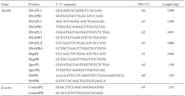
Table 1- Primers used for porcineMyd88isolation, SNPs detection and mRNA tissue distribution analysis.
Gene Primers 5’-3’ sequence TM (°C) Length (bp)
Myd88 DNAPL1 GGAAGCACAGGCCCACAAG 60 1380
DNAPR1 GGTGATGCCTGACATCCAAG
DNAPL2 AGCACGAGGCAGCTGAGAAG 61 1308
DNAPR2 TTGGTGCAGGGGTTGGTGTAG
DNAPL3 CGGATGGTAGTGGTTGTCTCTGA 62 1491
DNAPR3 CCTGTATAAGCGTCTCTGCGTG
DNAPL4 CCCAACTTCTGACATCTCCATC 61 1046
DNAPR4 CCTGCTAAGTTTGGTTCCTGTG
MapPL CCCAACTTCTGACATCTCCATC 61 1046
MapPR CCTGCTAAGTTTGGTTCCTGTG
QpcrPL CGGATGGTAGTGGTTGTCTCTGA 65 194
QpcrPR TTGGTGCAGGGGTTGGTGTAG
SNPPL AAAAATTCCTCAGGTTCCTAGAAAGTGCA 60 185
SNPPR GATCCACAGCTGATGTGAGCA
b-actin ControlPL GGACTTCGAGCAGGAGATGG 65 233
using Gene Expression Macro software (Bio-Rad, Rich-mond, CA) employing an optimized comparative Ct (DDCt) value method. Expression was considered undetect-able when the Ct value of the targeted gene exceeded 35.
SNPs identification and association analysis
DNA samples from seven breeds, including three Chinese indigenous (Wuzhishan, Laiwu, Bamaxiang and Guizhouxiang) and two foreign breeds (Landrace and Yorkshire), were used as PCR templates for Myd88
genomic DNA isolation. All PCR products were se-quenced. Subsequently, all sequenced information related to the porcineMyd88gene, this including our PCR results, the ESTs and genomic DNA fragments available on NCBI, was used to analyse potential SNPs. A potential SNP site was considered as that where different alleles appeared more than twice.
Genetic variation was studied in seven unrelated breeds of pigs, namely, Tongcheng, Wuzhishan, Laiwu, Bamaxing, Guizhouxiang, Yokshire and Landrace. The ex-perimental group underwent association analysis. This group consisted of three pure-blood populations, Tongcheng (T), Landrace (L) and Yorkshire (Y), and two crossbred populations, LYT (L male x YT female) and YLT (Y male x LT female). Six porcine immune-traits were examined. These were red blood cell count (RBC), hematocrit (HCT), mean corpuscular volume (MCV), IgG, blood cell distribution width (RDW) and delayed-type hy-persensitivity (DTH). In order to determine immune-traits, the blood from 20-weeks-old pigs was collected so as to de-tect RBC, HCT, MCV and RDW, by using a blood cell auto-analyzer (MEK-5216K). IgG concentration was as-certained through the radial immuno-diffusion method. The Delayed-type hypersensitivity (DTH) trait was de-tected by means of the phytohemagglutinin (PHA) skin test, according to the van Heugten method, with a minor modification (Van Heugtenet al., 1994).
A general linear model (GLM) was used to estimate the association between genotypes and immune traits. Ac-cording to the structure of the population, the model used for trait association analysis is described as follows:
Yijk=m+Pi+Gj+Bk+ (PG)ij+ (PB)ik+ (GB)jk+eijk
whereYijkl =lthtrait measured in the animal;m= overall
mean;Pi= fixed effect of theithpopulation (i= 1, 2, 3, 4, 5);
Gj= fixed effect of thejthgenotype (j= 1, 2, 3);Bk= fixed
effect of thekthbatch (phenotypic data were recorded in two periods,k= 1, 2); (PG)ij= effect of interactionithpopulation
jthgenotype; (PB)ik= effect of interactionithpopulation kth
batch; (GB)jk= effect of interactionjthgenotype kthbatch;
eijkl= error term.
Phylogenetic tree and structure analysis
Myd88 proteins from many species were collected for phylogenetic tree analysis and the homologues of the
se-quences analyzed by means of the ClustalW program. A phylogenetic tree was retrieved by using MEGA 3.1 soft-ware. Conserved residues of the functional domain of por-cine Myd88 were analyzed through multiple sequences alignment. The three-dimensional (3-D) model was pre-dicted through the 3djigsaw program. An image of the 3-D model was obtained by using software Raswin 2.7 soft-ware. Phosphorylation sites were predicted by the NetPhos program.
Results and Discussion
Isolation and chromosome assignment
The DNA segment isolated was 4464 bp, subse-quently deposited in to GenBank (GenBank no, EU056737). The isolated genomic sequence contained the complete ORF (882 bp) of the porcine Myd88 gene (NM_001099923). RH mapping results revealed that this gene was assigned to the long arm of the pig chromosome 13 (SS13q), the closest linked marker being S0288 (dis-tance = 40 cR; LOD = 8.66). In humans,Myd88has been mapped on 3p22 (Bonnert et al., 1997). Comparative genomic analysis results confirmed that pig chromosome 13 is homologous with human chromosome 3 (Sunet al., 1999). Thus, our mapping results conformed to those from comparative genomic analysis.
Detection of tissue distribution
Real time-PCR analysis was performed to determine the mRNA expression profile of the Myd88 gene in Wuzhishan mini-pigs. The data revealedMyd88gene ex-pression in all examined tissues, this exex-pression being rela-tively low in skeletal muscle tissue (Figure 1). Previous studies also showed wideMyd88gene expression in diges-tive tissues, the spleen and mesenteric lymph nodes (Tohno
et al., 2007). In humans,Myd88was found to be constitu-tively expressed in many tissues (Hardimanet al., 1996), this thus implying the similarity of the tissue distribution profile of the porcineMyd88gene to that in humans. TLRs
are widely expressed in many tissues (Zarember and Godowski, 2002), and Myd88 functions as the adaptor pro-tein of TLRs. Moreover, sub-cellular localization results confirmed that Myd88 found in cytoplasm was not a secre-tory protein (Nishiyaet al., 2007). Therefore,Myd88needs to be widely expressed in order to participate in TLR signal transduction.
Polymorphism detection and association analysis
PorcineMyd88gene polymorphisms were detected by multiple sequence comparison. According to our results, the 4464 bp genomic DNA ofMyd88contained 16 poten-tial SNPs which were 797T/C, 813A/g, 1721T/g, 1755C/A, 2130T/A, 2461C/T, 2468G/A, 2519G/A, 2743A/g, 2757C/T, 3076A/g, 3258A/g, 3291T/A, 3298C/T, 3345A/g and 3485G/A. None of these resulted in residual changes, this indicating that the porcine Myd88 protein was highly conserved. 797T/C polymorphism of the porcine Myd88
gene, which can be detected by the PCR-RFLP method, was further studied. The genotypes of this site were identi-fied by using the restriction enzymeApaL I (TT 185 bp, CC 160/25 bp, TC 185/160/25 bp) (Figure 2). Allele frequency analysis revealed a much higher frequency of allele T in five Chinese indigenous breeds than in Landrace and York-shire (Table 2). We performed a preliminary association study to determine whether this polymorphism had affected any immune-traits in the pig. The data showed that there was no significant association between this SNP and
im-mune traits RBC, HCT, MCV, IgG, DTH and RDW (p > 0.05) (Table 3).
Phylogenetic tree and structural characterization
The porcine Myd88 protein contained 293 residues with an overall sequence similarity to Myd88 in human (88%), chicken (70%), clawed frog (65%), zebrafish (62%) and sea urchin (40%). Phylogenetic tree analysis also showed that Myd88 was conserved during evolution (Fig-ure 3). Highly conserved residues of Myd88 were detected through sequence comparison with the five species men-tioned above. Porcine Myd88 contained two functional do-mains, DD (residues: 19-109) and TIR (residues: 157-293) (Tohnoet al., 2007), the most abundant amino acid in DD being Leu (19.8%). 17 DD residues were conserved in all the species examined. Among the conserved residues, there were 7 Leu residues and 12 hydrophobic residues (A, I, L, Figure 2- RFLP analysis of porcineMyd88gene polymorphism. 797T/C polymorphism was detected byApaL I(TT 185 bp, CC 160/25 bp, TC 185/160/25 bp). M: DNA ladder.
Table 2- Genotypes and allelic frequencies for the polymorphism 797 T/C ofMyd88in several pig breeds
Breeds N Genotypes Allele frequencies
TT TC CC T% C%
Wuzhishan 35 35 0 0 100 0
Bamaxiang 33 33 0 0 100 0
Guizhouxiang 38 38 0 0 100 0
Tongcheng 42 40 1 1 96.4 3.6
Laiwu 37 19 18 0 75.7 24.3
Yorkshire 38 14 17 7 59.2 40.8
Landrace 31 7 14 10 45.2 54.8
Table 3- Association analysis of 797 T/C polymorphism inMyd88with porcine RBC, HCT, MCV, IgG, DHA and RDW traits.
Genotypes N RBC HCT MCV IgG DTH RDW
CC 24 6.34±0.32 37.32±2.02 59.20±1.40 50.50±3.27 8.73±0.39 18.12±0.42
TC 65 6.57±0.29 37.26±1.83 56.11±1.27 43.95±2.96 8.96±0.35 18.85±0.38
TT 68 6.61±0.19 37.64±1.18 56.37±0.82 45.44±1.92 8.76±0.23 18.46±0.25
p-value* 0.777 0.984 0.171 0.274 0.876 0.402
*Means the probability of F-test for the genotype effect. Phenotypic value = mean±SE.
F, W, and V belong to hydrophobic amino acid). These re-sults indicate that hydrophobic residues, especially of Leu, may play important roles in maintaining DD structure and functioning.. The 3-D model of the porcine Myd88 death domain was predicted using the 3djigsaw program. Ac-cording to the model, the hydrocarbon chains of Leu resi-dues were packed in the inner part of the DD, thus forming a hydrophobic interior. The highly conserved Leu residues (Leu33, 75, 89, 90, 105) formed a Leu plane (Figure 4). These results also indicate that Leu residues may play im-portant roles in the death domain.
The other functional domain of porcine Myd88 is a TIR containing 137 residues. A BB-loop, which was found in the TIR domain of TLRs (Xuet al., 2000), was also found in the TIR of the porcine Myd88 in our studies. This loop contained the motif (RDxLPG, x represents L or V), and was found to be highly conserved among all the species studied. Previous research has confirmed that the BB-loop was essential for maintaining theTLR4 function. Substitu-tion of the Pro residue in the BB-loop of TLR4 by His abol-ished the TLR4 immune-response to lipopolysaccharide (Poltoraket al., 1998). The BB-loop, highly conserved dur-ing evolution, may be very important for the porcine Myd88 signaling pathway. In addition, another conserved motif (CDFQTKFAxSL, x represents L or V) was found in the TIR of porcine Myd88 (Box 2). This motif contains a conserved Ser which may be a phosphorylation site pre-dicted through using the NetPhos program (p = 0.959), and may be related to phosphorylation of porcine Myd88. A 3-D model of the TIR of porcine Myd88 was predicted by using the 3djigsaw program (Figure 5), the conserved do-mains being labeled in yellow and the Ser in Box 2 in blue.
Acknowledgments
We are grateful to Dr. Yerle for supplying the RH panel. This research was supported by the State Platform of Technology Infrastructure (2005DKA21101), the Key Pro-ject of National Basic Research and Developmental Plan of China (G2006CB102105), the National High Science and Technology Foundation of China (20060110Z1039), the National Natural Science Foundation of China (30571300), the National Scientific and Technology Mainstay Project (2006BDA13B08) and the Project of Science and Technol-ogy Innovation Team for “Research and Improvement of Domestic Animal Germplasm” of IAS, CAAS.
Figure 5- A 3-D model of the Toll/IL-1 receptor domain (TIR) of porcine Myd88. The first residue (Glu156) and the last residue (Leu292) are la-beled in green. The highly conserved BB-loop and Box2 are lala-beled in yel-low. The Ser239 residue, the possible phosphorylation site, is labeled in blue.
Figure 3- The phylogenetic tree of theMyd88gene. Bootstrap confidence values, shown at the nodes of the tree, are based on 1000 bootstrap repli-cates. Horizontal branch lengths are proportional to the estimated diver-gence of the sequence from the branch point. GenBank accession numbers are: Human, AAB449967; Monkey, XP_001088062; Dog, XP_534223; Cattle, NP_001014404; Pig, ABM90642; Rat, AAH9726; Mouse, AAC53013; Chicken, NM_001030962; Clawed frog, NP_001016837, Zebrafish, AAQ90476; Sea urchin, XP_780590; Fruit fly, NP_610479; Red flour beetle, XP_973419.
References
Adachi O, Kawai T, Takeda K, Matsumoto M, Tsutsui H, Saka-gami M, Nakanishi K and Akira S (1998) Targeted disrup-tion of the MyD88 gene results in loss of IL-1- and IL-18-mediated function. Immunity 9:143-150.
Bonnert TP, Garka KE, Parnet P, Sonoda G, Testa JR and Sims JE (1997) The cloning and characterization of human MyD88: A member of an IL-1 receptor related family. FEBS Lett 402:81-84.
Hardiman G, Rock FL, Balasubramanian S, Kastelein RA and Bazan JF (1996) Molecular characterization and modular analysis of human MyD88. Oncogene 13:2467-2475. Lemaitre B, Nicolas E, Michaut L, Reichhart JM and Hoffmann
JA (1996) The dorsoventral regulatory gene cassette spatzle/Toll/cactus controls the potent antifungal response in Drosophila adults. Cell 86:973-983.
Li C, Zienkiewicz J and Hawiger J (2005) Interactive sites in the MyD88 Toll/interleukin (IL) 1 receptor domain responsible for coupling to the IL1beta signaling pathway. J Biol Chem 280:26152-26159.
Li L, Cousart S, Hu J and McCall CE (2000) Characterization of interleukin-1 receptor-associated kinase in normal and endotoxin-tolerant cells. J Biol Chem 275:23340-23345. Medvedev AE, Lentschat A, Wahl LM, Golenbock DT and Vogel
SN (2002) Dysregulation of LPS-induced Toll-like receptor 4-MyD88 complex formation and IL-1 receptor-associated kinase 1 activation in endotoxin-tolerant cells. J Immunol 169:5209-5216.
Medzhitov R and Janeway Jr CA (1997) Innate immunity: The virtues of a nonclonal system of recognition. Cell 91:295-298.
Milan D, Hawken R, Cabau C, Leroux S, Genet C, Lahbib Y, Tosser G, Robic A, Hatey F, Alexander L, et al.(2000) IMpRH server: An RH mapping server available on the Web. Bioinformatics 16:558-559.
Nishiya T, Kajita E, Horinouchi T, Nishimoto A and Miwa S (2007) Distinct roles of TIR and non-TIR regions in the subcellular localization and signaling properties of MyD88. FEBS Lett 581:3223-3229.
Palladino MA, Johnson TA, Gupta R, Chapman JL and Ojha P (2007) Members of the toll-like receptor family of innate immunity pattern-recognition receptors are abundant in the male rat reproductive tract. Biol Reprod 6:958-964. Poltorak A, He X, Smirnova I, Liu MY, Van Huffel C, Du X,
Birdwell D, Alejos E, Silva M, Galanos C,et al.(1998) De-fective LPS signaling in C3H/HeJ and C57BL/10ScCr mice: Mutations in Tlr4 gene. Science 282:2085-2088.
Scanga CA, Aliberti J, Jankovic D, Tilloy F, Bennouna S, Denkers EY, Medzhitov R and Sher A (2002) Cutting edge: MyD88 is required for resistance to Toxoplasma gondii in-fection and regulates parasite-induced IL-12 production by dendritic cells. J Immunol 168:5997-6001.
Sun HF, Ernst CW, Yerle M, Pinton P, Rothschild MF, Chardon P, Rogel-Gaillard C and Tuggle CK (1999) Human chromo-some 3 and pig chromochromo-some 13 show complete synteny con-servation but extensive gene-order differences. Cytogenet Cell Genet 85:273-278.
Takeda K and Akira S (2004) TLR signaling pathways. Sem Immunol 16:3-9.
Tohno M, Shimazu T, Aso H, Kawai Y, Saito T and Kitazawa H (2007) Molecular cloning and functional characterization of porcine MyD88 essential for TLR signaling. Cell Mol Immunol 4:369-376.
Uematsu S and Akira S(2006) Toll-like receptors and innate im-munity. J Mol Med 84:712-725.
Van Heugten E, Spears JW, Coffey MT, Kegley EB and Qureshi MA (1994) The effect of methionine and aflatoxin on im-mune function in weanling pigs. J Anim Sci 72:658-664. Xu Y, Tao X, Shen B, Horng T, Medzhitov R, Manley JL and
Tong L (2000) Structural basis for signal transduction by the Toll/interleukin-1 receptor domains. Nature 408:111-115. Yamamoto M and Akira S (2004) TIR domain-containing
adap-tors regulate TLR-mediated signaling pathways. Nippon Rinsho 62:2197-2203.
Yerle M, Pinton P, Robic A, Alfonso A, Palvadeau Y, Delcros C, Hawken R, Alexander L, Beattie C, Schook L,et al.(1998) Construction of a whole-genome radiation hybrid panel for high-resolution gene mapping in pigs. Cytogenet Cell Genet 82:182-188.
Zarember KA and Godowski PJ (2002) Tissue expression of hu-man like receptors and differential regulation of Toll-like receptor mRNAs in leukocytes in response to microbes, their products, and cytokines. J Immunol 168:554-561.
Internet Resources
The IMpRH mapping tool, http://IMpRH.toulouse.inra.fr/. The clustal w program, http://www.ebi.ac.uk/clustalw/.
The 3djigsaw program, http://www.bmm.icnet.uk/serv-ers/3djigsaw/.
The NetPhos program, http://www.cbs.dtu.dk/services/NetPhos/.
Associate Editor: Luiz Lehmann Coutinho