DOI 10.1007/s00280-014-2663-x
ORIGINAL ARTICLE
Regulatory role of Lactobacillus acidophilus on inflammation
and gastric dysmotility in intestinal mucositis induced
by 5-fluorouracil in mice
Priscilla F. C. Justino · Luis F. M. Melo · Andre F. Nogueira · Cecila M. Morais · Walber O. Mendes · Alvaro X. Franco · Emmanuel P. Souza · Ronaldo A. Ribeiro · Marcellus H. L. P. Souza · Pedro Marcos Gomes Soares
Received: 21 July 2014 / Accepted: 22 December 2014 / Published online: 9 January 2015 © Springer-Verlag Berlin Heidelberg 2015
group. Furthermore, 5-FU significantly (p < 0.05) increased
cytokine (TNF-α, IL-1β, and CXCL-1) concentrations and
decreased IL-10 concentrations compared with the
con-trol group. 5-FU also significantly (p < 0.05) delayed
gas-tric emptying and gastrointestinal transit compared with the control group. All of these changes were significantly
(p < 0.05) reversed by treatment with L. acidophilus.
Conclusions Lactobacillus acidophilus improves the
inflammatory and functional aspects of intestinal mucositis induced by 5-FU.
Keywords Intestinal mucositis · 5-Fluorouracil · Lactobacillus acidophilus · Probiotics · Gastrointestinal dysmotility
Introduction
Probiotics are “live microorganisms which, when admin-istered in adequate amounts, confer benefits to the health
of the host” [1]. Probiotics promote crypt cell proliferation,
prevent cytokine-induced apoptosis [2], reduce
pro-inflam-matory cytokine production, and regulate the intestinal
immune system [3]. Among probiotics, Lactobacillus
aci-dophilus stands out for being a widely used, thermophilic,
nonpathogenic bacteria [4]. Lactobacillus acidophilus is
used for the treatment and prevention of gastrointestinal
disorders associated with diarrhea of varying etiology [5].
Intestinal mucositis (IM) is inflammation associated with the cytotoxicity of chemotherapy and radiotherapy for can-cer. Intestinal mucositis usually accompanies cell loss in the
epithelial barrier of the lining of the gastrointestinal tract [6,
7]. Symptoms of IM include nausea, dyspepsia, dysphasia,
vomiting, and diarrhea [8]. Intestinal mucositis development
can be separated into three stages of increased epithelial
Abstract
Purpose Lactobacillus acidophilus is widely used for gastrointestinal disorders, but its role in inflammatory con-ditions like in chemotherapy-induced mucositis is unclear.
Here, we report the effect of L. acidophilus on
5-fluoroura-cil-induced (5-FU) intestinal mucositis in mice.
Methods Mice weighing 25–30 g (n= 8) were separated
into three groups, saline, 5-FU, and 5-FU +L. acidophilus
(5-FU-La) (16 × 109 CFU/kg). In the 5-FU-La group, L.
acidophilus was administered concomitantly with 5-FU on the first day and alone for two additional days. Three days
after the last administration of L. acidophilus, the animals
were euthanized and the jejunum and ileum were removed for histopathological assessment and for evaluation of lev-els of myeloperoxidase activity, sulfhydryl groups, nitrite,
and cytokines (TNF-α, IL-1β, CXCL-1, and IL-10). In
addition, we investigated gastric emptying using spectro-photometry after feeding a 1.5-ml test meal by gavage and euthanasia. Data were submitted to ANOVA and
Bonfer-roni’s test, with the level of significance at p < 0.05.
Results Intestinal mucositis induced by 5-FU significantly
(p < 0.05) reduced the villus height–crypt depth ratio and
GSH concentration and increased myeloperoxidase activ-ity and the nitrite concentrations compared with the control
P. F. C. Justino · L. F. M. Melo · A. F. Nogueira · C. M. Morais · W. O. Mendes · A. X. Franco · R. A. Ribeiro · M. H. L. P. Souza · P. M. G. Soares
Department of Physiology and Pharmacology, Medical School, Federal University of Ceara, Rua Cel. Nunes de Melo 1315, Rodolfo Teofilo, Fortaleza, Ceara CEP 60.430-270, Brazil E. P. Souza · P. M. G. Soares (*)
Department of Morphology, Medical School, Federal University of Ceara, Rua Delmiro de Farias s/n, Rodolfo Teofilo, Fortaleza, Ceara CEP 60.416-030, Brazil
dysfunction: an inflammatory stage, epithelial degradation stage, and an ulceration/bacterial stage. This dysfunction is
rescued by restoration of the functional epithelia [9].
5-Fluorouracil (5-FU), widely used as
chemotherapeu-tic for colorectal and breast cancer [10, 11], is known to
induce intestinal damage through IM [12] and stem cell
apoptosis [13]. Our group has shown that IM induced by
5-FU promotes infiltration of neutrophils, increases pro-inflammatory cytokine levels, and significantly delays
gas-tric emptying [8]. Furthermore, IM induced by 5-FU
pro-motes dysbiosis [14]. The effect of restoring the normal
microbiota in IM has not been studied.
Therefore, the aim of the present study was to evaluate the
effect of administration of L. acidophilus on the
inflamma-tory and functional outcomes of 5-FU-induced IM in mice.
Materials and methods
Animals
Male Swiss mice weighing 25–30 g (supplied by the Department of Physiology and Pharmacology, UFC Medi-cal School) were kept in a temperature-controlled room with ad libitum access to water and fasted for 24 h prior to all experiments. The study was previously approved by the local research ethics committee (protocol#34/10), and all procedures involving animals were performed in accordance with the Guide for the Care and Use of Laboratory Animals of the US Department of Health and Human Services.
Model of IM induced by 5-FU
Twenty-four male Swiss mice were randomly divided into a
saline group (n= 8), a 5-FU group (saline + 5-FU 450 mg/
kg, single dose, i.p. n= 8), and a 5-FU-La group (5-FU
450 mg/kg, single dose, i.p. +L. acidophilus 16 × 109 CFU/
kg for 3 days, n= 8). Saline served as control for the 5-FU
group, and 5-FU served as a control for the 5-FU +L.
aci-dophilus group. The animals in 5-FU + L. acidophilus
received 5-FU and L. acidophilus simultaneously. The mice
were euthanized three days after completing treatment with L. acidophilus. Blood samples were collected, and the jeju-num and ileum were removed for morphological and histo-pathological analyses and for evaluation of myeloperoxidase activity (MPO), sulfhydryl groups, and levels of nitrite and
cytokines (TNF-α, IL-1β, CXCL-1, and IL-10). The animals
were weighed daily throughout the experiment.
Intestinal morphometry and histopathology
Segments of jejunum (a 3-cm segment immediately distal to the ligament of Treitz) and distal ileum (a 6-cm segment
adjacent to the ileocecal valve) were collected, fixed, and stained with hematoxylin and eosin for the measurement of villus height and crypt depth. Ten intact and well-oriented villi and crypts were measured and averaged for each sam-ple. The microscopy analysis was double-blinded. Mucosal inflammation was assessed using a modification of the
his-topathological scores described by [15].
Intestinal MPO activity
Myeloperoxidase is found in azurophilic neutrophil granules and has been extensively used as a biochemi-cal marker of granulocyte infiltration into various tissues, including the gastrointestinal tract. The extent of neutro-phil accumulation in the intestinal mucosa was
quanti-fied using an MPO activity assay kit [16]. Briefly,
intes-tinal tissue (50 mg/ml) was homogenized in HTAB buffer (Sigma-Aldrich, St Louis, MO). The homogenate was centrifuged at 4,500 rpm for 7 min at 4 °C. MPO activ-ity in the resuspended pellet was assayed by measuring the change in absorbance at 450 nm using o-dianisidine dihydrochloride (Sigma-Aldrich, St Louis, MO) and 1 % hydrogen peroxide (Merck, Whitehouse Station, NJ). The results were expressed as MPO units/mg tissue. A unit of MPO was defined as the amount of enzyme required to
convert 1 µmol/min of hydrogen peroxide into water at
22 °C.
Glutathione assay
Intestinal tissue levels of glutathione (GSH) were assessed
with an assay for nonprotein sulfhydryl content [17]. In
summary, 100 mg/ml frozen intestinal tissue was
homog-enized in 0.02 M EDTA. Aliquots of 400 µl of
homogen-ate were mixed with 320 µl distilled water and 80 µl 50 %
trichloroacetic acid (TCA) to precipitate proteins. The material was centrifuged (3,000 rpm) for 15 min at 4 °C.
Aliquots of 400 µl of supernatant were mixed with 800 µl
0.4 M Tris buffer (pH 8.9) and 20 µl
5.5-dithiobis-(2-ni-trobenzoic acid) (DTNB) (Fluka, St Louis, MO), followed by shaking for 3 min. Within 5 min of addition of DTNB, the absorbance was read at 412 nm against a blank reagent
without homogenate. The results were expressed as µg
GSH/mg tissue.
Determination of nitrite levels
The production of NO was determined indirectly by
measuring nitrite levels using the Griess reaction [18].
Briefly, 100 µl intestinal tissue homogenate was incu-bated with 100 µl Griess reagent (1 % sulfanilamide in
1 % H3PO4/0.1 % N-(1-naphthyl)ethylenediamine
temperature for 10 min. A microplate reader measured the absorbance at 540 nm. Nitrite levels were determined from
a standard nitrite curve generated using NaNO2.
Detection of cytokines (TNF-α, IL-1β, CXCL-1,
and IL-10)
Cytokine (TNF-α, IL-1β, CXCL-1, and IL-10)
concentra-tions in jejunum and ileum samples were determined using enzyme-linked immunosorbent assay (ELISA) using proto-cols supplied by the manufacturer (R&D Systems, Minne-apolis, USA). The results were expressed as pg/ml.
Gastric emptying and intestinal transit
Gastric emptying and intestinal transit were measured using
the modified technique of Reynell and Spray [19]. Initially,
the animals received a 300-µl test meal by gavage, consist-ing of a nonabsorbable marker (0.75 mg/ml phenol red in 5 % glucose). After 20 min, the animals were euthanized by cervical dislocation. After laparotomy, the stomach and bowels were exposed and the esophageal–gastric, gastrodu-odenal, and ileocecal junctions were immediately isolated using ligatures. The specimens were removed and divided into stomach and proximal, medial and distal bowel. Each segment was placed in a graduated cylinder, and the total volume was measured by adding 10 ml of 0.1 N NaOH. Then, the samples were cut into small pieces and homog-enized for 30 s. Twenty minutes later, 1 ml supernatant was removed and centrifuged for 10 min at 2,800 rpm. The pro-teins in the homogenate were precipitated by adding 20 % TCA and centrifuged again for 20 min at 2,800 rpm. Then, 150 ml of supernatant was collected and added to 200 ml 0.5 N NaOH. The absorbance of the samples was deter-mined using spectrophotometry at 540 nm and expressed as optical density.
The fractional dye retention was expressed in percent-age, according to the following equation: gastric dye
reten-tion = amount of phenol red recovered in stomach/total
amount of phenol red recovered from two segments (stom-ach and small intestine). Intestinal transit was calculated for each bowel segment by dividing the amount of phenol red recovered from a given segment by the amount of phe-nol red recovered from all three segments and is expressed as a percentage.
Statistical analysis
The results were reported as the mean values ± standard
error of the mean (SEM) for each group. The data were submitted to analysis of variance (ANOVA) followed by Bonferroni’s test. The level of statistical significance was
set at p < 0.05.
Results
Effect of L. acidophilus on the weight of mice with IM
induced by 5-FU
The average weight of the animals in the saline group
increased throughout the study period (6.13 ± 1.22 %),
with the highest weight registered on the last day. In con-trast, weight in the 5-FU group decreased considerably
(−14.54 ± 2.03 %) by day 3 after 5-FU administration
in relation to that of the saline group. The weight of the
5-FU +L. acidophilus group decreased −5.48 ± 0.66 % in
relation to that of the 5-FU group.
Effect of L. acidophilus on histopathological changes in the
intestinal mucosa
The animals in the 5-FU group presented the following his-topathological changes in the jejunum and ileum: mucosa with shortened villi with vacuolated cells, intense
inflam-matory infiltrate and vacuolization (Fig. 1c, d) compared to
those in the control group (Fig. 1a, b). The 5-FU +L.
aci-dophilus group experienced a significant improvement of histopathological changes, as shown by photomicrographs
(Fig. 1e, f).
The 5-FU group showed a significant decrease in villus
height (Fig. 2a), an increase in crypt depth (Fig. 2b), and a
decrease in the villus/crypt ratio (Fig. 2c) of the jejunum
and ileum, compared to the control group. Panel b, Fig. 2,
shows that treatment with L. acidophilus significantly
(p < 0.05) reversed the 5-FU-induced increase in crypt
depth in both segments. Likewise, the decrease in the vil-lus/crypt ratio observed in the 5-FU group was significantly
reversed in the 5-FU +L. acidophilus group (Fig. 2c).
Effect of L. acidophilus on MPO activity and GSH
and nitrite concentrations
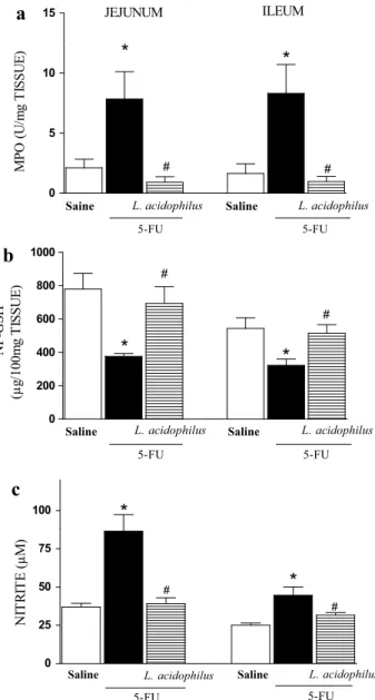
Following administration of 5-FU, the animals
expe-rienced a significant (p < 0.05) increase in neutrophil
infiltration in the jejunum (7.84 ± 2.28 UMPO/mg)
and ileum (8.31 ± 2.40 UMPO/mg) compared to the
saline group (jejunum: 2.11 ± 0.71 UMPO/mg; ileum:
1.64 ± 0.81 UMPO/mg). Treatment with L.
acidophi-lus significantly (p < 0.05) reduced neutrophil infiltration
in the jejunum (0.90 ± 0.46 UMPO/mg) and in the ileum
(0.97 ± 0.42 UMPO/mg) compared to those of the 5-FU
group (Fig. 3a).
5-FU significantly (p < 0.05) reduced GSH
con-centrations in the jejunum (376.30 ± 16.79 µg/mg)
and in the ileum (322.30 ± 37.01 µg/mg) compared
to those in the saline group (jejunum: 779.30 ± 94.60
L. acidophilus significantly (p < 0.05) reversed GSH
reduc-tions in the jejunum (693.00 ± 100.05 µg/g) and in the
ileum (514.80 ± 51.91 µg/g) (Fig. 3b).
Figure 3c shows that administration of 5-FU significantly
(p < 0.05) increased nitrite concentrations in the jejunum
(86.43 ± 10.93 µM) and in the ileum (44.66 ± 5.46 µM)
compared to those in the saline group (37.00 ± 2.93 and
25.08 ± 1.38 µM, respectively). Treatment with L. acidophilus
significantly (p < 0.05) reduced nitrite concentrations in both
the jejunum and ileum (39.13 ± 3.91 and 31.65 ± 1.67 µM,
respectively) compared to those in the 5-FU group.
Effect of L. acidophilus on TNF-α, IL-1β, CXCL-1,
and IL-10 production
Figure 4 shows that the animals with IM present
sig-nificantly (p < 0.05) increased levels (p < 0.05) of
TNF-α, IL-1β, and CXCL-1 in the jejunum (56.3, 350.6,
and 163.5 %, respectively) and in the ileum (195.4, 77.6, and 535.7 %, respectively) compared to those of the
con-trols (panel a, b, and c). IL-10 showed significant (p < 0.05)
reductions in the jejunum and ileum (39.1 and 52.2 %,
respectively). In contrast, these increases in TNF-α, IL-1β,
and CXCL-1 and decreases in IL-10 concentrations were
significantly (p < 0.05) reversed by treatment with L.
aci-dophilus (panel d). Fig. 1 Administration of Lactobacillus acidophilus reduced
intes-tinal damage in mice caused by exposure to 5-fluorouracil. Photo-micrographs (×200) of the jejunum and ileum of mice treated with saline (a, b), 5-FU + saline (c, d), and 5-FU +L. acidophilus for 3 days (e, f). Note that treatment with L. acidophilus reversed 5-FU-induced shortening of the villi (c arrowhead) and reduced the inten-sity of inflammatory infiltrates (c, d arrow)
a
b
Saline Saline
0 200 400 600
*
*
#
#
JEJUNUM ILEUM
5-FU 5-FU
L. acidophilus L. acidophilus
Vi
llus
hi
gh
t
(µ
m)
Saline Saline
0 50 100 150 200
*
#
#
5-FU 5-FU
L. acidophilus L. acidophilus
*
Crypts depth (µm)
Saline Saline
0 2 4 6
*
#
*
#
5-FU 5-FU
L. acidophilus L. acidophilus
Villus hight / Crypt depth (µm
)
c
Effect of L. acidophilus on gastric emptying and gastrointestinal transit
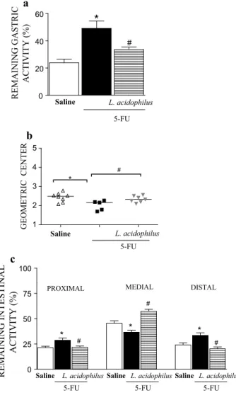
Gastric retention was significantly (p < 0.05) higher in
the 5-FU group (49.06 ± 5.42 %) than in the saline group
(23.78 ± 2.73 %) (Fig. 5a). This was reversed by treatment
with L. acidophilus (33.58 ± 1.85 %). Panel b shows that
gastrointestinal transit was significantly slower in the 5-FU group than in the saline group (median geometric center:
2.02 ± 0.11 vs. 2.43 ± 0.08) (p < 0.05), but was
normal-ized by treatment with L. acidophilus (2.34 ± 0.06).
Likewise, a significant level of retention in the proximal, medial, and distal bowel segments (5-FU vs. saline) was
associated with diarrhea. Treatment with L. acidophilus
significantly (p < 0.05) reversed these changes (Fig. 5c).
Discussion
Treatment with L. acidophilus improved the
histopathologi-cal changes in murine IM induced by 5-FU, possibly due to reductions in inflammatory markers (levels of nitrite, GSH,
cytokines, and neutrophil infiltration). In addition, L.
aci-dophilus reversed the gastrointestinal dysmotility induced by 5-FU.
Currently, some probiotics have achieved success in
the treatment of mucositis induced by 5-FU.
Streptococ-cus thermophilus TH-4 improved the mitotic count, cryptal
fissions, and histological deficits caused by 5-FU [20].
Lactobacillus fermentum BR11 showed a partial rescue of histological pathology while being ineffective in
com-bination with prebiotics [21]. Supernatant of Escherichia
coli Nissle 1917 partially reduced the damage caused by
5-FU [22]. However, L. rhamnosus GG, Bifidobacterium
lactis BB12, and skim milk were totally ineffective in
res-cuing the mucositis caused by 5-FU [23]. Several studies
have shown epithelial damage and neutrophil infiltration in the mucosa during the inflammatory stage of the
mucosi-tis induced by antineoplastic drugs [24]. Edens et al. [25]
reported changes in the permeability of the intestinal epi-thelium caused by migration of neutrophils to epithelial cells. This finding can explain the dyspepsia in animals
with 5-FU-induced IM. Lima et al. [26] found that
pentoxi-fylline and thalidomide inhibited the lesions and myelop-eroxidase activity in 5-FU-induced oral mucositis. Another study using methotrexate reported a significant increase in
villous atrophy in the rat intestinal mucosa [27].
Intesti-nal injury may include changes in brush border hydrolase activity, blunted villus height, deepening and increased cell
apoptosis in the crypt, and decreased proliferation [28, 29].
Few studies have assessed the effects of L. acidophilus
on inflammation. One of these studies found lower levels of
leukocyte migration in animals treated with L. acidophilus
in a model of IM induced by irinotecan [30]. Several
stud-ies have reported reduced inflammatory effects using other probiotic species.
Probiotics derived from E. coli and L. fermentum
low-ered the inflammation caused by 5-FU in the rat jejunum
[22]. Moreover, L. brevis reduced nitrite and nitrate
lev-els in patients with chronic periodontitis [31]. Matsumoto
et al. [32] evaluated the effect of L. casei strain Shirota on
murine chronic inflammatory bowel disease and reported a
a
b
c
Saine Saline
0 5 10 15
*
#
*
#
JEJUNUM ILEUM
5-FU 5-FU
L. acidophilus L. acidophilus
MP
O
(U
/m
g
TI
SS
UE
)
Saline Saline
0 200 400 600 800 1000
*
*
##
5-FU 5-FU
L. acidophilus L. acidophilus
NP
-G
SH
(µ
g/
100m
g
TI
SSU
E)
Saline Saline
0 25 50 75
100
*
#
*
#
5-FU 5-FU
L. acidophilus L. acidophilus
NI
TR
IT
E
(µ
M)
reduction in body weight loss, diarrhea, and occult blood. In
an earlier study, we reported that Saccharomyces boulardii
lowered pro-inflammatory cytokine levels (TNF-α, IL-1β,
and CXCL-1) in the rat jejunum and ileum induced by 5-FU [33]. Saccharomyces boulardii may have a protective effect against diarrheal pathogens by reducing the
pro-inflamma-tory response [34]. These authors reported lower secretion
of pro-inflammatory cytokines (IL-1β) and higher levels of
anti-inflammatory cytokines (IL-4 and IL-10) in animals
treated with S. boulardii. The mechanisms of S. boulardii
may be similar to the mechanisms of L. acidophilus action
in the model of IM induced by 5-FU used in our study.
In a clinical trial, L. acidophilus associated with B.
bifi-dum was satisfactory for diarrhea prophylaxis during pelvic
radiation therapy with concomitant cisplatin. Acute inflam-matory changes might play an important role in the
patho-genesis of these symptoms [35]. Furthermore, L. casei
strain Shirota may be a useful probiotic to manage
inflam-matory bowel disease. L. casei improves murine chronic
inflammatory bowel disease and is associated with a
down-regulation of pro-inflammatory cytokines such as IL-6 [32].
Strains of L. acidophilus, such as CBA4P, decrease
leptin-immunostimulated activity by lowering levels of
mac-rophage IL-1β and TNF-α in mice [36].
Other pathways to explain the L. acidophilus action
that we observed include the stimulation of apical Cl−
/
OH− exchange activity, corresponding to increased
sur-face expression of DRA in Caco-2 cells via the PI-3
kinase-mediated pathway. Thus, L. acidophilus may be
useful in treating diarrhea and other intestinal inflamma-tory disorders involving impairment of electrolyte
absorp-tion [37].
Moreover, 5-FU-induced IM is associated with delayed
gastric emptying/intestinal transit of liquids [38].
Hyper-contractility of the deep muscles of the stomach and duo-denum in both the inflammatory and post-inflammatory phase has also been reported. Patients receiving anticancer therapy experience gastrointestinal symptoms such as
dys-pepsia, dysphagia, and diarrhea [39], referred to as
cancer-associated dyspepsia syndrome (CADS).
In this study, L. acidophilus reversed 5-FU-induced
changes in gastrointestinal motility, enhancing intestinal transit and gastric emptying and decreasing retention in the distal bowel segment. This may partially account for the observed improvement in diarrhea and reduced weight loss.
Thus, we hypothesize that treatment with L. acidophilus
normalizes bowel function by reducing inflammation asso-ciated with 5-FU-induced IM.
a
b
c
Saline Saline
0 200 400 600 800
*
#
*
#
TN
F-α (p
g/
ml
)
JEJUNUM ILEUM
5-FU 5-FU
L. acidophilus L. acidophilus Saline Saline 0
200 400 600 800
*
*
#
JEJUNUM ILEUM
#
5-FU 5-FU
L. acidophilus L. acidophilus
IL
-1
β
(p
g/
ml
)
Saline Saline
0 200 400 600 800
*
*
# #
5-FU 5-FU
L. acidophilus L. acidophilus
CX
CL
-1
(p
g/
ml
)
Saline Saline
0 20 40 60 80
*
#
*
#
5-FU 5-FU
L. acidophilus L. acidophilus
IL
-1
0
(p
g/
ml
)
d
Fig. 4 Administration of Lactobacillus acidophilus reversed increased levels of TNF-α, IL-1β, CXCL-1, and IL-10 in 5-fluoroura-cil-induced intestinal mucositis in mice. Note the increased levels of TNF-α (a), IL-1β (b), and CXCL-1 (c) and decreased levels of IL-10 (d) due to exposure to 5-fluorouracil. Treatment with L.
acidophi-lus (a–d) normalized levels of cytokines. The results are presented as the mean ± SEM. The data were submitted to ANOVA followed by Bonferroni’s test. *p < 0.05 compared with Group I (saline only). #p
Few studies have evaluated the effects of probiotic
bac-teria on intestinal motility. One study, [40], suggested that
Bifidobacterium, Lactobacillus, and Streptococcus mediate relaxation in colonic motility. This relaxation could explain the observed improvement in diarrhea by reducing stool frequency and restoration of the microflora. Czerucka and
Rampal [41] demonstrated that S. boulardii restored
lumi-nal electrolyte transport in cholera toxin-induced diarrhea in the rabbit jejunum and suggested a mechanism involving
cAMP-dependent chloride secretion. Budriesi et al. [42]
used a mixture containing Castanea sativa and S.
boular-dii to induce antispasmodic and spasmolytic effects in
seg-ments of intestinal smooth muscle contracted by carbachol,
histamine, KCl, and BaCl2. These effects may be due to the
inhibition of voltage-dependent Ca2+ channels.
Dysmotility accompanies inflammatory bowel. Cells present in inflammatory conditions, such as macrophages, produce nitric oxide and prostaglandins, which lead to
dys-motility [43]. Inflammatory factors such as IL-1β and IL-6
[44], monocyte chemoattractant protein-1, and CCL-2 [45]
also induce dysmotility. These data corroborate the fact that L. acidophilus reduces inflammation and thus improves intestinal motility.
In summary, this study showed, for the first time, the
anti-inflammatory effects of L. acidophilus and its
regula-tory role in the dysmotility associated with IM induced by 5-FU. We hope that our findings will contribute to the dis-covery of new probiotic-based treatments of gastrointesti-nal toxicity associated with antineoplastic therapy.
Acknowledgments The authors would like to thank Conselho Nacional de Desenvolvimento Científico e Tecnológico (CNPq) and Fundação Cearense de Apoio ao Desenvolvimento Científico e Tec-nológico (FUNCAP) for financial support and Maria Silvandira Freire França for technical assistance. Dr. Ribeiro, Dr. Souza, and Dr. Soares are CNPq fellowship holders.
References
1. Fuller R (1991) Probiotics in human medicine. Gut 32(4):439–442 2. Laudanno O, Vasconcelos L, Catalana J, Cesolari J (2006)
Anti-inflammatory effect of bioflora probiotic administered orally or subcutaneously with live or dead bacteria. Dig Dis Sci 51(12):2180–2183. doi:10.1007/s10620-006-9175-4
3. Dieleman LA, Goerres MS, Arends A, Sprengers D, Torrice C, Hoentjen F, Grenther WB, Sartor RB (2003) Lactobacillus GG prevents recurrence of colitis in HLA-B27 transgenic rats after antibiotic treatment. Gut 52(3):370–376
4. Jespersen L (2003) Occurrence and taxonomic characteristics of strains of Saccharomyces cerevisiae predominant in African indigenous fermented foods and beverages. FEMS Yeast Res 3(2):191–200
5. Mack DR, Lebel S (2004) Role of probiotics in the modulation of intestinal infections and inflammation. Curr Opin Gastroenterol 20(1):22–26
6. van Vliet MJ, Harmsen HJ, de Bont ES, Tissing WJ (2010) The role of intestinal microbiota in the development and severity of chemotherapy-induced mucositis. PLoS Pathog 6(5):e1000879. doi:10.1371/journal.ppat.1000879
7. Sonis ST (2004) The pathobiology of mucositis. Nat Rev Cancer 4(4):277–284. doi:10.1038/nrc1318
8. Soares PM, Mota JM, Gomes AS, Oliveira RB, Assreuy AM, Brito GA, Santos AA, Ribeiro RA, Souza MH (2008) Gastroin-testinal dysmotility in 5-fluorouracil-induced inGastroin-testinal mucosi-tis outlasts inflammatory process resolution. Cancer Chemother Pharmacol 63(1):91–98. doi:10.1007/s00280-008-0715-9 9. Duncan M, Grant G (2003) Oral and intestinal mucositis—causes
and possible treatments. Aliment Pharmacol Ther 18(9):853–874 10. Longley DB, Harkin DP, Johnston PG (2003) 5-Fluorouracil:
mecha-nisms of action and clinical strategies. Nat Rev Cancer 3(5):330–338 11. Sonis ST (1998) Mucositis as a biological process: a new hypoth-esis for the development of chemotherapy-induced stomatotoxic-ity. Oral Oncol 34(1):39–43
a
b
c
Saline
0 20 40 60
#
*
5-FU
L. acidophilus
REMAINING GASTRIC
ACTIVITY (%)
Saline
1 2 3 4 5
*
#
5-FU
L. acidophilus
GEOMETRIC CENTER
Saline Saline Saline
0 25 50 75 100
#
* #
#
#
* *
PROXIMAL MEDIAL DISTAL
5-FU
L. acidophilus
5-FU
L. acidophilus
5-FU
L. acidophilus
REMAINING INTESTINAL
ACTIVITY (%)
Fig. 5 Administration of Lactobacillus acidophilus reversed delayed gastric emptying and gastrointestinal transit associated with 5-FU-induced intestinal mucositis. Note delayed gastric emptying (a), dis-placement of the geometric center (b), and intestinal transit (c) due to exposure to 5-fluorouracil. Treatment with L. acidophilus (a–c) improved gastrointestinal symptoms. The results are presented as the mean ± SEM. The data were submitted to ANOVA followed by Bonferroni’s test. *p < 0.05 compared with Group I (saline only). #p
12. Baerg J, Murphy JJ, Anderson R, Magee JF (1999) Neutropenic enteropathy: a 10-year review. J Pediatr Surg 34(7):1068–1071 13. Keefe DM, Gibson RJ, Hauer-Jensen M (2004) Gastrointestinal
mucositis. Semin Oncol Nurs 20(1):38–47
14. Stringer AM, Gibson RJ, Bowen JM, Logan RM, Yeoh AS, Keefe DM (2007) Chemotherapy-induced mucositis: the role of gastro-intestinal microflora and mucins in the luminal environment. J Support Oncol 5(6):259–267
15. MacPherson BR, Pfeiffer CJ (1978) Experimental production of diffuse colitis in rats. Digestion 17(2):135–150
16. Bradley PP, Christensen RD, Rothstein G (1982) Cellular and extracellular myeloperoxidase in pyogenic inflammation. Blood 60(3):618–622
17. Sedlak J, Lindsay RH (1968) Estimation of total, protein-bound, and nonprotein sulfhydryl groups in tissue with Ellman’s reagent. Anal Biochem 25(1):192–205
18. Chen SM, Swilley S, Bell R, Rajanna S, Reddy SL, Rajanna B (2000) Lead induced alterations in nitrite and nitrate levels in dif-ferent regions of the rat brain. Comp Biochem Physiol C Toxicol Pharmacol 125(3):315–323
19. Reynell PC, Spray GH (1958) Chemical gastroenteritis in the rat. Gastroenterology 34(5):867–873
20. Whitford EJ, Cummins AG, Butler RN, Prisciandaro LD, Fauser JK, Yazbeck R, Lawrence A, Cheah KY, Wright TH, Lymn KA, Howarth GS (2009) Effects of Streptococcus thermophilus TH-4 on intestinal mucositis induced by the chemotherapeutic agent 5-fluorouracil (5-FU). Cancer Biol Ther 8(6):505–511
21. Smith CL, Geier MS, Yazbeck R, Torres DM, Butler RN, How-arth GS (2008) Lactobacillus fermentum BR11 and fructo-oli-gosaccharide partially reduce jejunal inflammation in a model of intestinal mucositis in rats. Nutr Cancer 60(6):757–767. doi:10.1080/01635580802192841
22. Prisciandaro LD, Geier MS, Butler RN, Cummins AG, How-arth GS (2011) Probiotic factors partially improve parameters of 5-fluorouracil-induced intestinal mucositis in rats. Cancer Biol Ther 11(7):671–677
23. Mauger CA, Butler RN, Geier MS, Tooley KL, Howarth GS (2007) Probiotic effects on 5-fluorouracil-induced mucositis assessed by the sucrose breath test in rats. Dig Dis Sci 52(3):612– 619. doi:10.1007/s10620-006-9464-y
24. Sonis ST, Elting LS, Keefe D, Peterson DE, Schubert M, Hauer-Jensen M, Bekele BN, Raber-Durlacher J, Donnelly JP, Rubenstein EB (2004) Perspectives on cancer therapy-induced mucosal injury: pathogenesis, measurement, epidemiology, and consequences for patients. Cancer 100(9 Suppl):1995–2025. doi:10.1002/cncr.20162
25. Edens HA, Levi BP, Jaye DL, Walsh S, Reaves TA, Turner JR, Nusrat A, Parkos CA (2002) Neutrophil transepithelial migration: evidence for sequential, contact-dependent signaling events and enhanced paracellular permeability independent of transjunc-tional migration. J Immunol 169(1):476–486
26. Lima V, Brito GA, Cunha FQ, Reboucas CG, Falcao BA, Augusto RF, Souza ML, Leitao BT, Ribeiro RA (2005) Effects of the tumour necrosis factor-alpha inhibitors pentoxifylline and thalido-mide in short-term experimental oral mucositis in hamsters. Eur J Oral Sci 113(3):210–217. doi:10.1111/j.1600-0722.2005.00216.x 27. Carneiro-Filho BA, Oria RB, Wood Rea K, Brito GA, Fujii J,
Obrig T, Lima AA, Guerrant RL (2004) Alanyl-glutamine hastens morphologic recovery from 5-fluorouracil-induced mucositis in mice. Nutrition 20(10):934–941. doi:10.1016/j.nut.2004.06.016 28. Petschow BW, Carter DL, Hutton GD (1993) Influence of orally
administered epidermal growth factor on normal and dam-aged intestinal mucosa in rats. J Pediatr Gastroenterol Nutr 17(1):49–58
29. Orazi A, Du X, Yang Z, Kashai M, Williams DA (1996) Inter-leukin-11 prevents apoptosis and accelerates recovery of small
intestinal mucosa in mice treated with combined chemotherapy and radiation. Lab Investig 75(1):33–42
30. Sezer A, Usta U, Cicin I (2009) The effect of Saccharomyces boulardii on reducing irinotecan-induced intestinal mucosi-tis and diarrhea. Med Oncol 26(3):350–357. doi:10.1007/ s12032-008-9128-1
31. Riccia DN, Bizzini F, Perilli MG, Polimeni A, Trinchieri V, Amicosante G, Cifone MG (2007) Anti-inflammatory effects of Lactobacillus brevis (CD2) on periodontal disease. Oral Dis 13(4):376–385. doi:10.1111/j.1601-0825.2006.01291.x
32. Matsumoto S, Hara T, Hori T, Mitsuyama K, Nagaoka M, Tomiyasu N, Suzuki A, Sata M (2005) Probiotic Lactobacillus -induced improvement in murine chronic inflammatory bowel dis-ease is associated with the down-regulation of pro-inflammatory cytokines in lamina propria mononuclear cells. Clin Exp Immu-nol 140(3):417–426. doi:10.1111/j.1365-2249.2005.02790.x 33. Justino PF, Melo LF, Nogueira AF, Costa JV, Silva LM, Santos
CM, Mendes WO, Costa MR, Franco AX, Lima AA, Ribeiro RA, Souza MH, Soares PM (2014) Treatment with Saccharo-myces boulardii reduces the inflammation and dysfunction of the gastrointestinal tract in 5-fluorouracil-induced intestinal mucositis in mice. Br J Nutr 111(9):1611–1621. doi:10.1017/ S0007114513004248
34. Fidan I, Kalkanci A, Yesilyurt E, Yalcin B, Erdal B, Kustimur S, Imir T (2009) Effects of Saccharomyces boulardii on cytokine secretion from intraepithelial lymphocytes infected by Escherichia coli and Candida albicans. Mycoses 52(1):29–34. doi:10.1111/j.1439-0507.2008.01545.x
35. Chitapanarux I, Chitapanarux T, Traisathit P, Kudumpee S, Thar-avichitkul E, Lorvidhaya V (2010) Randomized controlled trial of live lactobacillus acidophilus plus bifidobacteriumbifidum in prophylaxis of diarrhea during radiotherapy in cervical cancer patients. Radiat Oncol 5:31. doi:10.1186/1748-717X-5-3136 36. Bleau C, Lamontagne L, Savard R (2005) New Lactobacillus
aci-dophilus isolates reduce the release of leptin by murine adipocytes leading to lower interferon-gamma production. Clin Exp Immu-nol 140(3):427–435. doi:10.1111/j.1365-2249.2005.02785.x 37. Borthakur A, Gill RK, Tyagi S, Koutsouris A, Alrefai WA, Hecht
GA, Ramaswamy K, Dudeja PK (2008) The probiotic Lactoba-cillus acidophilus stimulates chloride/hydroxyl exchange activity in human intestinal epithelial cells. J Nutr 138(7):1355–1359 38. Soares PM, Mota JM, Gomes AS, Oliveira RB, Assreuy AM,
Brito GA, Santos AA, Ribeiro RA, Souza MH (2008) Gastroin-testinal dysmotility in 5-fluorouracil-induced inGastroin-testinal mucosi-tis outlasts inflammatory process resolution. Cancer Chemother Pharmacol 63(1):91–98. doi:10.1007/s00280-008-0715-9 39. Riezzo G, Clemente C, Leo S, Russo F (2005) The role of
elec-trogastrography and gastrointestinal hormones in chemotherapy-related dyspeptic symptoms. J Gastroenterol 40(12):1107–1115. doi:10.1007/s00535-005-1708-7
40. Massi M, Ioan P, Budriesi R, Chiarini A, Vitali B, Lammers KM, Gionchetti P, Campieri M, Lembo A, Brigidi P (2006) Effects of probiotic bacteria on gastrointestinal motility in guinea-pig iso-lated tissue. World J Gastroenterol 12(37):5987–5994
41. Czerucka D, Rampal P (2002) Experimental effects of Sac-charomyces boulardii on diarrheal pathogens. Microbes Infect 4(7):733–739
42. Budriesi R, Ioan P, Micucci M, Micucci E, Limongelli V, Chi-arini A (2010) Stop Fitan: antispasmodic effect of natural extract of chestnut wood in guinea pig ileum and proximal colon smooth muscle. J Med Food 13(5):1104–1110. doi:10.1089/ jmf.2009.0210
signals. Am J Physiol Gastrointest Liver Physiol 302(5):G524– G534. doi:10.1152/ajpgi.00264.2011
44. Vilz TO, Overhaus M, Stoffels B, Websky M, Kalff JC, Wehner S (2012) Functional assessment of intestinal motility and gut wall inflammation in rodents: analyses in a standardized model of intestinal manipulation. J Vis Exp. doi:10.3791/40864086


