Contents lists available atScienceDirect
European Journal of Pharmaceutical Sciences
journal homepage:www.elsevier.com/locate/ejps
Role of regulatory T cells in irinotecan-induced intestinal mucositis
Camila Fernandes
a,⁎, Carlos Wagner Souza Wanderley
a, Camila Meireles Souza Silva
a,
Heitor Amorim Muniz
a, Maraiza Alves Teixeira
a, Nathália Ribeiro Pinho Souza
a,
André George Ferreira Cândido
a, Renata Brito Falcão
a, Marcellus Henrique Loiola Ponte Souza
b,
Paulo Roberto Carvalho Almeida
c, Lilia Maria Carneiro Câmara
c,
Roberto César Pereira Lima-Júnior
a,⁎aDepartment of Physiology and Pharmacology, Faculty of Medicine, Federal University of Ceará, Brazil bDepartment of Clinical Medicine, Faculty of Medicine, Federal University of Ceará, Brazil
cDepartment of Pathology and Forensic Medicine, Faculty of Medicine, Federal University of Ceará, Brazil
A R T I C L E I N F O
Keywords:
Intestinal mucositis Irinotecan Regulatory T cells Inflammation Intestine
A B S T R A C T
Intestinal mucositis (IM) is a common side effect of irinotecan-based chemotherapy. The involvement of in-flammatory mediators, such as TNF-α, IL1-β, IL-18 and IL-33, has been demonstrated. However, the role of adaptive immune system cells, whose activation is partially regulated by these cytokines, is yet unknown. Thus, we investigated the role of regulatory T cells (Tregs) in irinotecan-induced IM. C57BL/6 mice were injected with saline or irinotecan (75 mg kg−1, i.p.), once a day for 4 days, and euthanized at day 1, 3, 5 or 7 following the
first dose of irinotecan. For Treg depletion, the mice were pretreated with a low single dose of cyclophosphamide (100 mg kg−1, i.p). Intestinal lamina propria lymphocytes were harvested and purified by Percoll gradient. Treg
and Th17 cells were identified byflow cytometry. Blood leukocyte count was obtained and ileum samples were collected for histopathological analysis and myeloperoxidase assay. IM caused an accumulation of Tregs and Th17 cells over time. Treg depletion exacerbated intestinal damage, diarrhea, neutrophil infiltration and animal mortality, despite a reduction in Th17 cell number. The frequency of other Th cells increased and was positively correlated with neutrophil infiltration. Tregs showed a negative correlation with neutrophils and the frequency of non-regulatory Th cells. In conclusion, Tregs are important in the control of intestinal damage induced by irinotecan, and their depletion showed a deleterious effect on IM. Activation of these cells appears to be a compensatory mechanism for intestinal inflammation.
1. Introduction
Irinotecan is a semi-synthetic prodrug that inhibits topoisomerase I and is active as thefirst-line therapy for several cancer types, including colorectal cancer (Campbell et al., 2016). Intestinal mucositis is a common side-effect of cancer chemotherapy and is present in ap-proximately 50–80% of patients, depending on the treatment regime (Touchefeu et al., 2014). Diarrhea is the main sign of mucositis, which negatively affects the quality of life of patients and increases health care costs (Andreyev et al., 2014; Touchefeu et al., 2014; Van Sebille et al., 2015; Ribeiro et al., 2016). Among patients with cancer using ir-inotecan, 6–47% present with more severe forms of diarrhea, including grades 3 and 4 (Andreyev et al., 2014).
Unfortunately, the pathophysiology of intestinal mucositis has not been fully elucidated. It has been speculated that thefirst insult is due to a direct cytotoxic effect of Irinotecan and its active metabolite SN-38, which causes apoptosis of intestinal crypt cells (Ribeiro et al., 2016). Cell injury releases damage-associated molecular patterns (DAMPs), which are recognized by pattern-recognition receptors such as toll-like receptors (TLR) on innate immune cells and epithelial cells contributing to pro-inflammatory cytokine production (Newton and Dixit, 2012; Wong et al., 2015; Ribeiro et al., 2016). Furthermore, breakdown of the epithelial intestinal cell lining enables bacterial translocation to the intestinal lamina propria, which also activates immune and epithelial cells (Lee et al., 2014; Wong et al., 2015; Ribeiro et al., 2016), ampli-fying neutrophil influx and tissue damage (Wong et al., 2015; Ribeiro
https://doi.org/10.1016/j.ejps.2018.01.006
Received 22 May 2017; Received in revised form 27 November 2017; Accepted 3 January 2018
⁎Corresponding authors at: Departamento de Fisiologia e Farmacologia, Faculdade de Medicina, Universidade Federal do Ceará, Rua Cel Nunes de Melo, 1000, Rodolfo Teó
filo, 60430-270 Fortaleza, Ceará, Brazil.
E-mail addresses:camilafarmaco@gmail.com(C. Fernandes),robertocesar@ufc.br(R.C.P. Lima-Júnior).
Abbreviations:IM, intestinal mucositis; Treg, regulatory T cells; Th, T helper cell; DAMPs, damage-associated molecular patterns; TLR, Toll Like Receptors; DSS, dextran sodium sulfate; MPO, myeloperoxidase; FOXP3, Forkhead box P3; ROR, related orphan receptor
Available online 04 January 2018
0928-0987/ © 2018 Elsevier B.V. All rights reserved.
et al., 2016). Despite the role of inflammatory mediators, for instance, TNF-α, IL-1β, IL-18, IL-33, and the innate immune system in intestinal mucositis (Melo et al., 2008; Lima-Júnior et al., 2012; Lima-Júnior et al., 2014; Guabiraba et al., 2014; Wong et al., 2015), the involvement of adaptive immune cells in this model has not yet been evaluated.
In this context, the balance between the T helper 1 (Th1), Th2 and/ or Th17 inflammatory reaction and the T regulatory (Treg) cell anti-inflammatory response appears to be critical (Matricon et al., 2010). The contribution of T cells has been shown in other models of
in-flammatory bowel disease, such as Crohn's disease, ulcerative colitis, dextran sodium sulfate (DSS)-induced colitis and others (Podolsky, 2002; Hartog et al., 2015; Gálvez, 2014; Ueno et al., 2015). In Crohn's disease, the IL-18- and IL-12-driven Th1 cell response in the intestine plays a pathogenic role in the injury process. This finding is accom-panied by a decrease in the frequency of Tregs (Podolsky, 2002). However, the increase in Tregs is related to an amelioration of the disease mediated by dietary non-digestible polysaccharides (Hartog et al., 2015). In addition, the adoptive transfer of murine Tregs im-proves the symptoms of colitis induced by DSS (Hsu et al., 2013).
Considering that Tregs can suppress a wide range of immune cells, which is important for maintaining homeostasis (Sakaguchi et al., 2009), we investigated the role of Tregs in the pathogenesis of ex-perimental irinotecan-induced intestinal mucositis.
2. Materials and methods
2.1. Animals
C57BL/6 male mice, weighing 20–22 g, were randomly divided into experimental groups and maintained in a temperature-controlled room under a dark-light cycle, with water and food providedad libitum. This mouse strain was selected due to the adequate reproducibility of ex-perimental model of intestinal mucositis induced by irinotecan. Moreover, most of previous data on the pathophysiology of mucositis was obtained with this mouse strain (Júnior et al., 2012; Lima-Júnior et al., 2014; Wong et al., 2015). Mice were randomly divided into experimental groups (n= 10 animals/group). Due to the animal mortality of about 50% in the groups that were injected with irinotecan, the results are shown for 6–10 mice per group, which is represented in each figure. This paper adheres to the principles for transparent re-porting and scientific rigor of preclinical research as set out in the UK Concordat on Openness on Animal Research, the USA NIH Guidelines on reporting preclinical research, the ARRIVE Guidelines, and the Guidelines for the Care and Use of Laboratory Animals (McGrath et al., 2010). All efforts were made in order to minimize animal suffering. In the survival study, the animals were monitored twice daily for fourteen days following thefirst injection of irinotecan. During the experiment, the animals succumb due to the treatment and its consequences, in-cluding diarrhea. Those animals that showed signs of imminent death, including piloerection, reduced locomotion, inability to maintain up-right position, ataxia, tremor and altered breath frequency were eu-thanized by ketamine/xylazine overdose (> 100/10 mg kg−1, s.c., União Química, São Paulo, Brazil) followed by cervical dislocation. Pain relievers or anesthesia were not used in our experiments since those agents directly interfere with the production of inflammatory mediators and/or alter the gastrointestinal transit and mask the diar-rheic events in this animal model. At the end of the survival experiment, live animals were euthanized by ketamine/xylazine overdose (> 100/ 10 mg kg−1, s.c., União Química, São Paulo, Brazil) followed by cer-vical dislocation. The experimental protocol was approved by the Committee on the Ethics of Animal Experiments of the Federal Uni-versity of Ceará (Number: 75/2013).
2.2. Drugs
Irinotecan hydrochloride (Evoterin, Evolabis, São Paulo, Brazil,
100 mg ampoule), Cyclophosphamide (Genuxal, Baxter, Halle/ Westfalen, Germany, 200 mg ampoule) and sterile saline were used.
2.3. Induction of experimental intestinal mucositis
Experimental intestinal mucositis was induced according to the protocol ofIkuno et al. (1995), which was adapted for our experimental conditions. Briefly, the mice received irinotecan (75 mg kg−1/day, i.p) for four days or saline (3.5 mL kg−1, i.p.) as a control. For the depletion of regulatory T cells, the mice received a low single dose of cyclopho-sphamide (100 mg kg−1, i.p), 2 h before thefirst administration of ir-inotecan. The diarrhea score and body weight were measured on all days until the animal was euthanized. After anesthesia and blood col-lectionviathe retro-orbital plexus, the mice were euthanized on day 1, 3, 5 or 7 by cervical dislocation. The blood leukocyte count was per-formed using a cell counter and expressed as cells × 103/L of blood. The small intestine was removed for lymphocyte isolation, and ileum samples were stored for morphometric and histopathological analysis and myeloperoxidase (MPO) activity assay. To study animal survival, the mice were observed until the 14th day following thefirst injection of irinotecan.
2.4. Diarrhea assessment
Diarrhea was blinded evaluated on all days and scored as described byKurita et al. (2000): 0-normal stool or absent; 1-slightly wet and soft stool; 2-wet and unformed stool with moderate perianal staining of the coat; and 3-watery stool with severe perianal staining of the coat.
2.5. Isolation of lamina propria lymphocytes from the small intestine
The isolation of mouse lymphocytes from the small intestine was performed according to the protocol ofSheridan and Lefrançois (2012). To achieve this, the small intestine was removed and placed in a buffer containing Hanks balanced salt solution, HEPES-bicarbonate and 5% fetal bovine serum (CMF buffer) at 4 °C. The remaining fat and con-nective tissues were carefully removed for optimal lymphocyte yield. Next, Peyer's patches were removed and discarded. After removal of fecal matter and mucus, the intestine was opened longitudinally along its entire length and cut into pieces of 0.5 mm. The intestinal pieces were washed in CMF buffer until the supernatants were clear. The re-maining tissue was incubated twice with 25 mL EDTA buffer 1 (Hanks balanced salt solution, HEPES-bicarbonate, 10% fetal bovine serum and EDTA 0.1 Mm) in 50-ml Erlenmeyer flask on a magnetic stirrer for 20 min at 37 °C and ~ 220 rpm. The supernatant was discarded, and the tissue was then incubated twice with 25 mL EDTA buffer 2 (Hanks balanced salt solution, HEPES, L-glutamine, penicillin/streptomycin, and gentamycin EDTA 1.3 mM) on a magnetic stirrer for 30 min at 37 °C and ~ 220 rpm. The supernatant was discarded, and after washing, the tissue was digested enzymatically with collagenase solu-tion for 45 min at 37 °C and ~ 220 rpm.
The entire contents of the Erlenmeyerflask were decanted into a 70-μm cell strainer, and the pellet resultant was washed twice with 10 mL of culture medium. Lymphocytes were obtained in the 44% to 67% interface of the Percoll gradient. The viable cells were quantified using Trypan blue dye exclusion.
2.6. Immunophenotyping
clone) (all of BD Biosciences, USA) and CCR6 Alexa 647 (29-2L17 clone, Biolegend, USA) or with their isotype controls for 30 min at 4 °C in the dark. Intracellular labeling of FOXP3 and ROR-γt was performed according to the specifications of the manufacturer. After several wa-shes, the cells werefixed in 1% paraformaldehyde and stored at 4 °C for up to 24 h. The data were collected using a FACSCaliburflow cytometer (BD Biosciences), with a measurement of up to 100,000 events per sample in the lymphocyte gate. The results were analyzed using the FlowJo program (version 7.6.5; Treestar US, Ashland, OR). The quad-rants were defined based on isotype controls.
2.7. Myeloperoxidase activity assay (MPO)
Ileum samples were resuspended in a buffer containing 0.02 M NaPO4, 0.1 M NaCl and 0.015 M Na2EDTA (pH 4.7), homogenized and centrifuged at 800 ×g for 15 min at 4 °C. The pellet was lysed with hypotonic solution (0.2% NaCl buffer). After a centrifugation step, the pellet was suspended in 300μL of 0.05 M NaPO4 buffer, pH 5.4, con-taining 0.5% hexadecyltrimethyl-ammonium bromide (HTAB, Sigma) and homogenized. The reaction was developed with the addition of a color reagent tetramethylbenzidine (1.6 mM) and enzyme substrate H2O2(0.5 mM). The reaction was stopped with a 2 M H2SO4solution, and the reading was performed at 450 nm in a spectrophotometer. The absorbance was plotted in a standard curve of mouse peritoneal neu-trophils processed in the same manner. The data obtained were ex-pressed as the number of neutrophils/mg of tissue.
2.8. Histopathological and morphometric analyses
The ileum samples werefixed in 10% neutral buffered formalin, dehydrated, and embedded in paraffin. Sections of 5μm thickness were obtained for hematoxylin-eosin staining (H&E) and subsequent ex-amination by light microscopy for histopathological and morphometric analyses. The length of the intestinal villi and crypt was measured using ImageJ 1.4 software (NIH–National Institute of Health, Bethesda, MD, USA) for the villus/crypt ratio. Ten villi were measured per slice. Mucosal damage was also assessed in a blind manner using the histo-pathological score system described byMacPherson and Pfeiffer (1978) (Score 0-normal histologicalfindings; Score 1-mucosa: villus blunting, loss of crypt architecture, sparse inflammatory cell infiltration, vacuo-lization and edema. Muscle layer: normal; Score 2-mucosa: villus blunting with fattened and vacuolated cells, crypt necrosis, intense
in-flammatory cell infiltration, vacuolization and edema. Muscle layer: normal; Score 3-mucosa: villus blunting with fattened and vacuolated cells, crypt necrosis, intense inflammatory cell infiltration, vacuoliza-tion and edema. Muscle layer: edema, vacuolizavacuoliza-tion and neutrophilic infiltration).
2.9. Statistical analysis
was used to determine differences between survival curves. Statistical significance was accepted whenP < 0.05. The statistical analysis was performed with the GraphPad Prism software version 6.0 (La Jolla, CA, USA).
3. Results
3.1. Irinotecan injection induced intestinal mucositis accompanied by an accumulation of regulatory T cells and Th17 lymphocytes over time
The induction of intestinal mucositis was characterized by a sig-nificant body weight change starting from the second day, with an approximate 30% reduction observed on the seventh day compared to the control group (Fig. 1A). Diarrhea was also observed mainly on the seventh day of mucositis development (Fig. 1B). However, neutrophil infiltration peaked at day 5, which was reduced at day 7 (Fig. 1C). In addition, irinotecan-injected animals showed sustained leukopenia over the entire experimental period when compared to saline (Fig. 1D).
Treg frequency in intestinal lamina propria, which is characterized by positivity for CD3, CD4, CD25 and FOXP3 markers, showed a three-fold and a seven-three-fold increase, respectively, on days 5 and 7 in ir-inotecan-injected animals compared to mice that received saline (Fig. 1E). The increase in Treg frequency on the seventh day occurred only in the CD25+subset of cells. As observed inFig. 3A, no differences between the frequency of CD3+CD4+FOXP3+Tregs, which included CD25+and CD25−cells, compared to saline were found. Furthermore, we observed a reduction in the percentage of CD25−Tregs (Fig. 3C). The meanfluorescence intensity of CD25 also increased compared to the control group (Fig. 3B, D and E).
The percentage of Th17 cells increased on thefifth day (a two-fold increase) and on the seventh day of mucositis development compared to saline (three-fold increase) (Fig. 1F). Intestinal damage also increased over time, peaking on the seventh day, as confirmed by histopathology (Fig. 2A), morphometric analysis (Fig. 2B) and histopathological scores (Fig. 2C).
3.2. Low dose of cyclophosphamide depletes Tregs in the small intestine
As shown inFig. 4, a single injection of CYC at a low dose 2 h before thefirst dose of irinotecan caused a significant reduction (92%) in la-mina propria intestinal Tregs on the seventh experimental day com-pared to mice receiving IRI only (Fig. 4A and B). In addition, CY-C + IRI-treated animals showed a partial reduction (approximately 40%) in the frequency of Th17 cells (Fig. 4A and C). We also observed a significant depletion of systemic Tregs seven days after CYC adminis-tration, as demonstrated in spleen samples of mice pretreated with CYC compared to mice receiving irinotecan only (CYC + IRI: 1.4 ± 0.6vs.
IRI: 7.3 ± 2.8,P < 0.01, data not shown).
3.3. Treg depletion enhances irinotecan-induced intestinal mucositis
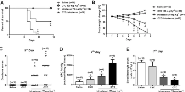
Fig. 5A shows that Treg-depleted animals receiving irinotecan pre-sented a reduced survival (100% mortality on the ninth day) compared to non-depleted mice that were also injected with irinotecan (50% survival until the end of the follow-up period). In addition, signs of clinical irinotecan-related intestinal mucositis were more prominent in mice pretreated with CYC (Fig. 5B–E). The CYC + IRI-treated mice showed a more pronounced weight loss than those receiving IRI only (Fig. 5B). In addition, the severity of diarrhea and neutrophil accu-mulation was enhanced in the CYC + IRI group (Fig. 5C and D, re-spectively). Furthermore, all of the groups that received irinotecan presented marked leukopenia compared with the saline-injected group (Fig. 5E). The enhancement of mucositis severity was confirmed by histopathological analysis (Fig. 6). IRI-treated mice presented a sig-nificant reduction in the villus/crypt ratio compared with the saline group, which was potentiated in CYC + IRI-injected animals (Fig. 6A–C). The injection of CYC alone did not cause a loss of body weight, alterations in systemic leukocyte count, animal mortality, diarrhea, inflammatory infiltration or histopathological damage (Figs. 5 and 6) compared to the saline group.
3.4. Depletion of regulatory T cells increases the frequency of other T helper cell subsets
As described inFig. 7, a negative correlation between the percen-tage of Tregs and the number of neutrophils (Fig. 7B), but not between Th17 and neutrophils (Fig. 7D), in the intestine on the seventh ex-perimental day was observed in mice receiving irinotecan. In contrast, no correlation was found between Tregs or Th17 and neutrophils in
saline-treated mice (Fig. 7A and C). The frequency of non-regulatory CD3+CD4+CD25−FOXP3−cells was also investigated. As shown in
Fig. 7I, the frequency of the lymphocyte subset was reduced on the seventh day of mucositis development compared to saline. However, the frequency of non-regulatory T cells increased in CYC-treated ani-mals (Fig. 7J). These cells showed a positive correlation with
in-filtrating neutrophils in mice with intestinal mucositis (Fig. 7F), which was not observed in the control group (Fig. 7E). We also observed a Fig. 3.Irinotecan increases membrane CD25 ex-pression in intestinal regulatory T cells. Mice re-ceived irinotecan (75 mg kg−1, i.p, once a day for four days) to induce mucositis and were eu-thanized on day seven (D7). The control group received only saline as a vehicle. Lamina propria lymphocytes were isolated, and the frequencies of total Tregs (CD3+CD4+FOXP3+), which in-cludes the CD25+and CD25−fraction (A), and CD25−Tregs (CD3+CD4+CD25−FOXP3+) (C) were evaluated. The increase in CD25 expression (Mean of Fluorescence Intensity-MFI) is observed in Panels B and D, with the dotted line and solid line representing saline and irinotecan, respec-tively. Panel E summarizes this information, with saline represented by the blue line and irinotecan by the red line. (For interpretation of the refer-ences to color in thisfigure legend, the reader is referred to the web version of this article.)
Fig. 4.Low dose cyclophosphamide depletes regulatory T cells. Mice were injected with saline or irinotecan (75 mg kg−1, i.p, once a day for four days) to induce mucositis and were euthanized on the seventh day. Tregs were depleted by the administration of cyclophosphamide (CYC, 100 mg kg−1, i.p, single dose) 2 h before the
strong negative correlation between the frequency of Tregs and non-Treg cells on the seventh day of intestinal mucositis (Fig. 7H), which was not observed in mice that received saline (Fig. 7G).
4. Discussion
In the present study, we showed for thefirst time the role of Tregs in controlling intestinal mucositis induced by irinotecan, since depletion of these cells exacerbated tissue damage and increased animal mor-tality.
Tregs have been described to suppress immunity against several stimuli, including commensal bacteria and self-antigens (Kanamori et al., 2016). These cells are frequently found in the intestinal mucosa and impair excessive effector T-cell responses and maintain organ
homeostasis (Pabst, 2013; Omenetti and Theresa, 2015). In the present study, a higher frequency of Tregs was found in the lamina propria of the small intestine of mice with mucositis, which correlates with disease severity and diarrhea. Several previous studies have reported a higher frequency of Tregs in intestinal inflammatory diseases (Reikvam et al., 2011; Lord, 2015; Sznurkowska et al., 2016). However, in murine models of colitis induced by DSS or trinitrobenzenicsulfonic acid (TNBS), the frequency of Tregs in the gut was reduced and correlated with tissue damage (Kang et al., 2015; Zou et al., 2015). In our study, the increase in frequency of Tregs can be a compensatory mechanism to counterbalance the inflammation mediated by neutrophils and other Th cells.
We observed, particularly in the saline group, an important popu-lation of FOXP3+ T cells that did not express the marker CD25. Fig. 5.Cyclophosphamide enhances the severity of irinotecan-induced intestinal mucositis. Mice were injected with saline or irinotecan (75 mg kg−1, i.p, once a day for four days) to induce mucositis and were euthanized on the seventh day. Tregs were depleted by the administration of cyclophosphamide (CYC, 100 mg kg−1, i.p, single dose) 2 h before thefirst dose of irinotecan. The percentage of animal survival was monitored until the 14th day. CYC injection enhanced animal mortality (Panel A), loss of body weight (Panel B), severity of diarrhea (Panel C), MPO activity (D) and leukopenia (Panel E). *P< 0.05vs.Saline;#P< 0.05vs.CYC + Irinotecan; °P< 0.05vs.CYC only.
However, Tregs of mice with intestinal mucositis showed an upregu-lation of CD25 expression. According to Zelenay et al. (2005), CD25−FOXP3+T cells are a reservoir of committed regulatory T cells that upregulate CD25 expression upon changes in tissue homeostasis. The study reported that expression of CD25 occurs as a signature of Treg activation and not only of T effector cells (Zelenay et al., 2005). Here, we propose that intestinal Tregs are activated after administra-tion of irinotecan and recover expression of CD25.
injection of only cyclophosphamide did not change the leukocyte count, gut integrity or animal weight gain, suggesting no detectable toxic ef-fect of the drug. Interestingly, Treg-depleted mice injected with ir-inotecan showed aggravated intestinal mucositis, as detected by wor-sened intestinal inflammation and diarrhea and 100% mortality. Consistent with thisfinding, the protective role of Tregs in other animal models of intestinal inflammation was previously demonstrated. In colitis induced by DSS, depletion of Tregs with anti-CD25 antibodies also induced more severe intestinal damage (Wang et al., 2015). In addition, adoptive transfer of Tregs prevented the development of co-litis (Liu et al., 2003; Mottet et al., 2003).
A negative correlation between the number of Tregs and neutrophils suggested that the activation of Tregs might limit the accumulation of these cells in the intestine, in an attempt to control the tissue damage. Indeed, depletion of Tregs caused an increase in neutrophil infiltration and had a deleterious effect, potentially by activating a prominent
in-flammatory response. Atkinson et al. (2016) also observed intense neutrophil infiltrations in mice that were depleted of Tregs in an ar-thritis model. In colitis induced by DSS, depletion of Tregs increased cellular infiltration in the colon (Boehm et al., 2012).
In addition to the increase in the frequency of Tregs, in the present study, a higher number of Th17 cells on thefifth and seventh days of mucositis development were detected. Th17 cells are involved in the pathophysiology of several inflammatory bowel diseases, such as Crohn's disease, ulcerative colitis (Gálvez, 2014; Ueno et al., 2015) and different murine colitis models (Leppkes et al., 2009; He et al., 2012; Lim et al., 2016). These cells produce cytokines, such as 17A and IL-17F, which induce the expression of inflammatory mediators and re-cruitment of neutrophils to inflammatory sitesviadirect and indirect mechanisms (Pelletier et al., 2010). The involvement of Th17 cells in experimental mucositis induced by irinotecan has not been previously suggested. However, its role has not yet been clarified.
Unexpectedly, depletion of Tregs with cyclophosphamide did not increase Th17 cell frequency. However, a reduction of approximately 40% in the gut was detected, despite increased intestinal damage.Chen et al. (2011)showed that Treg depletion in mice expressing diphtheria toxin receptor under the control of the Foxp3 promoter resulted in a lower frequency of Th17 cells in blood and draining lymph nodes. The studies showed that Tregs promoted Th17 development by the con-sumption of IL-2, thereby reducing the upregulation of CD25 and ac-tivation of the STAT5 pathway (Chen et al., 2011). Furthermore, Tregs exhibit plasticity and could differentiate into IL-17-producing cells that express the related orphan receptor, ROR-γt, and CCR6 (Koenen et al., 2008). Most likely, the Treg repertoire also contributed to the devel-opment of a part of the Th17 cells in our model.
Although Treg depletion coincided with a reduction in Th17 cell number, we observed higher intestinal neutrophil infiltration. This
finding suggests the involvement of other immune cells in neutrophil recruitment to the intestine. Interestingly, we did not observe a corre-lation between the number of intestinal neutrophils and the percentage of Th17 cells. However, when other Th cells (CD3+CD4+, negative for Treg and Th17 markers) were evaluated, we found a positive correla-tion between those cells and neutrophils. In addicorrela-tion, Tregs showed a strong negative correlation with other Th cells and depletion of Tregs resulted in an increase in Th cell frequency, suggesting that non-reg-ulatory T cells could be controlled by Tregs in the context of mucositis. Importantly, in addition to alterations in the frequency of Tregs and Th17 cells, irinotecan-induced intestinal mucositis caused animal body weight loss, diarrhea, intestinal neutrophil infiltration, leukopenia, re-duction of the villus/crypt ratio and intestinal damage, as shown else-where (Lima-Júnior et al., 2012; Melo et al., 2008; Lima-Júnior et al., 2014; Wong et al., 2015). Interestingly, we showed the mucositis peak on the seventh experimental day with the exception of neutrophil
in-filtration, which peaks on day 5. However, it is still not clear which T cell subset mediates such a deleterious response during mucositis. Nevertheless, the participation of IL-18, a key cytokine involved in Th1
and Th2 differentiation, has been reported to mediate intestinal mu-cositis development (Lima-Júnior et al., 2014).
Notably, irinotecan-induced intestinal mucositis should not be re-garded as the consequence of a benign disease or simply another form of chemically-induced mucosal inflammation, such as colitis associated with the injection of dextran sodium sulfate (DSS) and trinitrobenzene sulfonic acid (TNBS). However, one important limitation of the present study is that mucositis was induced in non-tumor-bearing mice, since the presence of cancer cells may alter the immune response. Nevertheless, local and systemic effects of irinotecan injection on some cells of the immune system were shown, increasing the knowledge of the complex underlying mechanisms related to this life-threatening side effect.
Considering the possible involvement of adaptive immune cells in the pathogenesis of intestinal mucositis (Lima-Júnior et al., 2014), sti-mulation of Tregs might be a strategy to limit disease progression. Conversely, in cancer patients, such approach is not suitable, due to the potential enhancement of tumor progression and the occurrence of metastases by the inhibition of antitumor innate and adaptive immune responses (Curiel, 2008; Elkord et al., 2010; Mougiakakos et al., 2010). Suchfindings indicate that targeting the immune system is a double-edged sword.
In the clinical setting, low dose of cyclophosphamide, known as metronomic chemotherapy, has been used as adjuvant treatment in association with conventional chemotherapy for advanced tumors (Colleoni et al., 2016; Perroud et al., 2016). The advantages of me-tronomic cyclophosphamide-based regimens include the reduction of Tregs (Madondo et al., 2016) and the anti-angiogenic effect (Zhang et al., 2006), which impair tumor growth and show a low-toxicity profile. Taking into account that the low dose of cyclophosphamide aggravated preclinical irinotecan-related intestinal mucositis, caution is required in order to identify potential severe gastrointestinal toxicities in clinical trials that include the combination of these drugs in on-cology.
In conclusion, Tregs are critical cells for controlling irinotecan-re-lated intestinal mucositis, which might be associated to other T cell subsets. In our opinion, the modulation of the immune system to sy-nergize with antitumor chemotherapy is currently a milestone in on-cology, but efforts to the adequate management of side effects, such as mucositis, are to be specially considered to improve patients' quality of life. Understanding the pathogenesis of anticancer drug toxicities is essential for obtaining better clinical outcomes, which is the main contribution of this study.
Author contributions
Study design: CF, MHLPS and RCPLJ. Performed the Experiments: CF, CWSW, HAM, CMSS, MAT, NRPS, AGFC and RBF. Data analysis:CF, MHLPS, PRCA, LMCC and RCPLJ. Interpretation of the results: CF, MHLPS, PRCA, LMCC and RCPLJ. Wrote the paper: CF and RCPLJ. All the authors revised and approved the paper.
Conflicts of interest
The authors declare that there is no conflict of interest regarding the publication of this study.
Acknowledgments
References
Andreyev, J., Ross, P., Donnellan, C., Lennan, E., Leonard, P., Waters, C., et al., 2014. Guidance on the management of diarrhoea during cancer chemotherapy. Lancet Oncol. 15, 447–460.
Atkinson, S.M., Hoffmann, U., Hamann, A., Bach, E., Danneskiold-Samsøe, N.B., Kristiansen, K., et al., 2016. Depletion of regulatory T cells leads to an exacerbation of delayed-type hypersensitivity arthritis in C57BL/6 mice that can be counteracted by IL-17 blockade. Dis. Model. Mech. 9, 427–440.
Boehm, F., Martin, M., Kesselring, R., Schiechl, G., Geissler, E.K., Schlitt, H.J., et al., 2012. Deletion of Foxp3 + regulatory T cells in genetically targeted mice supports devel-opment of intestinal inflammation. BMC Gastroenterol. 12, 97.
Campbell, J.M., Stephenson, M.D., Bateman, E., Peters, M.D., Keefe, D.M., Bowen, J.M., 2016. Irinotecan-induced toxicity pharmacogenetics: an umbrella review of sys-tematic reviews and meta-analyses. Pharm. J.http://dx.doi.org/10.1038/tpj. 2016.58.
Chen, Y., Haines, C.J., Gutcher, I., Hochweller, K., Blumenschein, W.M., McClanahan, T., et al., 2011. Foxp3 + regulatory T cells promote T helper 17 cell development in vivo through regulation of interleukin-2. Immunity 34, 409–421.
Colleoni, M., Gray, K.P., Gelber, S., Láng, I., Thürlimann, B., Gianni, L., et al., 2016. Low-dose oral cyclophosphamide and methotrexate maintenance for hormone receptor-negative early breast cancer: international breast cancer study group trial 22-00. J. Clin. Oncol. 34, 3400–3408.
Curiel, T.J., 2008. Regulatory T cells and treatment of cancer. Curr. Opin. Immunol. 20, 241–246.
Elkord, E., Alcantar-Orozco, E.M., Dovedi, S.J., Tran, D.Q., Hawkins, R.E., Gilham, D.E., 2010. T regulatory cells in cancer: recent advances and therapeutic potential. Expert. Opin. Biol. Ther. 10, 1573–1586.
Gálvez, J., 2014. Role of Th17 cells in the pathogenesis of human IBD. ISRN Inflamm. http://dx.doi.org/10.1155/2014/928461.
Guabiraba, R., Besnard, A.G., Menezes, G.B., Secher, T., Jabir, M.S., Amaral, S.S., et al., 2014. IL-33 targeting attenuates intestinal mucositis and enhances effective tumor chemotherapy in mice. Mucosal Immunol. 7, 1079–1093.
Hartog, A., Belle, F.N., Bastiaans, J., de Graaff, P., Garssen, J., Harthoorn, L.F., et al., 2015. A potential role for regulatory T-cells in the amelioration of DSS induced colitis by dietary non-digestible polysaccharides. J. Nutr. Biochem. 26, 227–233.
He, Y., Lin, L.J., Zheng, C.Q., Jin, Y., Lin, Y., 2012. Cytokine expression and the role of Th17 cells in a mouse model of colitis. Mol. Med. Rep. 6, 1438–1442.
Hsu, H.Y., Kuan, Y.C., Lin, T.Y., Tsao, S.M., Hsu, J., Ma, L.J., et al., 2013. Reishi protein LZ-8 induces FOXP3 (+) Treg expansion via a CD45-dependent signaling pathway and alleviates acute intestinal inflammation in mice. Evid. Based Complement. Alternat. Med.http://dx.doi.org/10.1155/2013/513542.
Ikuno, N., Soda, H., Watanabe, M., Oka, M., 1995. Irinotecan (CPT–11) and characteristic
mucosal changes in the mouse ileum and caecum. J. Natl. Cancer Inst. 87, 1876–1883.
Kanamori, M., Nakatsukasa, H., Okada, M., Lu, Q., Yoshimura, A., 2016. Induced reg-ulatory T cells: their development, stability, and applications. Trends Immunol. 37, 803–811.
Kang, G.D., Lim, S., Kim, D.H., 2015. Oleanolic acid ameliorates dextran sodium sulfate-induced colitis in mice by restoring the balance of Th17/Treg cells and inhibiting NF-κB signaling pathway. Int. Immunopharmacol. 29, 393–400.
Koenen, H.J., Smeets, R.L., Vink, P.M., Van Rijssen, E., Boots, A.M., Joosten, I., 2008. Human CD25highFoxp3pos
regulatory T cells differentiate into IL-17–producing cells.
Blood 112, 2340–2352.
Kurita, A., Kado, S., Kaneda, N., Onoue, M., Hashimoto, S., Yokokura, T., 2000. Modified irinotecan hydrochloloride (CPT–11) administration schedule improves induction of
delayed-onset diarrhea in rats. Cancer Chemother. Pharmacol. 46, 211–220.
Lee, C.S., Ryan, E.J., Doherty, G.A., 2014. Gastro-intestinal toxicity of chemotherapeutics in colorectal cancer: the role of inflammation. World J. Gastroenterol. 20, 3751–3761.
Leppkes, M., Becker, C., Ivanov, I.I., Hirth, S., Wirtz, S., Neufert, C., et al., 2009. RORγ -expressing Th17 cells induce murine chronic intestinal inflammation via redundant effects of IL-17A and IL-17F. Gastroenterology 136, 257–267.
Lim, S.M., Jeong, J.J., Choi, H.S., Chang, H.B., Kim, D.H., 2016. Mangiferin corrects the imbalance of Th17/Treg cells in mice with TNBS-induced colitis. Int.
Immunopharmacol. 34, 220–228.
Lima-Júnior, R.C., Figueiredo, A.A., Freitas, H.C., Melo, M.L., Wong, D.V., Leite, C.A., et al., 2012. Involvement of nitric oxide on the pathogenesis of irinotecan-induced intestinal mucositis: role of cytokines on inducible nitric oxide synthase activation. Cancer Chemother. Pharmacol. 69, 931–942.
Lima-Júnior, R.C., Freitas, H.C., Wong, D.V., Wanderley, C.W., Nunes, L.G., Leite, L.L., et al., 2014. Targeted inhibition of IL–18 attenuates irinotecan-induced intestinal
mucositis in mice. Br. J. Pharmacol. 171, 2335–2350.
Liu, H., Hu, B., Xu, D., Liew, F.Y., 2003. CD4 + CD25 + regulatory T cells cure murine colitis: the role of IL-10, TGF-β, and CTLA4. J. Immunol. 171, 5012–5017.
Lord, J.D., 2015. Promises and paradoxes of regulatory T cells in inflammatory bowel disease. World J. Gastroenterol. 21, 11236–11245.
Lutsiak, M.E., Semnani, R.T., De Pascalis, R., Kashmiri, S.V., Schlom, J., Sabzevari, H.,
2005. Inhibition of CD4 + 25+ T regulatory cell function implicated in enhanced immune response by low-dose cyclophosphamide. Blood 105, 2862–2868.
MacPherson, B.R., Pfeiffer, C.J., 1978. Experimental production of diffuse colitis in rats. Digestion 17, 135–150.
Madondo, M.T., Quinn, M., Plebanski, M., 2016. Low dose cyclophosphamide: mechan-isms of T cell modulation. Cancer Treat. Rev. 42, 3–9.
Matricon, J., Barnich, N., Ardid, D., 2010. Immunopathogenesis of inflammatory bowel disease. Self Nonself 1, 299–309.
McGrath, J.C., Drummond, G.B., McLachlan, E.M., Kilkenny, C., Wainwright, C.L., 2010. Guidelines for reporting experiments involving animals: the ARRIVE guidelines. Br. J. Pharmacol. 160, 1573–1576.
Melo, M.L., Brito, G.A., Soares, R.C., Carvalho, S.B., Silva, J.V., Soares, P.M., et al., 2008. Role of cytokines (TNF-alpha, IL-1beta and KC) in the pathogenesis of CPT-11-in-duced intestinal mucositis in mice: effect of pentoxifylline and thalidomide. Cancer Chemother. Pharmacol. 61, 775–784.
Mottet, C., Uhlig, H.H., Powrie, F., 2003. Cutting edge: cure of colitis by CD4 + CD25 + regulatory T cells. J. Immunol. 170, 3939–3943.
Mougiakakos, D., Choudhury, A., Lladser, A., Kiessling, R., Johansson, C.C., 2010. Regulatory T cells in cancer. Adv. Cancer Res. 107, 57–117.
Newton, K., Dixit, V.M., 2012. Signaling in innate immunity and inflammation. Cold Spring Harb. Perspect. Biol.http://dx.doi.org/10.1101/cshperspect.a006049. Omenetti, S., Theresa, T.P., 2015. The Treg/Th17 axis: a dynamic balance regulated by
the gut microbiome. Front. Immunol.http://dx.doi.org/10.3389/fimmu.2015. 00639.
Pabst, O., 2013. Trafficking of regulatory T cells in the intestinal immune system. Int. Immunol. 25, 139–143.
Pelletier, M., Maggi, L., Micheletti, A., Lazzeri, E., Tamassia, N., Costantini, C., et al., 2010. Evidence for a cross-talk between human neutrophils and Th17 cells. Blood 115, 335–343.
Perroud, H.A., Alasino, C.M., Rico, M.J., Mainetti, L.E., Queralt, F., Pezzotto, S.M., et al., 2016. Metastatic breast cancer patients treated with low-dose metronomic che-motherapy with cyclophosphamide and celecoxib: clinical outcomes and biomarkers of response. Cancer Chemother. Pharmacol. 77, 365–374.
Podolsky, D.K., 2002. Inflammatory bowel disease. N. Engl. J. Med. 347, 417–429.
Reikvam, D.H., Perminow, G., Lyckander, L.G., Gran, J.M., Brandtzaeg, P., Vatn, M., et al., 2011. Increase of regulatory T cells in ileal mucosa of untreated pediatric Crohn's disease patients. Scand. J. Gastroenterol. 46, 550–560.
Ribeiro, R.A., Wanderley, C.W., Wong, D.V., Mota, J.M., Leite, C.A., Souza, M.H., et al., 2016. Irinotecan- and 5-fluorouracil-induced intestinal mucositis: insights into pa-thogenesis and therapeutic perspectives. Cancer Chemother. Pharmacol. 78, 881–893.
Sakaguchi, S., Wing, K., Onishi, Y., Prieto-Martin, P., Yamaguchi, T., 2009. Regulatory T cells: how do they suppress immune responses? Int. Immunol. 21, 1105–1111.
Sheridan, B.S., Lefrançois, L., 2012. Isolation of mouse lymphocytes from small intestine tissues. Curr. Protoc. Immunol.http://dx.doi.org/10.1002/0471142735.im0319s99. Sznurkowska, K.,Żawrocki, A., Sznurkowski, J., Zieliński, M., Landowski, P., Plata-Nazar, K., et al., 2016. Peripheral and intestinal T-regulatory cells are upregulated in chil-dren with inflammatory bowel disease at onset of disease. Immunol. Investig. 19, 1–10.
Touchefeu, Y., Montassier, E., Nieman, K., Gastinne, T., Potel, G., Bruley, des, Varannes, S., et al., 2014. Systematic review: the role of the gut microbiota in chemotherapy- or radiation-induced gastrointestinal mucositis - current evidence and potential clinical applications. Aliment. Pharmacol. Ther. 40, 409–421.
Ueno, A., Ghosh, A., Hung, D., Li, J., Jijon, H., 2015. Th17 plasticity and its changes associated with inflammatory bowel disease. World J. Gastroenterol. 21, 12283–12295.
Van Sebille, Y.Z., Stansborough, R., Wardill, H.R., Bateman, E., Gibson, R.J., Keefe, D.M., 2015. Management of mucositis during chemotherapy: from pathophysiology to pragmatic therapeutics. Curr. Oncol. Rep. http://dx.doi.org/10.1007/s11912-015-0474-9.
Wang, L., Ray, A., Jiang, X., Wang, J.Y., Basu, S., Liu, X., et al., 2015. T regulatory cells and B cells cooperate to form a regulatory loop that maintains gut homeostasis and suppresses dextran sulfate sodium-induced colitis. Mucosal Immunol. 8, 1297–1312.
Wong, D.V., Lima-Júnior, R.C., Carvalho, C.B., Borges, V.F., Wanderley, C.W., Bem, A.X., et al., 2015. The adaptor protein Myd88 is a key signaling molecule in the patho-genesis of irinotecan-induced intestinal mucositis. PLoS One.http://dx.doi.org/10. 1371/journal.pone.0139985.
Zelenay, S., Lopes-Carvalho, T., Caramalho, I., Moraes-Fontes, M.F., Rebelo, M., Demengeot, J., 2005. Foxp3+CD25−CD4 T cells constitute a reservoir of committed
regulatory cells that regain CD25 expression upon homeostatic expansion. Proc. Natl. Acad. Sci. U. S. A. 102, 4091–4096.
Zhang, Q., Kang, X., Zhao, W., 2006. Antiangiogenic effect of low-dose cyclophosphamide combined with ginsenoside Rg3on Lewis lung carcinoma. Biochem. Biophys. Res. Commun. 342, 824–828.