Bull Pan Am Health Organ 11(3), 1977.
SEROLOGIC
SURVEYS FOR ANTIBODIES
TO WESTERN,
EASTERN,
CALIFORNIA,
AND ST. LOUIS ENCEPHALITB
AND DENGUE 3
ARBOVIRUSES
IN MIDDLE
AMERICA,
1961-1975” ’
William F. Schemer,’ Robert W. Dickerman,
and Jose’ V. Ord&ez5
A survey of sera and plasmas collected during 1961-1975 has shown that many geofile and animals of Middle America are
suscefltible to infection by dengue 3 virus and the viruses of
eastern, western, St. Louis, and California encephalitis. @read
of any of these arboviruses in Middle America could cause
serious efiidemics, since vaccines are not currently available fo7 these virus infections and mosquito vector control efforts are not always successful.
Introduction
Historically, the arboviruses which have
produced significant human epidemics or
equine epizootics in the Americas are yellow
fever (YF), Dengue (DEN), and various
encephalitis viruses-including western
(WE), eastern (EE), St. Louis (SLE), Vene-
zuelan (VE), and California (CAL) enceph-
alitis. The presence or absence and the geo-
graphic distributions of these viruses are
fairly well known in the United States of
‘These investigations were performed in collabora-
rion with the Pan American Health Organization and
the Governments of Mexico, Guatemala, Belize, Hon-
duras, El Salvador, Nicaragua, and Costa Rica. They were also sponsored in part by the U.S. Army Medical Research and Development Command, Washington, D.C. 20314, under contract no. DADA-17.7% C-2140, and in part by U.S. Public Health Service re- search grant AI-06248 from the National Institute of Allergy and Infeceious Diseases.
2Also appearing in Spanish in the Boletcn de la
Oficina Sanitaria panamericana, 1978.
SProfessor, Department of Microbiology, Cornell University Medical College, New York, New York 10021, U.S.A.
4Associate Professor, Department of Microbiology, Cornell University Medical College.
6Professor, University of San Carlos Medical School, Guatemala.
America, Canada, and the Caribbean area,
but only YF and VE viruses have been
mapped extensively in Middle America
(1-3). EE virus has been isolated from
horses in Mexico, from a sentinel hamster
in Guatemala, and from a migrant bird in
Belize: and SLE virus has been recovered
from a bird and from mosquitoes in Mexico
(4-8). On the whole, however, little is
known of the geographic distributions of
EE and SLE viruses in Middle America. No
isolations of WE, CAL, or DEN viruses
have been reported from Middle America,
and published serologic surveys for anti-
bodies to WE, EE, SLE, and DEN viruses
have covered only limited regions (Y-IS).
Therefore, it seemed worthwhile to test
sera from Middle America more extensively
for antibodies to WE, EE, CAL, SLE, and
DEN arboviruses, and in this way to seek
areas with a high prevalence of infection
which could subsequently be studied to
isolate viruses and define disease incidences.
Prior investigations of VE virus in Middle
America during 1961-1971 provided us with
sera to examine. These were tested for
hemagglutination-inhibition (HI) anti-
bodies to WE, EE, CAL, and SLE viruses,
Scherer et al. * SURVEY OF ARBOVIRUSES FROM MIDDLE AMERICA 213
and for neutralizing(N) antibodies to type 3
DEN, WE, EE, and VE viruses. This article reports the results of those tests.
Materials and Methods
S#ecimen Sources and Collection Methods
Sera or plasmas were obtained from the
locations shown in Figure 1. These were
collected in the manner previously des-
cribed for investigation of VE virus (19) and
were stored at -20%.
The ages of human subjects and animals
providing samples were as follows: Three
per cent of the human subjects were 0 to 5
years of age, 11% were 6 to 10, 26% were 11 to 20, 35$& were 21 to 4O,lS’% were 41 to 60, and 7% were over 60. Wild birds sampled at
Tlacotalpan, Veracruz, Mexico, in 1963
and 1964 were juveniles and adults several
months to several years of age. Other wild
birds (mostly ardeids and associated marsh
birds) were either nestlings less than a
month old or (in a few cases) juveniles up to
six months old. Samples were also taken
from horses and burros ranging in age from
1 to 15 years; from pigs, some 1 to 6 months old but most in the 6 month to 2 year range; and from dogs 1 to 7 years of age. The ages of the wild mammals sampled are unknown.
HI Tests
These tests were performed with micro-
technics previously described, using goose
erythrocytes; serum was acetone-extracted
twice (20). Pipettes (1 ml and 0.2 ml) were
used to dilute sera 1:lO and to make
subsequent two-fold dilutions. The virus
strains employed were as follows: The WE
virus was strain 1985-60, which had been
isolated at the Rocky Mountain Laboratory
in Montana (15). The EE virus used in most
cases was strain 68U230 from Guatemala;
however the human sera obtained from
Sontecomapan, Veracruz, Mexico, in 1965
and pig sera collected in Belize in 1966 were
tested with strain Riche from Louisiana (5,
15). The CAL virus used was strain BFS283
from California (I). The SLE strain
employed was 65V310 from Mexico in most
cases, but strain TRVL9464 from Trinidad
was used for the sera tested with EE strain
Riche (7). The VE virus used was strain
63U2 from Mexico (21).
Hemagglutinins were made from infected
suckling mouse brains with the sucrose-
acetone technic (2.2). For CAL virus,
hemagglutinin was treated with 5 mg of
trypsin (Difco 1:250) per 10 ml of hemag-
glutinin solution, was kept at 22% for one
hour, and was subsequently mixed with 5
mg of soybean trypsin inhibitor. Four to
eight units of hemagglutinin were used in
most cases. The pH of the HI tests was 6.2,
except in the tests for SLE (pH 6.6) and for
VE (pH sometimes 6.4). Serum-hemagglu-
tinin mixtures were kept overnight at 5“C
and were incubated at 37’C following addi-
tion of erythrocytes. All HI results recorded
as positive in the accompanying tables were
confirmed by a repeat test.
N Tests
N tests were accomplished by plaque-
reduction in plastic plate wells with an area
of two square centimeters, using methods
similar to those previously described for VE
virus (21). Tests for WE, EE, and VE virus
N antibodies employed primary chicken
embryonic cells produced as described else-
where (21); tests for DEN type 3 antibody
were done in LLCMK2 cultures as previous-
ly described (23). Virus strains utilized were
as follows: WE strain 1985-60, EE strain
68U230, VE strain 63U2 or 69Z1, and DEN 3 strain 16562. Sera were tested at a 1:4 dilu-
tion after heating at 60% for 20 minutes.
Serum-virus mixtures containing about 100
plaque-forming units (pfu) were incubated
at 37% for one hour. Sera were considered
positive for antibody if the log neutraliza-
tion index (LNI) exceeded 1.6 (98% plaque
214 PAHO BULLETIN l vol. XI, no. 3, 1977
Matamoros
GULF OF MEXICO
Oaxaci
I I I
0 250 500 Km
PACIFIC OCEAN
I
105 100
I 95"
COSTA RICA &
a5
16” -,
16 5”
!5"
!O"
15"
10"
90’ aa- a6
Scherer et a/. l SURVEY OF ARBOVIRUSES FROM MIDDLE AMERICA 215
exceeded 1.5 (97oJ, plaque reduction) in
DEN 3 tests.
Results
WE Virus
Numerous samples taken on the Atlantic
side of Middle America yielded positive HI
results for WE antibody. Positive findings
were obtained with human sera from loca-
tions in Belize, Guatemala, Honduras, and
the Mexican State of Veracruz (see Table 1).
In addition, various animal plasmas col-
lected in Veracruz reacted with WE virus
hemagglutinin; these positive samples in-
cluded 18 from wild terrestrial mammals,
one from a bat, and four from wild nestling
herons, egrets, or associated marsh birds.
Positive results were also obtained with
equine sera collected from northern Vera-
cruz in 1971, from northern Tamaulipas in
1969, and from the Atlantic foothills of
Guatemala in 1969. Some pig sera from
Veracruz (Mexico), Belize City (Belize),
Cort&s (Honduras), and the Atlantic foot-
hills of Guatemala also reacted positively, as
did a few dog sera from Santa Lucia,
Guatemala.
On the Pacific side, positive HI tests for
WE virus occurred with human sera from
Guatemala, with wild mammalian sera
from Guatemala, with bird plasmas and pig
sera from Mexico and Guatemala, with
horse and dog sera from Guatemala, and
with pig sera from Costa Rica (Table 2).
Most sera were positive at dilutions in the
1:lO to 1:40 range. The few sera that reacted
at higher dilutions are cited in footnotes to
the tables.
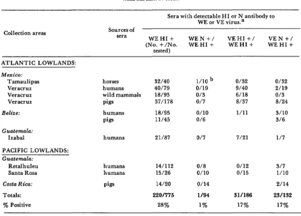
On the other hand, N tests with WE virus
showed that most of the positive HI re-
actions were not specific for WE (Table 3).
In fact, out of 94 HI-positive sera, only one
was N-positive and two others were possibly
N-positive. Furthermore, all three of these
sera came from the northern Atlantic coast
of Mexico next to southern Texas, where
WE virus is known to exist (24).
Another interesting point is that the
positive HI results with WE virus were not
entirely accounted for by coexistence in the
sera of VE virus antibodies, since only 31 of
186 sera that were HI-positive with WE
virus were also HI-positive with VE virus
(Table 3). Only in a few places, where sera
were obtained during or shortly after the
Middle American VE outbreak, were sera
found to be HI-positive with both WE and
VE viruses. These places (not shown in
Table 3) were in northern Veracruz near
Tampico during 1971 (34 of 45 horse sera
HI-positive with WE were also HI-positive
with VE); the Atlantic foothills of Guate-
mala during 1969 (8 of 9 horse sera and 3
of 3 dog sera); La Avellana and Guazaca-
pan in Santa Rosa, Guatemala, during
1970-71 (22 of 26 human sera); and Parce-
lamiento Montufar in J utiapa, Guatemala,
during 1969 (9 of 9 horse sera and 15 of 21
dog sera).
During 1972-1975, sera became available
from horses on the Pacific coasts of El
Salvador and Nicaragua. WE HI antibodies
were not detected in any of 18 sera from El
Salvador, but were detected in seven out of
63 Nicaraguan sera. However, six of the
seven positive sera had HI titers of only
l:lO, and no WE N antibody was found at a
1:4 dilution. One serum had a WE HI titer
of at least 1:20 and a N titer of at least 1:4;
this serum came from a three-year-old horse
bled during May 1975 in Nicaragua’s
Chinandega Department. Whether these
results indicated naturally-induced or vac-
cine-induced WE antibodies in this horse is
unknown.
EE Virus
Only four of 1,255 human sera from
Middle America gave positive HI results
with EE virus (Table 4). These sera came
216 PAHO BULLETIN l vol. XI, no. 3, 19 77
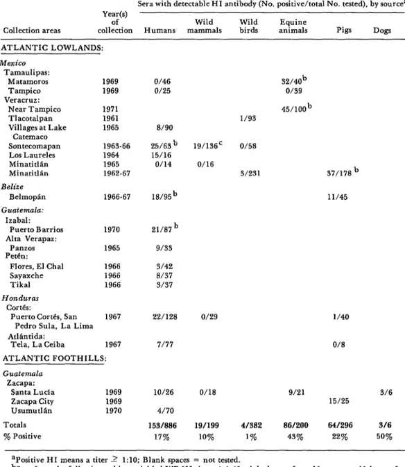
Table 1. Prevalences of serum HI antibodies reacting with WE virus in people and animals sampled on the Atlantic coastal lowlands of Mexico, Belize, Guatemala, and Honduras and the
Atlantic foothills of Guatemala during 1961-1971.
Collection areas
Sera with detectable HI antibody (No. positive/total No. tested), by sourcea Year(s)
of Wild Wild Equine
collection Humans mammals birds animals Pigs Dogs
ATLANTIC LOWLANDS: Mexico
Tamaulipas:
Matamoros 1969
Tampico 1969
Veracruz:
Near Tampico 1971
Tlacotalpan 1961
Villages at Lake 1965 Catemaco
Sontecomapan 1963-66
Los Laureles 1964
MinatitlLn 1965
Minatitlan 1962-67
Ed&?
Belmopan 1966-67
Guatemala: Izabal:
Puerto Barrios 1970
Alta Verapaz:
Panzos 1965
Pet&:
Flares, El Chal 1966
Sayaxche 1966
Tikal 1966
Honduras CortCs:
Puerto Cort&, San 1967 Pedro Sula, La Lima
O/46 o/25
8/90 25/6?jb 15/16
o/14
18/95b 11/45
21/87b 9/33 3/42 8/37 3137
22/128 o/29 l/40
AtlBntida:
Tela, La Ceiba 1967 ATLANTIC FOOTHILLS: Guatemala
Zacapa: Santa Lucia Zacapa City Usumutlln Totals y. Positive
1969 1969 1970
7/77
lo/26 4/70 153/886
17%
32/40b o/39 45/100b l/93
19/136’ O/58 O/16
3/231 37/178b
O/8
O/18 9/21 3/6
15/25
19/199 4/382 86/200 641296 3/6
10% 1% 43% 22% 50%
aPositive HI means a titer 2 1:lO; Blank spaces = not tested.
bSera from the following subjects yielded WE HI titers >1:40: eight horses from Matamoros, 11 horses from Tampico (1971), two people from Sontecomapan, two pigs from Minatitlkn, one person from Belize, and four people from Puerto Barrios.
Scherer et al. . SURVEY OF ARBOVIRUSES FROM MIDDLE AMERICA 217
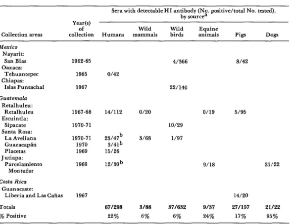
Table 2. Prevalences of serum HI antibodies reacting with WE virus in people and animals sampled on the Pacific coastal lowlands of Mexico, Guatemala, and Costa Rica during 1962-1971.
Collection areas
Sera with detectable HI antibody (No. positive/total No. tested), by sourcea
Year(s)
of Wild Wild Equine
collection Humans mammals birds animals Pigs Dogs
Mexico Nayarit:
San Bias Oaxaca:
Tehuantepec Chiapas:
IsIas Puntachal Guatemala
Retalhuleu: Retalhuleu EscuintIa:
Sipacate Santa Rosa:
La Avellana Guazacapti Placetas Jutiapa:
Parcelamiento Montufar Costa Rica
Guanacaste:
Liberia and LasCaEas Totals
% Positive
1962-65 1965 1967
1967-68 1970-71 1970-71 1970 1969 1969
1967
O/42
14/112
15/26 12/30b
671298
22%
4/366 8/42
22/140
9/18 21/22
14/20
3108 371632 9/37 27057 21122
6% ‘5% 24% 17% 95%
apositive HI means a titer _> 1:lO; Blank spaces = not tested.
bThe following human sera had WE HI titers > 1:40: five from La Awllana, one from Guazacapan and two from Montufar.
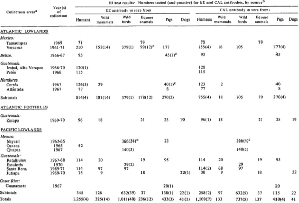
from the Pet&n area of Guatemala where EE
virus was isolated in 1968 (5). A few wild
terrestrial mammals and 40 nestling ardeids
and associated marsh birds were positive in
Veracruz, Mexico, and along the Pacific
coasts of Mexico and Guatemala. Horse sera
from Veracruz were positive (possibly due to
administration of inactivated EE virus
vaccine). Sera from pigs were negative
except in Costa Rica and in portions of
Belize and Honduras near areas where
positive human sera were obtained. A few
sera which were HI-positive to EE were also
HI-tested with VE virus; positive results
were found for 1 of 1 wild mammal serum
from Veracruz, Mexico; 1 of 1 human serum
from Izabal, Guatemala; 1 of 3 human sera
from Cort&, Honduras; 1 of 34 bird sera
from Nayarit, Mexico; 0 of 3 bird sera from
Chiapas, Mexico; 0 of 2 bird sera from
Escuintla, Guatemala; and 1 of 1 dog serum
from Jutiapa, Guatemala. VE N tests con-
firmed the positive VE HI result from
Cort& and the negative results from
Chiapas and Escuintla.
EE HI antibodies were found in 0 of 18
and 1 of 63 horse sera collected in El
Salvador and Nicaragua, respectively,
during 1972-1975; the one positive serum
218 PAHO BULLETIN . vol. XI, no. 3, 1977
Table 3. A comparison of WE and VE antibodies detected in Middle American sera with HI and N tests.
Sera with detectable HI or N antibody to WE or VE virus.a
Collection areas sources of sera
WEHI+ WEN +/ VEHI +/ VEN+/
W-&Jo. WEHI + WEHI + WEHI+
ATLANTIC LOWLANDS: Mexico:
Tamaulipas Veracruz Veracruz Veracruz Belize:
Guatemala: Izabal
PACIFIC LOWLANDS: Guatemala:
Retalhuleu Santa Rosa Costa Rica: Totals: % Positive
horses 32/40
humans 40/79
wild mammals 18/93
pigs 37/178
l/10 b o/19 O/3 o/7
humans 18/95 o/10
pigs 11/45 O/6
humans 21/87 o/7 7/21 l/7
humans humans pigs
14/112 O/8
15/26 o/10
14/20 o/14
220/775 l/94
28% 1%
O/32 O/32
9/40 2/19
6/18 o/3
8/37 8/24
l/11 3/10
3/6
o/12 o/15
31/186 17%
3/7 l/10 2/14 23/132
17% aPositive HI means a titer > 1:lO; positiveN signifies an LNI > 1.6 with a 1:4 dilution of serum. Blank spaces = not tested.
bTwo additional sera had LNI = 1.4 and 1.5.
CAL Virus
Only seven of 1,109 human sera gave a
positive HI response with CAL virus strain
BFS283. These positive seru came from
Veracruz, Mexico, and the Atlantic foot-
hills and Pacific coastal lowlands of Guate-
mala (see Table 4). Of 737 plasmas from
nestling marsh birds, only five from the
Pacific coast of Mexico were positive.
Similarly, of 410 pig sera, only four from
Veracruz were positive. No sera had titers
exceeding 1:40.
SLE, DEN, and Other Flaviviruses
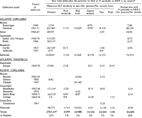
In all, 23% of 1,197 human sera reacted
with SLE virus hemagglutinin, and 20% of
405 sera responded positively in N tests with
DEN 3 virus (see Table 5). However, only
1% of 299 wild mammals, 5% of 900
nestling marsh birds, 6oJo of 236 horses: 5%
of 434 pigs, and 2% of 41 dogs gave a
positive HI response when their sera were
tested with SLE virus. The positive nestling
marsh birds were captured in Minatitl%n,
Veracruz, where SLE virus had been
isolated in 1965 (7), and at three colonies
along the Pacific coasts of Mexico and
Guatemala. Horse and pig sera from
Veracruz were also positive to SLE. A few
other positive pig sera came from Belize,
Honduras, the Atlantic foothills of Guate-
mala, and the Pacific lowlands of Mexico
and Costa Rica. The remaining positive
samples-six horse sera and one dog
Scherer et al. 0 SURVEY OF ARBOVIRUSES FROM MIDDLE AMERICA 219
Table 4. Prevalences of serum HI antibodies to EE and GAL viruses in people and animals sampled along the Atlantic and Pacific coasts of Mexico, Belize, Guatemala, Honduras, and
Costa Rica during 1961-1971.
A>ANTlC LOWlANDS
Mexico:
Tamauhpas 1969
wracruz 1961-71
&,,ZC 1966-67
th7remda.
lzabl. *,,a verapar 1966-70
Pet&, 1966
,%“d”,iX.
CO&S L967
Atllnbda 1967
Subtotals
ATLANTIC l=OOTHlLLS
Guoremdc7:
zacapa 1969-70
PAClFlC LOWLANDS
’ gl%5 1967
1967-68 1970 1969-71 1969.70
Subtotals Totals
71 79 70
210 152(14) 379(l) 99(12F 177 155(4) 16
95 45(l)C 95
120(L) 115
120 115
126(3) 29 40(l)C 123 2 40
77 8 77 8
814(4) 181(14) 379(l) 178Cl2) 270(2) 755C4) 18
96 18 21 25 19 96(l) 18
366(34)’ 23
42
14OL3)
114 20 19 95 114 20 19
114 97 GT2) ;;
75 9 18 22(l) H;(2) 6; 18
20(l)
345 126 632(39) 37 1380) 22(l) 258(2) 97
1,255(4) 3X(14) 1,011(40) 236(12) 433(3) 41(l) 1,109(7) 133
105 79
21
366(4)c
140(l)
632W 37
737(S) 137
270(4)
25 19
95
22
20
115 22
410(4) 41
aSPeclfic locatlans wthm states or departments are the same as those Wed m Tables 1 and 2. bPosttlve Hl means a titer 2 1:lO.
CSera wth EE t~tm > 1 40 were obtamed from the fallowmg animals’ 8 horses from Veracruz, 1 pg from Belize. 1 pig from Curt& (Honduras), and 10 buds from Nayarn, Mexico Three birds from Nay&t ytelded CAL titers Z 1.40.
DEN antibodies were found mostly in
older persons; antibody was never detected
in subjects under age 20 and was detected
infrequently in those under 40. The per-
centages of various age groups yielding posi-
tive sera were as follows: 1 l-20 years, 0% of
24 persons; 21-40 years, 9% of 172; 41-60 years, 30% of 149; over 60 years, 40% of 60.
Plasmas from two sentinel hamsters
exposed in Veracruz, Mexico, in 1969
yielded 1:lO HI titers with SLE virus
hemagglutinin. The positive samples were
taken after the animals had been exposed at
Tlacotalpan and Sontecomapan for 28 and
34 days, respectively. These two hamsters
were part of a sizable group used as sentinel
animals in Middle America to detect arbo-
viruses such as VE. Plasmas from 245
survivors exposed during 1967-1971 were
available for antibody tests, but only the
two just mentioned and five exposed at
Minatitlan, Veracruz, in 1967 (which
yielded 1:lO titers to WE virus) showed
positive results in HI tests with SLE, EE, or
WE viruses. The value of these hamster
plasmas for serologic surveys was obviously
limited because the exposures all occurred
in one season (July-August) and only lasted
for two to six weeks. Furthermore, one to
two weeks need to be subtracted from this
period because of the time required for
detectable HI antibody to develop after an
220 PAHO BULLETIN l vol. XI, no. 3, 1977
Table 5. Pmvalences of serum antibodies to flaviviruses (SLX and DEN 3) in people and animals sampled along Atlantic and Pacific coastal areas of Mexico, Belize,
Guatemala, Honduras, and Costa Rica during 1961-1971.
Collecllon are&
Year(s) of collection
Sera wth deteclable Hl antlbody to SLE or N dntlbody to DEN 3, by sourceb Flavwrus SLE antlbody m sera (No posltwe/No tested) from: Human sera wth Humans Wdd Wdd E9UlIltY N antlbody to DEN 3
mdmmdls buds ammals PlgS Dogs (No pos~t~ve/No tested)
ATLANTIC LOWLANDS: Mexico:
Tamauhpas Veracrtlz Belizc: Guatemala:
Izabal, Alta Verapaz P&l Honduras: tortes Atlintida 1969 1961.71 1966.67 1966.70 I966 1967 1967 Subtotals 1/74c 65/244c 28/95c 51/120c 3611 lSc 26/l 10c 6177 213/835 ATLANTIC FOOTHILLS:
Guatemala:
.&Cap 1969.70 17/90 l/18 o/21
PAClFlC LOWLANDS: Mexico: Nayarit OaXKa Chiapas Guatemala: Retalhuleu Escuintla Santa Rosa Jutiapa Costa Rica: Guanacaste 1962.65 I965 1967 o/42 1967.68 1970 1969.71 1969.70
171114c l/20 26/l 10E O/87
l/6 O/9 1967
Subtotals 441272
Totals 27411.197
“10 Positive 23%
l/l52 1 l/268’ o/79 8/99’
o/13
11165 1 l/268 8/178
31366 241140 o/19 4129 o/97 6/18C l/l16 31299
31/632 6137 42/900 141236
5 @A 6% 1 o;o 81178 2/45 l/40 O/S 111271
5125 o/19
l/23
o/95 l/22 S/20 61138 l/22 221434 l/41 5vo 2 4’0
7140 381158 14136 8/73 6/12 2156 751375 S/l6 3/ 14 8/30 83/405 20% aSpecific locations wthm states or departments are the same as those bsted m Tables 1 and 2
bPositive HI means a titer 1 I IO. pos~tlve N slgmfies an LNI > I 5 (97% plaque reduction) wth d I 4 dllutlon of serum heated to 60°C for 20 minutes and tested m LLCMKZ monkey kidney cell cultures
CThe followmg human sem had SLE HI titers > I 40 1 from Tamaubpds, 21 from Veracrur, 10 from Belne, 5 from lzdbdl, 9 from P&n. 3 from CortCs. 2 from Retalhuleu and 3 from Santa Rosa Two birds and I horse tram Veracruz dnd I horse from JutupJ also yielded serum titers > 1:40.
DiSCUSSiOll
This serologic survey of Middle America
revealed that numerous sera from the
Atlantic coasts of Mexico, Belize, Guate-
mala, and Honduras, as well as many from
the Pacific coast of Guatemala, contained
HI antibodies that reacted with WE virus.
However, N tests showed that these anti-
bodies were not specific for WE virus. The
only virus antibodies that appeared specific
for WE were found in horses from Mata-
moros, Mexico, a town bordering portions
of southern Texas where WE virus is known
to be active (24). In other regions, WE HI
antibodies did not always coexist with other
alpha-virus (VE or EE) antibodies. Only in
a few places that were heavily involved in
America did positive WE HI tests correlate
with positive VE HI tests. Thus in some
regions the antibodies that reacted in
Scherer et al. * SURVEY OF ARBOVIRUSES FROM MIDDLE AMERICA 221
induced by another alphavirus which was
not WE, EE, or VE. Future attempts should
therefore be made to isolate such an
alphavirus in Middle America and to learn
whether it causes disease in people or
animals.
Virtually no HI antibodies to EE virus
were found in sera from north-coastal
Middle America, except in sera obtained
near the base of the Yucatan Peninsula in
Belize, Guatemala, and Honduras. Since
EE virus was isolated in the rain forest of
Guatemala’s Pet&n region in 1968 (5), this
locale appears to be an enzootic focus.
Health personnel diagnosing equine and
human encephalitic disease there should
consider EE virus as a possible etiologic
agent. Also, the absence of EE virus anti-
body throughout much of north-coastal
Middle America should alert health person-
nel to the susceptibilities of people and
animals to EE virus diseases were the virus
to invade new regions.
HI tests for CAL virus antibodies
revealed no unequivocal CAL virus activity
in Middle America. Since the CAL virus
strain BFS283 hemagglutinin used for these
surveys seems to detect antibodies to at least
two other viruses of the CAL group (I), the negative HI results obtained with this strain
indicate the probable absence of La Crosse
virus, the major human pathogen of the
CAL group in the Americas, and also the
possible absence of Snowshoe Hare virus.
However, nothing definite can be con-
cluded about other CAL group viruses be-
cause HI tests with CAL strain BFS283
cannot be relied upon to detect their anti-
bodies.
HI tests with SLE virus detected anti-
bodies to SLE and other arboviruses of the
flavivirus genus. The results with Middle
American sera indicate moderate cycling of
SLE or other flaviviruses along the Atlantic
coasts of Mexico, Belize, Guatemala, and
Honduras, and also along the Pacific coasts
of Mexico, Guatemala, and Costa Rica.
Some of these flavivirus antibodies probably
resulted from infections with SLE, DEN,
Jutiapa, or Ilheus viruses, but some may
also have resulted from infections with as-
yet-undiscovered flaviviruses of the region.
Jutiapa virus was isolated from a cotton rat
(Sigmodon hi@idus) collected in the moun-
tains of Guatemala near the southeastern
Pacific coast during 1969, and Ilheus virus
was recovered from mosquitoes on the
Atlantic sides of Guatemala and Honduras
in 1956 (I). It is noteworthy that SLE virus
has recently caused a sizeable human
epidemic in the central upland plateau of
Mexico (17).
N tests revealed that DEN 3 virus anti-
bodies were present only in older people,
and that this virus had not cycled in
tropical Middle America for many years.
This observation fits with those of Rosen,
who found that DEN 2 and DEN 3 viruses
infected people in Panama between 1941
and 1954 (la). Suppression of DEN virus
activity in Middle America has undoubted-
ly resulted from programs for control of the
vector mosquito, A edes aegypti. However,
with DEN virus currently active in the
Caribbean region, vigilance should be
maintained in Middle America-so as to
prevent recurrence of enough Aedes aegy#ti
to transmit DEN viruses and cause human
disease.
ACKNOWLEDGMENTS
We are grateful for the able technical numerous persons in each counry of
assistance of F. A. Breakenridge, E. Du Middle America who contributed to the col-
Casse, K. Anderson, J. Chin, and B. lection of sera. In Mexico, studies were done
222 PAHO BULLETIN 0 vol. XI, no. 3, 1977
National Institute of Virology and the collecting sera in that country. Pig sera Institute of Health and Tropical Diseases. were collected in Costa Rica when Dr.
In Guatemala, Belize, Honduras, and Robert W. Dickerman participated in
Nicaragua the studies were greatly facili- teaching work for the Organization for
tated by health and agriculture officials Tropical Studies. Permits to collect wild and personnel. Drs. G. Godoy and J. E. birds and mammals in Mexico and Guate- Navarro M. of the Medical School of the mala were provided by appropriate author- University of El Salvador participated in ities in those countries.
SUMMARY
A serologic survey for antibodies to dengue 3 that are without antibodies and are therefore and western, eastern, St. Louis, and California susceptible to these arboviruses should serve as a encephalitis arboviruses shows that coastal warning to health authorities. For example, Middle America was relatively free of infections eastern or St. Louis encephalitis viruses could by these or antigenically related arboviruses conceivably extend their geographic range in during 1961-1975. Aside from pointing out a Middle America and cycle between vertebrates postulated but unidentified alphavirus that and vector mosquitoes which bite humans or cross-reacts with western encephalitis virus, these horses. Should this happen, the result could be findings provide no basis for further studies in human or equine encephalitis outbreaks like the the immediate future that seek to isolate these recent epidemics of Venezuelan encephalitis in arboviruses from natural sources in Middle Middle America and St. Louis encephalitis in America. However, the presence of populations Mexico’s central upland plateau.
REFERENCES
(I) Berge, T. 0. (ed.). International Cata-
logue of Arbouiruses, Including Certain Other
Viruses of Vertebrates (Second edition). U.S.
Department of Health, Education, and Welfare; DHEW Publication No. (CDC) 75-8301, Wash- ington, 1975.
(2) Elton, N. W. Yellow fever in Central America: The imminent threat to Mexico and the United States. Am J Public Health 46:1259-
1265, 1956.
(3) Trapido, H. Geographic distribution and ecologic setting. In: Venezuelan Encefihalitis:
Proceedings of the Worksho@-Symposium on
Venezuelan Ence$halitis Virus. Pan American
Health Organization. PAHO Scientific Publica- tion 243, Washington, 1972, pp. 302-312.
(4) Tellez Girbn, A., and 0. Valdes Ornelas. La presencia de virus tipo Este de la encefalo- mielitis equina en la epizootia ocurrida en el Estado de Tamaulipas, Mgxico, durante el aiio de 1941. Rev Sot Mex Hist Nut 2:251-259, 1941. (5) Ordoiiez, J. V., W. F. Scherer, and R. W. Dickerman. Isolation of eastern encephalitis virus in Guatemala from sentinel hamsters exposed during 1968. Bol Of Sanit Panam 70:
‘371-375, 1971.
(6) Calisher, C. H., K.S.C. Maness, R. D.
Lord, and P. H. Coleman. Identification of two
South American strains of eastern encephalo- myelitis virus from migrant birds captured on the Mississippi delta. Am J Efiidemiol 94:172- 178, 1971.
(7) Dickerman, R. W., W. F. Scherer, B. A. Pancake, and C. M. Bonacorsa. St. Louis encephalitis virus isolated from a nestling Common Egret in southeastern Mexico. Bol Of
Sunit Punam (English edition) 6:26-30, 1972.
(8) Sudia, W. D., Z. Ferngndez L., V. F. Newhouse, B. Sanz R., and C. H. Calisher. Arbovirus vector ecology studies in Mexico during the 1972 Venezuelan equine encephalitis outbreak. Am J Epidemiol 101:51-58, 1975.
(9) Rosen, L. Observations on the epidemi- ology of dengue in Panama. Am J Hyg 66:45-58,
1958.
(10) Reeves, W. C., C. Ortiz Mariotte, H. N. Johnson, and R. E. Scrivani. Encuesta serolb-
gica sobre 10s virus transmitidos por artr6podos en la zona de Hermosillo, Mexico. Bol Of Sunit
Panam 52:228-230, 1962.
(II) Sosa-Martinez, J., M. DurPn, and L. Benavides. St. Louis virus antibody survey on sera of residents of Yucatan, Mexico. Bol Med
Ho@ Infant Mex (English edition) -.37-42,
1963.
Scherer et al. l SURVEY OF ARBOVIRUSES FROM MIDDLE AMERICA 223
virus arbor: Estudios realizados en el Instituto National de Virologia de la SecretarIa de Salu- bridad y Asistencia. Gac Med Mex 93415-420, 1963.
(13) Mucha-Macias, J . de. Estudios epidemio- ldgicos sobre virus arbor en el sureste de Mexico. Salud Ptiblica Mex 5:523-527, 1963.
(14) Sosa-Martinez, J., L. Benavides, and M.
D. Vidaurri. Distribucibn de 10s virus arbor en las AmCricas. Gac Med Mex 8:855-867, 1964.
(Ii) Scherer, W. F., C. Campillo Sainz, J. de Mucha MacIas, R. Rubio-Brito, T. Miura, R. W. Dickerman, D. W. Warner, and M. Dyer. Serologic survey for neutralizing antibodies to eastern equine and western equine encephalitis viruses in man, wild birds, and swine in southern Mexico during 1961. Am J T~ofi Med
Hyg 15:211-218, 1966.
(16) Mucha Maclas, J. de. Enfermedades por arbovirus: Anticuerpos para la encefalitis de San Luis en enfermos mentales. Rev Znv S&d
Ptiblicu (Mexico) 34:169-174, 1974.
(17) Gonzalez CortCs, A., M. L. ZIrate Aquino, J . Guzman Bahena, J. Mir6 Abella, G. Cano Avila, and M. Aguilera Arrayo. St. Louis encephalomyelitis in Hermosillo, Sonora, Mex- ico. Bull Pan AmHealth Organ 9:306-316, 1975.
(18) Rosen, L. Dengue type 3 infection in Panama. Am J Trap Med Hyg 23:1205-1206,
1974.
(19) Scherer, W. F., J. V. Ordoiiez, P. B. Jahrling, B. A. Pancake, andR. W. Dickerman.
Observations of equines, humans, and domestic
and wild vertebrates during the 1969 equine epizootic and epidemic of Venezuelan encephali- tis in Guatemala. Am J Efiidemiol 95:255-266,
1972.
(20) Zarate, M. L., R. H. Geiger, R. E. Shope, and W. F. Scherer. Intergroup antigenic relationships among arboviruses manifested by a Mexican strain of Patois virus and viruses of the Bunyamwera, C, California, Capim and Guama groups. Am J Epidemiol 88:273-286, 1968.
(21) Scherer, W. F., C. Campillo-Sainz, J. de Mucha-Ma&as, R. W. Dickerman, C. Wong Chia, and M. L. Zarate. Ecologic studies ?f Venezuelan encephalitis virus in southeastern Mexico: VII. Infection of man. Am J Trap Med Hyg 21:79-85, 1972.
(22) Clarke, D. H., and J. Casals. Techniques for hemagglutination-inhibition with arthro- pod-borne viruses. Am J TTO# Med Hyg 7:561- 573, 1958.
(23) Scherer, W. F., F. A. Breakenridge, and R. W. Dickerman. Cross-protection studies and search for subclinical disease in New World monkeys infected sequentially with different immunologic types of dengue viruses. Am J
Epidemiol95:67-79, 1972.
(24) Sudia, W. D., V. F. Newhouse, L. D. Beadle, D. L. Miller, J. G. Johnston Jr., R. Young, C. H. Calisher, and K. Maness. Epidemic Venezuelan equine encephalitis in North America in 1971: Vector studies. Am J