Bull Pan Am Health Organ 14(l), 1980
FASTERN ENCEPHALITIS VIRUS FROM VIRGIN M)RESTS OF NORTHERN BRAZIL ’ b2
Robert W. Dickerman,S Francisco P. Pinheiro,4 Otavio F. P. Oli~a,~ Jorge F. Travassos da Rosa,” and Charles H. Calisher5
The following contribution describes isolation of eastern encephalitis (EE) virus from sentinel hamsters exposed to wet tropical forest at an isolated settlement in Brazil’s Amazon
region. This discovery of EE virus activity, together with indi- cations that human exposure to EE virus in the test area was slight, Providesfurther evidence of a truly sylvatic EE transmis- sion cycle outside man’s sphere of influence.
Introduction
In conjunction with studies carried out by the Phelps Ornithological Collection, under the auspices of the Joint Brazilian- Venezuelan Border Demarcation Commis- sion on Mount Urutani along the Vene- zuelan-Brazilian frontier, the senior au- thor was able to carry out preliminary arbovirus investigations employing sentinel hamsters at the Commission’s base camp at Uaica on the Uraricoera River in northern Brazil. This article presents the results of those field investigations, which were made during March and April 1977.
lAlso appearing in Spanish in the Bolet~n de la Oficina Sanituria Panamericana,
2This research was performed in collaboration with the Pan American Health Organization and was sup- ported in part by research grant Al-06248 from the U.S. Public Health Service, National Institute of Al- lergy and Infectious Diseases. The senior author’s transportation to Venezuela was provided by the American Museum of Natural History for ornithol- ogical studies, and field studies within Venezuela were supported by William H. Phelps, Jr. The work at the Evandro Chagas Institute was partially sup- ported by the Programa do Tr6pico Unido, CNPq, Brazil.
SDepartment of Microbiology, Cornell University Medical College, New York, N.Y., 10021, U.S.A,
41nstituto Evandro Chagas, Fundacgo Services de Saiide Pbblica, Ministerio da Saude, Belem, Brazil.
5Center for Disease Control, Vector-Borne Disease Division, Fort Collins, Col. 80522, U.S.A.
Materials and Methods
1 Study Site and Field Techniques
Uaica, the main supply camp for the Commission, was located in wet virgin tropical forest along the Uraricoera River, at an elevation of about 260 meters (Figure 1). The camp was serviced by a Venezuelan military helicopter that shuttled supplies in from Boa Vista, Brazil, to working camps of surveyors on Mount Urutani. When the Uaica base was established in December 1976-January 1977, large trees of the genus Cecro#ia (that had grown up since missionaries abandoned the camp 10 years before) were left standing; all shrubs were removed. The forest floor within the camp was swept at least twice a week. The staff, which ranged in size from 30 to 100 men, made use of toilet facilities located on cleared paths a short distance inside the undisturbed forest bordering the camp.
Syrian golden hamsters from the Lake- view Hamster Colony in New Jersey were exposed, when about 5 weeks old, in open wire mesh cages suspended about a meter above ground under protective roofs within the forests surrounding the Uaica camp and the adjacent landing strip. The hamsters had food and water available ad Zibidum. Animals that did not become
16 PAHO BULLETIN . vol. 14, no. I, 1980
Figure 1. A map showing the location of the Uaica, Brazil, study area and places in northern and central South America (indicated by letters) from which EE virus
has been isolated, by source of the isolate.
E = EQUINE M = MoSPlJlTO
s = SENTINEL /wItMLS
v = WILD VERTEBRATE
BRAZIL
FM sv
i--j
E
VENEZUELA .--- ,a- -_v.: I
moribund or die during the study period grew normally and developed extensive deposits of fat. Moribund and dead hamsters were labeled and stored at - 5 to 5% until autopsied, or were autopsied im- mediately-with tissue pools being frozen in vapor over liquid nitrogen. Three tissue samples were obtained under sterile condi- tions from each animal. These consisted of brain tissue alone: pooled heart, lung, liver, and kidney tissues: and pooled tissues from both the brain and the four organs.
Variations in conditions of preservation resulted from recall of the supply heli-
Dickerman et al. l EASTERN ENCEPHALITIS VIRUS FROM NORTHERN BRAZIL 17
Virus Isolation
At Cornell, 10 per cent suspensions of brain tissue samples were prepared in Hanks’ balanced salt solution containing 1 per cent bovine albumin, and were inocu- lated intracranially into suckling albino white mice 1 to 4 days of age that had been obtained from Taconic Farms in German- town, N.Y. Similar preparations were made from all mice that died after being inoculated with the hamster brain suspen- sions. Aliquots of first mouse passages of strains isolated at Cornell, as well as all unopened combined pools of brain and organ tissues from the field, were packed in dry ice and shipped to the Evandro Chagas Institute in Belem, where all were tested for the presence of virus by similar procedures employing suckling mice.
Virus Identification at the Evandro Chagas Institute
Both hemagglutination-inhibition (HI) and neutralization (N) tests were used for virus identification. The HI test procedure employed has been described before (1,Z). The N test was performed in 96 microplate wells (Linbro Chemical Co., New Haven, Conn.) using Vero cells, as described earlier (3). Basically, virus and heat-inac- tivated serum were mixed in the microplate wells and were incubated at 37OC for 1 hour. At this point, approximately 2 x lo4 cells were added to the contents of each well. Medium 199-containing 5 per cent fetal bovine serum and buffered with HEPES, sodium bicarbonate, and sodium hydroxide-was used to dilute the reagents and provide nutrients suitable for cell growth. Fresh rhesus monkey serum was added to the medium at a final dilution of 1:8. On some occasions the constant serum- varying virus method was used, whereas at other times fixed amounts of virus were mixed with several serum dilutions. The
presence of a cytopathic effect was taken as an indicator of virus growth.
Antibody Survey
Serum samples collected from 48 Yano- mama Indians in August 1977 were tested against eastern encephalitis (EE) virus by the HI and N tests. The procedures for these tests were similar to those cited above.
Virus Strain Identification
Four strains isolated at Cornell were sent to the Bureau of Laboratories, Vector- borne Disease Division, of the U.S. Center for Disease Control in frozen 10 per cent brain suspensions from the first or second passages in suckling mice. There they were tested with the short incubation hemagglu- tination test, using previously described methods (4).
Results
Viruses from Hamsters
Twenty-one hamsters were exposed in wet tropical forest at the base camp or along the landing strip for a total of 424 hamster-days between 7 March and 11 April 1977. Of these, 17 became moribund or died. The first animal died on the fourth day following placement in the field; seven hamsters died 4 to 10 days after exposure. The mean time between exposure and death for the 17 animals dying after exposure along the edge of the forest was approximately 19 days. No hamsters died during a total of 106 hamster-days of exposure in the middle of the camp area.
18 PAHO BULLETIN l vol. 14, no. 1, 1980
kidney, and liver tissues. No virus was isolated from the tissues of one hamster.
All 29 of the isolated virus strains were indistinguishable in HI tests from EE virus Bel&m prototype BeAn 7526. Also, two strains from Uaica (77U43 and 77U48) were found to be identical to BelCm strain BeAn 7526 when compared by complement fixation (CF) and cell culture N tests (Table 1).
Human Antibodies
As noted before, serum samples from 48 Yanomama Indians collected in August
1977 were tested for antibodies to EE virus by HI and N tests (Table 2). These Indians had lived in the Uaica site up to approxi- mately 10 years before collection of the samples. At the time of bleeding they had resided for several years at the village of Parimiu near the headwaters of the Uraricoera River. Serum from one subject over 15 years of age yielded HI and N test results positive for antibodies to EE virus.
CDC Tests
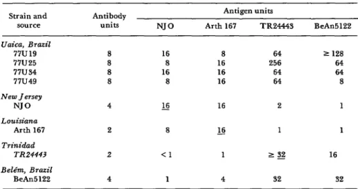
Four isolates from Uaica test by the short incubation HI test appeared similar if not
Table 2. HI and N antibodies to EE virus (Uaica strain 77WS) found amoq Indians who
formerly lived at Uaica.
sex Age No. positive/
(years) No. tested
M < 15 o/4
M > 15 1/19*
F < 15 O/l
F > 15 o/24
Total l/48
*Subject positive for both HI and N antibodies.
identical to one another and two other South American EE isolates, while yielding results different from two North American EE isolates (Table 3).
Discussion
The discovery of EE virus at Uaica, an isolated site in northern Brazil, adds to the data documenting the extremely wide- spread distribution of EE virus in northern South America (see Figure 1). EE virus is known to have caused human disease in Brazil (5) and Trinidad (6, not shown in figure), as well as equine disease in Panama (7), Venezuela (8), Guyana (7), Argentina (9), and the Brazilian States of
Table 1. HI, CF,a and Nb tests comparing EJZ virus strains from Uaica (77U433 and Bel6m (BeAn 7526), Brazil.
Virus strains - Test serum
BeAn 7526 77u45 77U48
BeAn 1:160 1:160 1:160
HI 77U43 1:160 1:160 1:160
77U48 I:80 I:80 1:80
BeAn 16/64 16/16 16/64
CF 77U43 32/64 32/16 32/64
77U48 8/16 16/16 16/64
BeAn 4.25 4.37 5.5
N 77U43 4.25 4.0 4.0
77U48 4.25 4.37 p.0
?Serum/antigen CF titers.
Dickerman et al. . EASTERN ENCEPHALITIS VIRUS FROM NORTHERN BRAZIL 19
Table 3. Results of short incubation tests comparing four EE viros strains from Uaica witb each other, two other South American strains, and two North American straim.
Strain and source Uaica, Brazil
77u19 77U25 77u34 77u49 New Jersey
NJ0 Louisiana
Arth 167 Trinidad
TR24443 Belt%, Brazil
BeAn
Antibody units
8 8 8 8 4 2 2 4
NJ0
16 8 16 8 s
8 <l
1
Antigen units
Arth 167 TR24443 BeAn
8 64 2 128
16 256 64
16 64 64
16 64 8
16 2 1
L!2 1 1
1 1 g 16
4 32 32
Par5 (IO), SIo Paulo, Rio de Janeiro, Bahia, and Pernambuco (II). It has been isolated from sentinel animals (hamsters, mice, monkeys, or chickens) in Colombia (12,13), Peru (14), and Venezuela (8); from sentinel animals, wild vertebrates, and mosquitoes in the Brazilian States of Par5 (10,14,15) and S5o Paulo (16); and from mosquitoes in Trinidad (7).
Although many of these isolations have been made in disturbed habitats, where human and domestic animal populations have impinged upon wild areas, numerous isolates have also been obtained from little-disturbed forests near BelCm. Shope et al. (15) found evidence of enzootic forest cycles of EE virus, the virus being isolated from mosquitoes and sentinel animals, with involvement of birds being infrequent. Also, EE virus has been isolated from Culex (Melanoconion) taeniopus mosqui- toes collected in 1975 in virgin forests of Aripuana in the north Brazilian State of Mato Gross0 (17).
Within this context, identification of an intensely active focus of EE virus at Uaica, inside a region of hundreds of square kilo- meters of virgin forest, further indicates a truly sylvatic cycle in the absence of
human influence, Especially interesting is the high rate of EE isolation from sentinel rodents-indicating active transmission of this usually avian-based arbovirus by mammalian-feeding vectors, a situation similar to that found by Shope et al. (15). No avian studies were carried out at U aica.
Casals (18) and more recently Calisher et al. (5) have demonstrated that timed HI tests can be used to distinguish Central and South American EE virus strains from North American strains. In addition, published studies by Walder, J ahrling, and Eddy (19) have employed hydroxylapatite chromatography to substantiate and en- large upon this work. Although, as noted above, EE virus has caused disease in humans and equine animals in South America, the relative virulence of the two groups of strains (Central-South and North American) is unknown.
20 PAHO BULLETIN l vol. 14, no. 1, 1980
a theory supported by the low incidence of EE antibody among Indians who previous- ly lived at Uaica. A further possibility is that the twilight-nocturnal flight ranges of the mosquito vectors may have restricted them to the confines of uncleared forest containing dense tree cover and abundant litter. (In contrast, human twilight and nocturnal activity was mostly restricted to
the cleared open airstrip and camp areas.) In general, however, there is little doubt that as Brazil and Venezuela extend their highway systems into unopened regions of the Amazon River Basin, ever-increasing numbers of people and domestic animals will be exposed to the risk of infection by this widely distributed enzootic pathogen.
ACKNOWLEDGMENTS
The senior author was invited by the Phelps Ornithological Collection of Cara- cas, through the American Museum of Natural History, to participate in ornitho- logical studies on Cerro Urutani under the auspices of the Joint Brazilian-Venezuelan Border Demarcation Commission (Comisi6n
Mixta Brasilefia- Venezohna Demarcadora de Lfmites). Mr. William H. Phelps, Jr., through the generous cooperation of Dr. Georges Pantchenko of the Border Admin- istration (Direccio’n de Fronteras) of Vene- zuela arranged for him to conduct these virological studies. Their success is largely
due to the unstinting help provided by Ram&r Aveledo and Luis Perez of the Phelps Ornithological Collection, by Engi- neer Dilermando de Murais Mendez of the
Brazilian Commission, and by Heinz Cardona of the Venezuelan Commission. The Pan American Health Organization facilitated the importation of experimental animals and equipment.
In addition, Eugene Di Paola provided expert assistance in the laboratory. We are also grateful to Dr. Mario A. P. Moraes of the Evandro Chagas Institute for providing the Indian sera.
Eastern encephalitis (EE) virus was success- Tests run on sera from Yanomama Indians fully isolated from 16 sentinel hamsters exposed who had lived in the area indicated local human to a wet tropical forest environment in the far- exposure to the virus was very slight. Therefore, northern reaches of the Brazilian Amazon. The the detection of intense EE virus activity in this place of exposure, a settlement named Uaica area provides further evidence of a truly sylvatic near the Brazil-Venezuela border, is surrounded EE cycle outside the present sphere of human by hundreds of square kilometers of virgin influence.
forest.
REFERENCES
(1) Clarke, D. H., and J. Casals. Techniques in a community of forest animals. Ann for hemagglutination and hemagglutination- Microbial (Paris) 9(A):167-171, 1963.
inhibition with arthropod-borne viruses. Am J (3) Pinheiro, F. P. Aplicacgo de uma micro- Tro# Med Hyg 7:561-573, 1958. tecnica no estudo do teste de neutralizacgo corn
Dickerman et al. l EASTERN ENCEPHALITIS VIRUS FROM NORTHERN BRAZIL 21
Lord, and P. H. Coleman. Identification of two South American strains of eastern equine encephalomyelitis virus from migrant birds captured on the Mississippi Delta. Am J Efiidemiol 94:172-178, 1971.
(5) Alice, F. J. 1nfeccZ.o humana pelo virus “Leste” da encefalite equina. Bol Inst Biol Bahia 3:3-9, 1956.
(6) Corniou, B., P. Ardoin, C. Bartholomew, W. Ince, and T. Massiah. First isolation of a South American strain of eastern equine virus from a case of encephalitis in Trinidad. Top Geogr Med 24(2):162-167, 1972.
(7) Theiler, M., and W. G. Downs. The Arthropod-borne Viruses of Vertebrates. Yale University Press, 1973, 578 pp.
(8) Walder, R., and 0. M. Sulrez. Primera evidencia en Venezuela de la encefalitis equina de1 este (EEE) en circunstancias silentes. Bol Di7 Malariologia y San Ambiental 14:119-125, 1976. (9) Mettler, N. E. Identification de cepas de encefalitis equina aislados en la Repfiblica Ar- gentina. Rev Sot Argent Biol 38:55, 1962.
(IO) Causey, 0. R., C. E. Causey, 0. M. Ma- roja, and D. G. Macedo. The isolation of arthropod-borne viruses, including members of two hitherto undescribed serological groups, in the Amazon region of Brazil. Am J Trap Med Hyg 10:227-249, 1961.
(II) Cunha, R. Viroses neurotrbpicas. Anais
V Congr Brazil Vet (Sdo Pa&o) 1:197-220, 1950. (12) SanmartIn, C., H. Trapido, P. Barreto, and C. I. Lesmes. Isolations of Venezuelan and eastern equine encephalomyelitis viruses from sentinel hamsters exposed in the Pacific lowlands of Colombia. Am J TroP Med Hyg 20:469-473, 1971.
(13) De Groot, H. Personal communication. (14) Scherer, W. F.. J. Madalengoitia, W. Flores, and M. Acosta. The first isolations of eastern encephalitis, group C, and Guama group arboviruses from the Peruvian Amazon region of Western South America. Bull Pan Am Health
Oqqzn 9:19-26, 1975.
(IS) Shope, R. E., A. Homobono Paes de Andrade, G. Bensabath, 0. R. Causey, and P. S. Humphrey. The epidemiology of EEE, WEE, SLE and Turlock viruses, with special references to birds in a tropical rain forest near Beltm, Brazil. Am J Epidemiol 84~467-477, 1966.
(16) Souza Lopes, O., and L. A. Saccheta. Epidemiological studies on eastern equine en- cephalitis virus in SZo Paulo, Brazil. Rev Inst Med Trol, &io Paul0 16:253-258, 1974.
(17) Instituto Evandro Chagas. Unpublished data.
(18) Casals, J. Antigenic variants of eastern equine encephalitis virus. J Exp Med 119:547- 566, 1964.
(19) Jahrling, P. B. Personal communication.
MOTOR ACCIDENT DEATH RATES*
According to a recent study of 26 developed and 4 developing coun- tries reported in WHO’s World Health Statistics Quarterly (volume 32, number 3, 1979). mortality rates from motor vehicle accidents are con- tinuing to climb for all age groups. But the increases are disproportion- ately high among the 15- to 24-year old age groups, with rises of more than 50 per cent among females in three-fourths of the countries. The abuse of alcohol is blamed for much of the increase.
Thus far, public health authorities generally consider auto accidents as “acts of God,” and confine their responsibility to the treatment of victims. The study urges a shift of concern to accident prevention.