ISSN 1806-3713 © 2016 Sociedade Brasileira de Pneumologia e Tisiologia
http://dx.doi.org/10.1590/S1806-37562016000000168
An uncommon tomographic association:
amiodarone pulmonary toxicity and
adenocarcinoma
Arthur Soares Souza Jr1,2, Gláucia Zanetti3, Edson Marchiori3
1. Faculdade de Medicina de São José do Rio Preto, São José do Rio Preto (SP) Brasil.
2. Clínica Ultra X, São José do Rio Preto, São José do Rio Preto (SP) Brasil.
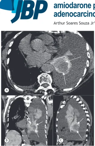
3. Universidade Federal do Rio de Janeiro, Rio de Janeiro (RJ) Brasil. Figure 1. Axial (in A), coronal (in B), and sagittal (in C) chest CT scans showing left pleural effusion and a high-density collapsed lung containing a round hypodense mass (arrows). Note also small right pleural effusion and liver hyperdensity (the liver is denser than the heart).
A 73-year-old woman, a current smoker, presented with progressive dyspnea. She had a history of ven-tricular tachyarrhythmia treated with amiodarone. A
chest X-ray demonstrated diffuse opaciication of the
left hemithorax. Chest CT showed left pleural effusion and a high-density collapsed lung containing a round hypodense mass (arrows). There was also small right pleural effusion and liver hyperdensity (the liver was
denser than the heart; Figure 1). Percutaneous ine-needle
aspiration biopsy of the mass revealed adenocarcinoma.
The histopathological indings of the dense pulmonary
parenchyma were compatible with amiodarone-induced pulmonary toxicity (APT). The patient died one month after the examination. Amiodarone is associated with a wide range of adverse effects, including APT.(1-3) The
diagnosis of APT can be suggested on the basis of a combination of clinical, radiological, and pathological
indings, and is conirmed by improvement after dis -continuation of amiodarone therapy.(3) The high iodine
content of the medication enables the detection of amiodarone deposits in the lung by CT as high-attenuation parenchymal opacities. The association of dense lung consolidations with high liver density is characteristic of amiodarone impregnation. (2,3) In the case described
here, the dense pulmonary parenchyma caused by amiodarone impregnation allowed the tomographic
identiication of the tumor. A
B C
REFERENCES
1. Hudzik B, Polonski L. Amiodarone-induced pulmonary toxicity. CMAJ. 2012;184(15):E819. http://dx.doi.org/10.1503/cmaj.111763
2. Hochhegger B, Soares Souza A Jr, Zanetti G, Marchiori E. An enlarged heart with hyperdense consolidation. Neth J Med. 2013;71(6):317, 321.
3. Jarand J, Lee A, Leigh R. Amiodaronoma: an unusual form of amiodarone-induced pulmonary toxicity. CMAJ. 2007;176(10):1411-3. http://dx.doi.org/10.1503/cmaj.061102
J Bras Pneumol. 2016;42(6):465-465