J Bras Pneumol. 2012;38(5):679-680
Urine in the pleural cavity: an unexpected finding
Urina em cavidade pleural: um achado inesperado
Melike Demir, Gülistan Karadeniz, Ebru Uz
To the Editor:
Urinothorax is an infrequent cause of pleural effusion and is caused by urine leakage into the pleural cavity through an anatomical defect in the diaphragm or via the lymphatic system.(1)
Urinothorax is usually secondary to obstructive uropathy or traumatic (typically iatrogenic) injury of the urinary system.(2)
A 61-year-old female with a history of nephrolithiasis was admitted to our institution with high-grade fever, decreased urine output, progressive shortness of breath, and right-sided chest pain for two days. The patient had undergone right-sided percutaneous nephrolithotomy three days prior to admission and had no history of pulmonary disease.
Physical examination revealed pallor and a body temperature of 38.4°C. Her chest movements were decreased. In the right hemithorax, there was a dull percussion note and breath sounds were absent, both of which are findings consistent with pleural effusion. No abnormalities were found regarding the other systems.
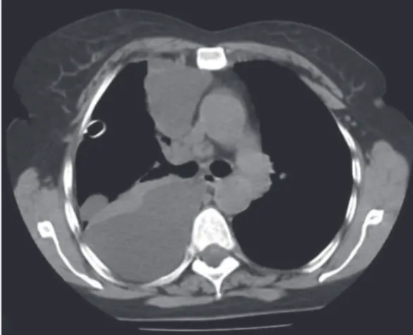
A chest X-ray demonstrated significant right-sided pleural effusion (Figure 1), and a CT scan of the chest revealed right pleural effusion, thin septa, and atelectasis of the underlying lung (Figure 2). Serum levels of urea and creatinine were 132 mg/dL and 3.9 mg/dL, respectively. Because the respiratory distress developed suddenly and soon after percutaneous nephrolithotomy, urinothorax was suspected. The diagnosis was confirmed after thoracocentesis, which yielded approximately 1,100 mL of pleural fluid, with a ratio between the creatinine content in pleural fluid and that in serum of 1.78 (normal, < 1). A 28 Fr chest tube was inserted into the right fifth intercostal space in order to drain the effusion.
The patient was started on broad spectrum antibiotics and carefully observed. Intravenous urography did not reveal any obstructive uropathy caused by kidney stones. Because the diagnosis of urinothorax had been confirmed, we did not perform radionuclide imaging in order to show
a connection between the urinary system and the pleural cavity. There was a gradual increase in urine output, accompanied by a simultaneous decrease in serum levels of urea and creatinine. The urine drainage from the chest tube stopped seven days after the procedure, and the tube was withdrawn. On post-admission day 9, the
Figure 2 - CT scan of the thorax revealing right pleural effusion, thin septa, and atelectasis of the underlying lung.
Figure 1 - Posteroanterior chest X-ray showing pleural effusion in the right hemithorax.
680 Demir M, Karadeniz G, Uz E
J Bras Pneumol. 2012;38(5):679-680
Melike Demir Pulmonologist, Sanatoryum Cd, Kecioren,
Ankara, Turkey
Gülistan Karadeniz Pulmonologist, Sanatoryum Cd, Kecioren,
Ankara, Turkey
Ebru Uz Nephrologist, Sanatoryum Cd, Kecioren, Ankara, Turkey
References
1. Handa A, Agarwal R, Aggarwal AN. Urinothorax: an unusual cause of pleural effusion. Singapore Med J. 2007;48(11):e289-92. PMid:17975679.
2. Garcia-Pachon E, Padilla-Navas I. Urinothorax: case report and review of the literature with emphasis on biochemical diagnosis. Respiration. 2004;71(5):533-6. PMid:15467335. http://dx.doi.org/10.1159/000080642 3. Hooper C, Lee YC, Maskell N; BTS Pleural Guideline Group.
Investigation of a unilateral pleural effusion in adults: British Thoracic Society Pleural Disease Guideline 2010. Thorax. 2010;65 Suppl 2:ii 4-17.
patient was asymptomatic and was discharged, remaining asymptomatic throughout a four-week follow-up period. Clinical characteristics and chest X-ray findings, as well as serum levels of urea and creatinine, were normal.
Urinothorax is probably more common than is supposed, because it is not usually included in the differential diagnosis of pleural effusion. For the definitive diagnosis of urinothorax, creatinine content should be determined in pleural fluid and in serum, a pleural/serum creatinine ratio of > 1 being considered diagnostic of the condition.(3)
Because creatinine is not routinely measured in pleural fluid, some patients with urinothorax might recover without having been correctly diagnosed. The history of percutaneous nephrolithotomy in our patient led us to determine the creatinine levels, and the diagnosis was established. It was our good fortune that the condition developed only a few days after that procedure.
In summary, given that urinothorax can develop several weeks after urologic procedures or without any apparent cause in cases of obstructive uropathy, we emphasize the importance of the clinical suspicion of urinothorax for its early diagnosis and treatment. Once urinothorax has been definitively diagnosed, we recommend that, with certain exceptions, radionuclide imaging should not be performed, thereby avoiding unnecessary radiation exposure and costs.