Arq Neuropsiquiatr 2006;64(3-A):672-675
Neurosurgical Unit Hospital Unimed, João Pessoa PB, Brazil: 1Neurosurgeon;2Neurologist;3Neuroanesthesist.
Received 8 December 2005, received in final form 7 March 2006. Accepted 24 April 2006.
Dr. José Alberto Gonçalves da Silva - Avenida Minas Gerais 1150 - 58030-092 João Pessoa PB - Brasil.
IMPACTED CISTERNA MAGNA WITHOUT
SYRINGOMYELIA ASSOCIATED WITH
SPASTIC PARAPARESIS
Case report
José Alberto Gonçalves da Silva
1, Maria do Desterro Leiros da Costa
2,
Maurus Marques de Almeida Holanda
1, Luiz Ricardo Santiago Melo
1,
Antônio Fernandes Almeida de Araújo
1, André Pachelli Bezerra Viana
3ABSTRACT - We re p o rton a 49 year old man with impacted cisterna magna without the presence of syringo-h y d romyelie (SM). Tsyringo-he clinical picture was csyringo-haracterized by spastic paraparesis. Magnetic resonance imag-ing depicted a cisterna magna filled by the cerebellar tonsils. Six months after osteodural-neural decom-p ression of the decom-posterior fossa there was resolution of neurological symdecom-ptoms and signs with the excedecom-p- excep-tion of hyperactive patellar and Achilles reflexes.
KEY WORDS: tight cisterna magna, impacted cisterna magna, Chiari malformation, posterior fossa decom-pression, spastic paraparesis.
C i s t e rna magna impactada sem siringomielia associada com paraparesia espástica: relato de caso
RESUMO - Hans Chiari (1891, 1895) descreveu 4 tipos de anomalias cere b e l a res que, ulteriormente, foram denominadas de malformação de Chiari (MC). Iskandar et al. (1998) relataram 5 casos de hidro s i r i n g o m i e l i a (SM) sem herniação rombencefálica, nos quais as tonsilas cere b e l a res preenchiam a cisterna magna. To d o s os casos melhoraram após a descompressão da fossa posterior com redução do tamanho da cavidade siringomiélica. Relatamos o caso de paciente de 49 anos com cisterna magna impactada sem a pre s e n ç a de siringo-hidromielia (SM). O quadro clínico caracterizava-se por paraparesia espástica. A re s s o n â n c i a magnética evidenciou a cisterna magna preenchida pelas tonsilas cerebelares. Seis meses após a descom-p ressão osteodural-neural da fossa descom-posterior, houve resolução dos sintomas e sinais neurológicos, com exceção da hiperatividade dos reflexos patelares e aquileus.
PA L AV R A S – C H AVE: cisterna magna impactada, malformação de Chiari, descompressão da fossa posterior, paraparesia espástica.
Hans Chiari1 , 2described four types of cere b e l l a r
anomalies (CM). Type I characterized by downward displacement of the cerebellar tonsils and the medi-al portions of the inferior cerebellar lobes which ac-companied the medulla into the cervical spinal canal. Type II showed downward displacement of port i o n s of the cerebellum (1891), and portions of the inferi-or vermis (1895), pons, medulla oblonga and, at least, p a rt of lengthened fourth ventricle, which re a c h e d the disc C 4-C 5, into the enlarged cervical spinal ca-nal. In type III, the hydrocephalic cerebellum, pons and medulla oblonga were inside a cervical
meningo-cele (hydroencephalomeningo-celes cerebellaris cerv i c a l i s ) , t h rough a spina bifida of the first three cervical ver-tebrae. In type IV, there was hipoplasia of the cere-bellum without herniation of cerebellar stru c t u res
in-to the spinal canal. Iskandar et al.3(1998) related
Arq Neuropsiquiatr 2006;64(3-A) 673
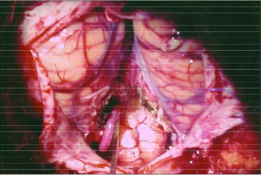
Fig 1. Impacted cisterna magna without SM.
The present publication is based on the rareness of the clinical picture characterized by spastic para-p a resis, hypara-popara-pallesthesia of the lower limbs and sex-ual impotency associated with impacted cistern a magna without SM. We did not find a similar re p o rt in the consulted literature (Medline and Lilacs).
CASE
A 49-year old male with a five-year history, of pro g re s-sive loss of muscular strength of the lower limbs causing s e v e re gait diff i c u l t y, sexual impotency and paraesthes ia on the feet, occasionally ascending up to the knees. Neu-rological exam showed spastic paraparesis, marked hyper-active patellar and Achilles reflexes, bilateral Hoff m a n n
and Babinski signs, inexhaustible knee and ankle clonus associated with diminished pallesthesia of the lower limbs. The patient was infected by hepatitis C virus (HCV) and sin-ce 1999 has been resin-ceiving i nterf e ron therapy. Magnetic resonance imaging (MRI) depicted a cisterna magna filled by the cerebellar tonsils (Fig 1).
The osteodural-neural decompression of the posterior fossa using the Gonçalves da Silva technique, was carr i e d out with the patient in sitting position. A large craniecto-my of the posterior fossa was perf o rmed and after the dur-al opening in Y format the following abnormdur-alities were o b s e rved (Fig 2): the cerebellar tonsils filled the cistern a magna without herniation into the spinal canal, the fourt h ventricle and the foramen of Magendie were compressed by the cerebellar tonsils and we could identify only the left
674 Arq Neuropsiquiatr 2006;64(3-A)
posterior inferior cerebellar art e ry (PICA). After dissection of the arachnoid membrane, we perf o rmed intrapial aspi-ration of the cerebellar tonsils and made a large opening of the fourth ventricle, and sutured the residual pial sac upwards to the dura-mater in cranial lateral position.
The postoperative MRI depicted the large created cis-t e rna magna and also cis-thacis-t cis-the cerebellar cis-tonsils did nocis-t c o m p ress the fourth ventricle and the foramen of Magendie a n y m o re (Fig 3). Six months after posterior fossa decom-p ression the disadecom-pdecom-pearance of neurological symdecom-ptoms and signs was observed, with the exception of the hyperactive patellar and Achilles reflexes and amelioration of sexual impotency.
This study was approved by the appropriate Bioethics Research Commitee.
DISCUSSION
Five cases of impacted cisterna magna without
hindbrain herniation were related by Iskandar et al.3,
n e v e rtheless these cases were accompanied by SM. All cases improved after posterior fossa decompres-sion with reduction in the size of the syrinx. The dra-matic response to decompression indicates that this entity has a Chiari like pathophysiology. Kyoshima
et al.4related four similar cases with impro v e m e n t
in symptoms and a reduction in syrinx size in thre e patients, and a reduction in ventricle size in two. The authors named “tight cisterna magna”, the impact-ed cisterna magna by the cerebellar tonsils, and callimpact-ed
the description according to Iskandar et al.3“ C h i a r i
“0” malformation”.
A c c o rding to Wi l l i a m s5, the cerebellar tonsils
her-niation may compress brainstem stru c t u res and con-tribute to bulbar and a long tract dysfunction.
In a similar way the impactation of the cere b e l-lar tonsils in the cisterna magna, without hern i a t i o n into the cervical spinal canal causes disturbances in the CSF flow at the foramen magnum and can devel-op neurological symptomatology by compression of the brainstem. In the present case during the oper-ation the fourth ventricle, brainstem and the fora-men of Magendie were compressed by the impact-ed cerebellar tonsils. Probably this pathophysiology could explain the neurological symptoms and signs of this patient.
Spastic tetraparesis and paraparesis are fre q u e n t-ly described in patients with basilar impression (BI)
and/or CM and SM6-17, in cases of cervical
spondilot-ic myelopathy1 8, constrictive arachnoiditis1 9, among
others.
SM is absent in the present case, despite an evi-dent obstruction to the CSF flow in the posterior fos-sa. This finding lead us to hypothesize wether in the f u t u rea SM would develop, since obstruction to the CSF flow in the posterior fossa is considered the con-dition for it is development.
R e g a rding the HCV, some authors described neu-rological complications like seizures, hemiparesis, he-mianopsia, and urinary re t e n t i o n2 0, vasculitic
neuro-pathy21, neuropsychiatric symptoms associated with
c h ronic HCV2 2. Highleyman2 3suggested that HCV
coin-fection adversely affects neuropsychological function in patients with HIV but does not seem to contribute to peripheral sensory neuro p a t h y. The disappearance of the clinical symptomatology after the posterior fossa decompression excludes the HCV as part of the pathophysiology of this case.
M o re studies should be carried out re g a rding the impacted cisterna magna to elucidate its pathophys-iology and the correct diagnosis for the surgical tre a t-ment. The study of CSF flow is a very important me-thod for the diagnosis of the tight cisterna magna but unfortunately this exam is not at our dispose.
Acknowledgements- This paper is dedicated with thanks to Prof. Dr. med h.c. Mario Brock
Arq Neuropsiquiatr 2006;64(3-A) 675
REFERENCES
1. Chiari H.Über Ve r ä n d e rungen des Kleinhirns infolge von Hydro c e-phalie des Grosshirns. Dtsch med Wschr 1891;17:1172-1175. 2. Chiari H. Über Ve r ä n d e rungen des Kleinhirns, des Pons und der
Medulla Oblongata in Folge von congenitaler Hydrocephalie dês Grosshirns. Dtsch Akd Wiss 1895;63:71-85.
3. Iskandar BJ, Hedlund GL, Grabb PA, Oakes WJ. The resolutions of s y r i n g o h y d romyelia without hindbrain herniation after posterior fos-sa decompression. J Neurosurg 1998; 89:212-216.
4. Kyoshima K, Kuroyanagi T, OYA F, Kamijo Y, El-Noamany H, Ko-bayashi S. Syringomyelia without hindbrain herniation: tight cisterna magna. J Neurosurg (Spine 2) 2002;96:239-249.
5. Williams B. Surgery for hindbrain related syringomyelia. In A d v a n c e d and Technical Standards in Neurosurgery. Wien, New York: Springer, 20:107-164.
6. A r ruda JAM. Tratamento da siringomielia associada à malformação de Chiari: análise de 30 casos. Tese. São Paulo, 1996.
7. A r ruda JAM. Resultados do tratamento cirúrgico da siringomielia asso-ciada à malformação de Chiari: análise de 60 casos operados. Tese. São Paulo, 2001.
8. A r ruda JAM, Costa CMC, Tella OI Jr. Results of the treatment of syringomyelia associated with Chiari malformation: analysis of 60 cas-es. Arq Neuropsiquiatr 2004;62:237-244.
9. Caetano de Barros M. Contribuição ao estudo da impressão basilar associada à malformação de Arnold-Chiari. Tese, Recife,1959. 10. Caetano de Barros M, Farias W, Ataíde L, Lins S. Basilar impre s s i o n
and Arnold-Chiari malformation: a study of 66 cases. J Neurol Neu-rosurg Psychiatr 1968;31: 596-605.
11. Canelas HM, Zaclis J, Tenuto RA. Contribuição ao estudo das malfor-mações occípito-cervicais, particularmente da impressão basilar. A rq Neuropsiquiatr 1952;10:407-476.
12. Canelas HM, Zaclis J, Tenuto RA, Cruz OR. Malformações
occípito-cervicais: a propósito de vinte novos casos. A rq Neuro p s i q u i a t r 1956;14:1-26.
13. C a r n e i ro GS Filho. Tratamento cirúrg i c o - c i rc u n f e rencial da invaginação basilar. Tese. Recife, 2001.
14. Gonçalves da Silva JA. Resultados do tratamento cirúrgico da impre s s ã o basilar e malformação de Arnold-Chiari: estudo de 72 casos. Tese. João Pessoa, 1977.
15. Gonçalves da Silva JA, Basilar impression and Arnold-Chiari malfor-mation: surgical findings in 209 cases. Neuro c h i ru rgia 1992;35:189-195. 16. Gonçalves da Silva JA, Holanda MMA. Basilar impression, Chiari mal-formation and syringomyelia: a re t rospective study of 53 surg i c a l l y treated patients. Arq Neuropsiquiatr 2003;61:368-375.
17. Taricco MA. Tratamento cirúrgico da siringomielia asociada à malfor-mação de Chiari do tipo I. Tese, São Paulo, 1994.
18. P e reira CAB. A rcocristectomia múltipla com foraminotomia: contri-buição ao tratamento da mielopatia cervical espondilótica. Tese. São Paulo, 1991.
19. Gonçalves da Silva JA, Taricco MA, Brito JCF, Neves VD, Farias RL. Aracnoidite constrictiva causada por pantopaque resultando em sirin-gomielia e paraparesia: relato de caso. A rq Neuropsiquiatr 2001;59: 619-622.
20. Sacconi S, Salviati L, Merelli E. Acute disseminated encephalomyelitis associated with hepatitis C virus infection. A rch Neurol 2001;58:1679-1681.
21. Heckmann JG, Kayser C, Heuss D, Manger B, Blum HE, Neundorfer. N e u rological manifestations of chronic hepatitis C. J Neurol 1999; 246:486-491.
22. Dieperink E, Willenbring M, Ho SB. Neuropsychiatric symptoms asso-ciated with hepatitis C and interferon alpha: a re v i e w. Am J Psychiatry 2000;157:867-876.