UNIVERSIDADE DE LISBOA
FACULDADE DE CIÊNCIAS
DEPARTAMENTO DE QUÍMICA E BIOQUÍMICA
MODULATION OF INVARIANT NATURAL KILLER T
(INKT)-CELL FUNCTION IN IMMUNE-MEDIATED
DISEASES
Catarina Filipa dos Santos Sá e Almeida
Mestrado em Bioquímica
Especialização em Bioquímica Médica
1/48
UNIVERSIDADE DE LISBOA
FACULDADE DE CIÊNCIAS
DEPARTAMENTO DE QUÍMICA E BIOQUÍMICA
MODULATION OF INVARIANT NATURAL KILLER T
(INKT)-CELL FUNCTION IN IMMUNE-MEDIATED
DISEASES
Dissertação orientada pelas
Prof. Doutora Marta Monteiro, Faculdade de Medicina da Universidade de Lisboa
Prof. Doutora Margarida Telhada, Faculdade de Ciências da Universidade de Lisboa
Catarina Filipa dos Santos Sá e Almeida
Mestrado em Bioquímica
Especialização em Bioquímica Médica
Acknowledgements
I would like to thank to:
Marta Monteiro for giving me the opportunity to work with her and for introducing me to science. Thank you for your patience and guidance.
Luis Graça for taking me in his already crowded laboratory Margarida Telhada for captivating my interest in immunology
Ana Água-Doce for the precious help in many big experiments, for the many taught procedures, including animal handling, but most of all, for bringing such a good mood to the lab.
Joana Duarte for the team work spirit, for the long nights in the FACSCanto, and of course… for the peanuts!
Vanessa Oliveira for her wise advices, jokes and of course… chocolates! Ivonne Wollenberg for her company, for the long talks and advices…
Marta Caridade for caring more than she should, for not letting me starve, for presenting me to the lab and for being there in the end
All members of UNICEL - Thank you for taking care of the kid in the lab! Francisca Almeida for presenting me to Marta Caridade
Ana Pena for the company during the long nights of writing
UNIMOL and UIB Malaria unit and personnel for borrowed reagents and constant socialization UCF members for cell sorting and other flow cytometry issues and company during data analysis Tatiana Leitão for every moment, smiles and tears… thank you for being the young sister I never had Isabel Zico e Carolina Leitão for taking me as one of their own and for catching me everytime i fall Rita Barroso for the long stimulating conversations, for making me laugh, for making me think, for making me listen, for reminding me that the sun will always shine, and for showing me that along the way some people are just meant to find each other. Thank you for finding me!
Joao Ferreira for his company, good mood, energy and positive thinking. Thank you for being there all the way to the end!
Marisa, Amélia, Sara, Patricia and Maggie for the unconditional support big laughs and many good times shared and constant boosts of confidence
Sofs Ilda Fernando and Carlos for letting me know that friends are not always age matched
Vicente Pinheiro for the long talks in the middle of the river, for teaching me how to behave in shaky waters and for never leaving me behind
Rute and Tó for being in my life for so many years now Bárbara for believing in me and for not letting me give up Kayaking for giving balance to my life
Solplay, for giving me a home and a family
My grandparents for teaching me to deal with ups and downs, thank you for your support My oncle who taught me so much
My mom without whom I would not exist. Thank you for not giving up!
Table of contents
Abbreviations... 4 List of Figures ... 4 Abstract ... 5 Abstract ... 5 Resumo ... 6 Introduction... 10The immune System ... 10
CD4 T-cell responses... 11
TGF-β as a master regulator of Th differentiation... 11
NKT cells... 12
iNKT cell function ... 14
iNKT cells in immune mediated diseases ... 14
iNKT cells in allergic asthma... 15
iNKT cells in multiple sclerosis ... 15
Therapeutical aims for iNKT cells ... 16
Aims of the project ... 17
Materials and Methods ... 18
Mice... 18
In vivo disease models... 18
Bronchoalveolar Lavage (BAL) ... 18
Organ processing... 19
Magnetic cell enrichment and cell sorting ... 19
In vitro cell cultures ... 19
Immunoglobulin and cytokine detection... 21
Statistical analysis... 21
Results... 22
NK1.1/CD4 iNKT-cell subpopulations induce de novo expression of Foxp3 and IL-9 upon activation in the presence of TGF-β... 22
NK1.1/CD4 iNKT-cell subpopulations express IL-17 in the presence of IL-6, IL-1β, anti-IFN-γ and TGF-β... 26
Co-receptor blockade with non-depleting anti-CD4 prevented allergic sensitization in BALB/c mice... 28
Co-receptor blockade with non-depleting anti-CD4 in AAD reduced iNKT cell infiltrates in BAL and lung and altered the phenotype of iNKT cells in the medLNs... 29
α-GalCer administration prevented EAE and led to an increase in Foxp3+ iNKT cells ... 35
Concluding remarks and Future perspectives... 41
Bibliography... 42
Abbreviations
α-GalCer AAD AHR BAL CervLNs CNS EAE FACS GFP HDM IFN-γ IL iNKT MAb MedLNs MOG NK NKT OVA TCR TGF-β Th Treg α-galactosylceramide Allergic airway disease Airway hyperreactivity Bronchoalveolar lavage Cervical lymph nodes Central nervous systemExperimental autoimmune encephalitis Fluorescence-activated cell sorting Green fluorescent protein
House dust mite Interferon-gamma Interleukin
Invariant natural killer T Monoclonal antibody Mediastinal lymph nodes
Myelin oligodendrocyte glycoprotein Natural killer
Natural killer T Ovalbumin T cell receptor
Transforming growth factor beta T helper
List of Figures
Fig. 1 Naive CD4+ T cells (Th0) can differentiate to different subtyes--- 12
Fig. 2 iNKT cells can induce Foxp3 expression in vitro --- 22
Fig. 3 Sorting strategy of NK1.1/CD4 iNKT-cell subpopulations --- 23
Fig. 4 Different NK1.1/CD4 iNKT cells are susceptible to Foxp3 and “Th9” polarization
in vitro in the presence of TGF-β and IL-2 --- 26
Fig. 5 Different NK1.1/CD4 iNKT cells express IL-17 in the presence of IL-1β IL-6 anti-IFN-γγγγ and TGF-β--- 28 Fig. 6 Non-depleting anti-CD4 MAb tolerization to AAD correlates with a reduction in
iNKT-cell infiltrates in the airways and alteration of phenotype in the draining LNs-- 32
Fig. 7 Prevention of EAE following non-depleting anti-CD4 MAb treatment correlates
with an increase of Foxp3+ iNKT cells --- 34
Fig. 8 Prevention of EAE by α-GalCer treatment correlates with increased Foxp3+ iNKT cells--- 36
Supplemental Fig. Administration of non depleting anti-CD4 MAb prevents AAD in mice--- 48
Abstract
Invariant natural killer T (iNKT) cells are innate-like lymphocytes that respond to glycolipids with rapid cytokine release, including IFN-γ, IL-4 or IL-17, thus recapitulating many features of T-helper (Th) responses. Furthermore, the host laboratory recently showed that iNKT cells can induce de novo expression of Foxp3 in presence of TGF-β, thereby adopting a regulatory phenotype similar to Treg cells.
Specific Th-like cytokine patterns have been associated to distinct iNKT-cell NK1.1/CD4 subsets. We investigated the plasticity of these four subpopulations in response to the same stimuli driving the polarization of conventional CD4 T cells towards a Treg, “Th9” or Th17-like phenotype in vitro. Polarization was achieved within all iNKT-cell subsets, although with different efficiencies. Importantly, NK1.1/CD4 were down-modulated, suggesting their expression probably define functional states rather than fixed lineages.
Using an allergic airways disease model where iNKT cells play a pathogenic role, we showed that anti-CD4 monoclonal antibody (MAb) treatment decreased iNKT-cell infiltrates in the airways and modified iNKT-cell phenotype in the draining lymph nodes, which correlated with disease prevention.
Our results also show that, in a model of experimental autoimmune encephalomyelitis where a protective role is attributed to iNKT cells, anti-CD4 MAb treatment, as well as
α-GalCer (a specific iNKT agonist) administration can prevent the disease. This correlated with increased iNKT-cell content in cervical LNs and spleen compared to sick mice and, more importantly, with higher numbers of Foxp3+ iNKT cells.
Collectively, our results reveal that all NK1.1/CD4 iNKT-cell subsets share a similar plasticity in response to environmental stimuli, thus challenging the general assumption that cytokine expression is restricted to certain iNKT subpopulations. Finally, we also show that iNKT cells can be subject to immunomodulation protocols that prevent diseases, which renders this population a new potential target for pharmacological intervention aiming to control immune mediated disorders.
Key-words
Invariant Natural Killer T cells; In vitro polarization; TGF-β; Allergic Airways disease; Encephalomyelitis Autoimmune Experimental; Regulatory T cells; T helper subtypes; Tolerance
Resumo
As células T “natural killer” invariantes (iNKT) constituem uma população heterogénea de linfócitos capaz de expressar receptores típicos de células NK (como NKG2D e NK1.1 - CD161 em humanos) e de linfócitos T convencionais (como CD3, CD4, CD8 ou TCR αβ). Estas células desenvolvem-se no timo e reconhecem glicolípidos apresentados por CD1d, uma molécula não clássica do complexo principal de histocompatibilidade (MHC). Apesar de representarem uma pequena população dentro do sistema imunitário (menos de 1% no sangue periférico humano), têm sido implicadas na modulação de diferentes patologias, incluindo na asma alérgica e na esclerose múltipla. A sua activação leva à rápida secreção de citocinas características de respostas Th1, Th2, “Th9” ou Th17. Estes tipos de resposta têm sido associados a diferentes linhagens de linfócitos iNKT, identificadas de acordo com a expressão de NK1.1 e do co-receptor CD4: respostas do tipo Th1 têm sido associadas a subpopulações CD4-, respostas do tipo Th2 atribuem-se principalmente a subpopulações CD4+ e, mais recentemente, dois estudos diferentes restringiram a secreção de IL-17 às subpopulações NK1.1-. Para além disso, resultados obtidos no nosso laboratório demonstram que, na presença de TGF-β, é possível induzir um fenótipo T regulador (Treg) associado à expressão do factor de transcrição Foxp3, tanto em células iNKT de ratinho, como humanas. Note-se que todas as células iNKT em que é induzida a expressão de Foxp3 perdem NK1.1 na sua superfície, embora possam expressar ou não o co-receptor CD4.
Para avaliar a plasticidade de cada uma das quatro subpopulações NK1.1/CD4 de células iNKT em resposta a diferentes estímulos, estas células foram isoladas de baços de ratinhos C57BL/6, as diferentes subpopulações identificadas com base na expressão de NK1.1 e CD4 e, então, separadas por citometria de fluxo. Após 3 a 5 dias de cultura nas condições usadas para induzir a expresão de Foxp3 ou IL-17 em células T CD4 convencionais, verificou-se que todas as subpopulações NK1.1/CD4 de células iNKT têm a capacidade de induzir a expressão destes genes. A expressão dos mesmos, no entanto, não foi detectável por citometria de fluxo quando as células foram analisadas ex vivo, antes da cultura. Curiosamente, foi detectada nas culturas que conduzem à indução de Foxp3 a expressão de elevados níveis IL-9. Sabe-se que a polarização para um fenótipo do tipo “Th9” é dependente de TGF-β e IL-4. Contudo, esta última citocina não constitui um componente do cocktail de polarização adicionado às culturas de indução de Foxp3. No entanto, as células iNKT são capazes de secretar IL-4 após activação, tendo a sua presença sido detectada por ELISA nos sobrenadantes das culturas. Esta observação sugere que as células iNKT em cultura,
ao serem estimuladas secretam IL-4 que, em sinergia com o TGF-β adicionado exogenamente, actua de forma autócrina sobre as células iNKT levando à indução da expressão de IL-9. Esta hipótese foi confirmada num ensaio em que diferentes concentrações de anticorpo neutralizante contra IL-4 foram adicionadas às culturas, o que não afectou a expressão de Foxp3, mas diminuíu de forma significativa a secreção de IL-9. Note-se que, no entanto, as eficiências de polarização para os diferentes fenótipos foram diferentes nas quatro subpopulações de células iNKT. A expressão máxima de Foxp3 foi observada na subpopulação NK1.1-CD4- (41%), sendo as restantes semelhantes entre si (20%). A expressão de IL-9 intracelular, por sua vez, variou entre os 5 e os 15% em todas as subpopulações, parecendo haver uma tendência para uma expressão mais elevada nas subpopulações NK1.1+CD4+ e NK1.1+CD4-. A expressão de IL-17 foi máxima nas subpopulações NK1.1-CD4+ e NK.1+CD4- (aproximadamente 30%) não ultrapassando os 10% nas restantes subpopulações. De notar que as quantidades de IL-17 secretadas por estas subpopulações são cerca de oito vezes superiores às produzidas por células T CD4 convencionais polarizadas nas mesmas condições. Apesar de, no caso da indução de 17, os resultados obtidos não serem suficientes para excluir expansão de células IL-17+ pré-existentes, estes indicam porém que a expressão de IL-17, bem como de IL-9 ou Foxp3, não é restrita a uma subpopulação específica, como previamente sugerido. Em todas as condições e subpopulações de células iNKT houve uma perda da expressão de NK1.1 e/ou CD4 após polarização, o que sugere que estas moléculas não definem linhagens dentro da população de linfócitos iNKT, reflectindo provavelmente um estado funcional.
A elevada plasticidade apresentada pelas células iNKT e a capacidade de apresentar um fenótipo regulador sugere a utilidade da sua manipulação para fins terapêuticos. Para testar esta hipótese, tiramos partido de dois modelos animais de resposta do tipo Th2 (asma alérgica) ou Th17 (esclerose múltipla), nos quais se sugere, respectivamente, um papel de agravamento ou protecção da doença por parte das células iNKT. Estudos efectuados pelo nosso grupo demonstraram que em ambas as patologias é possível reprogramar o sistema imunitário de modo a induzir tolerância através da administração de um anticorpo monoclonal anti-CD4 não depletante. Uma vez que as células iNKT podem expressar este co-receptor, colocámos a hipótese de que o estado de tolerância induzido pela administração do anticorpo pode dever-se também a alterações induzidas nesta população, reflectindo-se numa alteração nos números, fenótipo ou função das células iNKT.
No modelo de asma alérgica induzida mediante a administração intra-nasal de ovalbumina, o grupo experimental de ratinhos em que a doença foi prevenida com o anticorpo anti-CD4 apresentou uma diminuição nos infiltrados de células iNKT nos
lavados bronco-alveolares e nos pulmões, mas não nos gânglios linfáticos do mediastino. Apesar de os números de células iNKT nos gânglios drenantes não serem significativamente alterados, observámos alterações no seu fenótipo: a expressão de CD62L, CD103, e CD4 foi diminuída. Assim, para investigar se as mesmas alterações ocorrem na presença de um alergéneo mais fisiológico, a patologia foi induzida com ácaros. O mesmo protocolo de tolerância com anti-CD4 revelou-se capaz de prevenir a doença, levando a uma diminuição da infiltração de células iNKT nas vias aéreas.
No modelo de esclerose múltipla, observámos que os números de células iNKT no baço e gânglios linfáticos cervicais de ratinhos doentes eram inferiores aos do grupo controlo saudável, sendo a maioria destas células CD4-. O tratamento com o anticorpo monoclonal anti-CD4 teve um elevado sucesso na prevenção das manifestações clínicas da doença, impedindo a infiltração linfocitária do sistema nervoso central e levando a um aumento das células iNKT nos gânglios linfáticos cervicais e no baço, atingindo níveis semelhantes aos dos controlos saudáveis. De salientar que, neste modelo, detectámos pela primeira vez a expressão de Foxp3 nas células iNKT in vivo, estando o número destas células aumentado em animais que receberam o tratamento com o anticorpo.
Em conclusão, os resultados obtidos nos modelos de asma alérgica e esclerose múltipla sugerem que o tratamento com anticorpo monoclonal específico do co-receptor CD4 tem também impacto na população de células iNKT. Ainda que a acção deste anticorpo possa ser directa, dado que muitas células iNKT expressam CD4, não é possível excluir a possibilidade de que a acção seja indirecta, através linfócitos T CD4 convencionais.
Considera-se que a activação de células iNKT no modelo de esclerose múltipla contribui para a prevenção dos sintomas, estando associada à inibição da proliferação de células T convencionais agressivas do tipo Th17. Contudo, a relação desta protecção com um fenótipo regulador associado à expressão de Foxp3 nunca tinha sido investigada. Para testar esta hipótese, num grupo experimental de ratinhos em que a doença foi induzida, procedeu-se à activação in vivo das células iNKT com o ligando específico α-GalCer, em paralelo com a imunização. O estado de tolerância induzido por este protocolo foi semelhante ao induzido pelo anticorpo monoclonal anti-CD4, levando não só ao aumento do número de células iNKT nos gânglios linfáticos drenantes e no baço, mas principalmente a um aumento de células iNKT reguladoras que expressam Foxp3. Estudos adicionais são necessários para compreender os mecanismos subjacentes a este fenómeno.
A capacidade das células iNKT de mimetizar diferentes subtipos de células T CD4, nomeadamente a capacidade de adquirir um fenótipo regulador, associada à rápida e elevada produção de factores solúveis, sugere a possibilidade de utilização de terapias
dirigidas a esta população de linfócitos. Observámos que é possível expandir estas células em resposta a α-GalCer, o que sugere que é possível a sua utilização em terapias autólogas. A compreensão do grau de implicação das células iNKT em diferentes patologias humanas é importante para seleccionar as melhores abordagens de intervenção terapêutica. Em suma, os nossos estudos sugerem que a população iNKT pode ser um alvo promissor de imunoterapia.
Palavras Chave
Células T “natural killer” invariantes; Polarização in vitro; TGF-β; Asma Alérgica; Encefomielite autoimune experimental; Células T reguladoras; subtipos T helper; Tolerância.
Introduction
The immune System
A huge variety of pathogenic microbes, such as viruses, bacteria, fungi, protozoa, etc. have the ability to invade host organisms, causing disease or even leading to death. Such threats acted as selective pressures among evolution driving species to evolve multiple defensive mechanisms generally known as the immune system. This protective approach not only protects organisms from infectious agents, but also monitors altered cells, thus preventing tumor development within the host.
The immune response starts with the recognition of an antigen, which triggers a series of events involving the cooperation of soluble mediators and immune cells that ultimately lead to its neutralization or elimination [1]. In vertebrates, immune responses fall into two categories: innate immune responses and adaptive immune responses. Innate immunity is associated with the recognition of a broad spectrum of conserved structures among pathogens leading to a prompt response that does not change upon repeated exposure to the same agent. On the other hand, adaptive immune responses are highly specific, recognizing specific structures of pathogens and allowing immunological memory to emerge after the first encounter with an antigen [2]. Lymphocytes play a major role in adaptive immunity. They can be classified in two categories: B cells, which bear a B-cell receptor (BCR) and are responsible for antibody secretion; and T cells, which express an αβ/γδ T-cell receptor (TCR) and are responsible for cell-mediated immunity. αβ T cells, can further be divided according to the type of co-receptor they express: CD8 or CD4. CD8 T cells recognize antigens presented by the major histocompatibility complex (MHC) class I on the surface of any nucleated cell to which they can directly bind, inducing programmed cell-death of target cells through a variety of cytotoxic mechanisms. CD4 T cells are activated by peptides presented by MHC class II molecules on the surface of antigen presenting cells (APCs), providing cellular interactions with other lymphocytes important for the differentiation of their effector functions, as well as secreting soluble factors that mediate the recruitment, proliferation and differentiation of other cells involved in the immune response [3]. Since the cellular and soluble signals provided by CD4 T cells are vital to help other lymphocytes to accomplish their functional maturation, CD4 T cells were also designated as “helper” T cells (Th).
CD4 T-cell responses
Activation of naïve T cells following the encounter of a specific antigen induces their proliferation and differentiation of effector functions. The nature of such effector functions is used to classify the distinct CD4 T-helper subsets, which differentiate in response to different cytokines and are associated to characteristic transcription factors [4] (Fig 1). Type-1 T-helper (Th1) cells express the transcription factor T-bet and secrete IFN-γ and tumor necrosis factor (TNF) in response to IL-12. These effector cytokines play a major role in immune responses against intracellular pathogens and mediate tissue inflammation. Th2 cells are associated to GATA-3 expression, produce IL-4, IL-5 and IL-13, and differentiate in response to IL-4. These cells are involved in responses to extracellular pathogens, as well as in allergic reactions. Described more recently, Th17 cells require the transcription factor RORγt to differentiate and secrete IL-17A and IL-17F, being involved in the development of autoimmune and allergic diseases and host defense against a group of extracellular bacteria and fungi [5]. Another distinct population of helper–effector T cells that promotes tissue inflammation and lacks suppressive function, was recently identified as IL-9+IL-10+[6, 7]. However the transcription factor that associated to this “Th9” subtype remains yet to be identified.
To control exacerbated immune responses, which can cause severe damage to the host organism, several regulatory mechanisms have evolved. CD4+CD25+Foxp3+ regulatory T cells (Treg) and IL-10-producing type 1 regulatory T cells (Tr1) play a major role in this regulation. Naturally occurring Treg express the transcription factor Foxp3 and are generated in the thymus[2, 8]. More recently, evidence came up that Treg cells could also be generated in the periphery upon suboptimal activation of naïve T cells, depending on the balance of transforming growth factor β (TGF-β) and other soluble factors [9, 10].
TGF-
β
as a master regulator of Th differentiation
TGF-β, which was first discovered as a growth factor for non-immune cells, is now consensually recognized as an important factor controlling immune responses. TGF-β is a pleiotropic cytokine that prevents macrophage activation, dendritic cell (DC) maturation, B-cell antibody production and Th1 or Th2 differentiation[11]. As mentioned above, TGF-β was shown to be required for the generation of Treg cells in the periphery (also called acquired or induced Tregs) in both mice and humans. Some
studies show that TGF-β increases the activation threshold of naïve T cells through the Ca2+-calcineurin cascade, thus contributing to a suboptimal activation state that induces a genetic program leading to the upregulation of Foxp3 [12].
Interestingly, in vitro studies have shown that in presence of pro-inflammatory cytokines, TGF-β-dependent induction of Foxp3 is abrogated [6, 7]. For instance: in the presence of IL-4, the differentiation program of naïve CD4 T cells is driven towards a “Th9” phenotype characterized by IL-9 and IL-10 expression in both mice [6, 7] and humans [13]. In the presence of 6 and 1β, CD4 T cells are induced to express IL-17 [14-16].
Fig. 1
Naive CD4+ T cells (Th0) can differentiate to different subtypes. The signal generated by TCR engagement with the antigen presenting cells (APC) and the cytokine milieu that in the surrounding environment induce the expression of the transcription factors and signature cytokines. These are responsible for the different functions of each subtype.NKT cells
NKT cells, first described in the late 1980s, represent a distinct subset of T lymphocytes with regulatory capacity that recognizes glycolipid antigens presented by the non-classical MHC class I-like molecule CD1d[17]. The key factors leading to NKT
development and activation have been intensively studied[17-19]. Despite some endogenous antigens, such as isoglobotrihexosylceramide (iGb3), have already been identified, the existence of a ligand in the thymus responsible for their development is still a matter of controversy [17, 18, 20].
Like conventional T cells, NKT cells express an αβ TCR and the CD3-signalling complex. They also express receptors from the NK lineage, such as NKG2D and NK1.1 (NKR-P1C and CD161 in humans) [17]. More recently PLZF was identified as the specific transcription factor of the NKT lineage [21]. Although recognition of CD1d molecules by NKT cells’ TCR does not require co-receptors, such as CD4 or CD8, which are essential for successful activation of conventional T cells, NKT cells can express these co-receptors[17]. In mice, most NKT cells are CD4+, and a smaller subset lacks both CD4 and CD8 expression (referred as double negative or DN). In contrast, human NKT cells can be CD4+, CD8+ or DN [17, 19, 22, 23]. A recent report, where iNKT-cell enriched mice were used, suggests that CD8+ iNKT cells might also exist in mice, although in extremely reduced frequency, thus preventing its detection by flow cytometry [24].
Type I NKT cells (also known as classical or invariant NKT (iNKT) cells) have a semi-invariant TCR comprising an semi-invariant α-chain (Vα14-Jα18 in mice, Vα24-Jα18 in humans) and a restricted TCR-β chain repertoire (Vβ8.2, Vβ7, Vβ2 in mice, Vβ11 in humans), which recognize glycolipids presented by CD1d. These cells recognize the synthetic glycosphingolipid α-galactosylceramide (α-GalCer), isolated from the marine sponge Agelas mauritianus. This ligand, and its analogs are now commonly used to unambiguously identify iNKT cells through the binding to fluorescently-labeled CD1d dimmers, tetramers or pentamers [25].
Type II NKT cells are also CD1d dependent but express a broader range of Vα chains. Much less is known about this subset due to the lack of specific reagents for their unambiguous identification [19].
Although iNKT-cell lineage can be distinguished from conventional T lymphocytes at molecular level through the expression of PLZF, no commercial antibody is yet available for this transcription factor [21]. Many studies done so far rely on the expression of NK markers, such as NK1.1, and TCR to identify iNKT cells. However this approach is not formally correct for several reasons. First, activated T cells can up regulate NK receptors such NK1.1 and, thus, be mistakenly identified as iNKT cells. Secondly, not all iNKT-cell subpopulations express NK1.1 thus incurring the risk of being considered conventional CD4+ T cells. For instance, activated iNKT cells tend to downmodulate NK1.1. Finally, some mouse strains lack the expression of several NK markers, such as NK1.1 in the Balb/c strain [26].
In spite of many of the molecules expressed by iNKT cells being shared with other cell types, they have unique identity following a defined thymic selection process not shared with conventional T cells. Given the overlap in terms of markers, the best available strategy for unambiguous identification of iNKT cells still relies on their specificity to glycolipids presented in the context of CD1d.
iNKT cell function
Although iNKT represent less than 1% of human peripheral blood mononuclear cells (PBMC) or splenic cells in mice, after activation by CD1d-bound antigens, they respond within hours in an “innate-like” way by producing massive amounts of IL-4 and IFN-γ [26]. Such rapid response is explained by the presence in the cytosol of many pre-synthesized mRNA molecules coding for those cytokines, which were produced prior to antigen stimulation [27].
Importantly, iNKT cells share with conventional T cells several characteristics, such as the expression of CD8, CD4 and an αβ TCR as mentioned above. In addition, they are capable of secrete soluble factors, like chemokines and cytokines, characteristic of different T helper subsets [22, 28-33], including IFN-γ, IL-4 and IL-17. These molecules are currently believed to be associated to fixed lineages within iNKT cells [19, 27, 32, 34]. In particular, Th2-like phenotypes are mainly attributed to CD4+ iNKT cells, while Th1 are associated to CD4- [31, 34, 35]. In addition, IL-17 expression by iNKT cells was first considered to be restricted to NK1.1-CD4-compartment [31, 32].
However, two CD4-effector type cells have no known parallel in iNKT cells. The first is the “Th9” phenotype, despite IL-9 secretion by these lymphocytes has already been demonstrated [32]; and the second is a regulatory phenotype associated to Foxp3 expression.
Taking advantage of several in vitro culture systems, the host team has observed it is possible to convert freshly isolated murine and human iNKT cells to different functional states. This strategy has been routinely used in the laboratory in the last years to study the plasticity of iNKT cells and understand its impact on the orchestration of immune responses.
iNKT cells have been shown to play a role in a broad range of responses, including graft rejection [36], allergy [37, 38], autoimmunity [39, 40] and other inflammatory pathologies[17, 41], of which allergic asthma and multiple sclerosis have been the most extensively studied.
iNKT cells in allergic asthma
Allergic asthma is characterized by chronic airways inflammation, with the participation of basophils, mast cells, eosinophils, IgE-producing B cells, CD4+T cells and iNKT cells [42, 43]. These lymphocytes secrete Th2-type cytokines such as IL-4, IL-5 and IL-13, whose role in asthma is well established [44]: IL-4 promotes isotype switch to IgE; IL-5 recruits eosinophils to the lungs and IL-13 induces goblet cell hyperplasia playing an important role in mucous secretion, airways hypereactivity (AHR) and tissue remodeling. These events, result in intermittent airway obstruction, which is responsible for asthma’s typical episodes of breathlessness and wheezing [44].
Th2-type cytokine production has been generally attributed to conventional CD4+ T cells. However, recent studies using mice deficient in iNKT cells, which fail to develop allergen-induced airways disease (AAD) and AHR, have convincingly demonstrated that adoptive transfer of wild-type iNKT cells could restore the capacity to induce the disease [45] and that such ability was dependent of Th2-type cytokines, as similar transfer experiments with iNKT cells from mice deficient in IL-5 or IL-13 failed to rescue the susceptibility to develop AHR. Furthermore, stimulation of pulmonary iNKT cells induced per se AHR in mice, even in the absence of B cells or conventional CD4 T cells [37].
NKT cells have been implicated in the pathology of allergic asthma in humans as well [35], although in a more controversial way. Indeed the presence of a relative high frequency (up to 80%) iNKT cells was described in the airways of patients with severe allergic asthma [35]. However these observations were not in agreement with a second report where less severe cases were studied [46]. Moreover, the data presented by these authors suggests that iNKT cells might have been mistakenly identified. Therefore, further research in this area is required.
iNKT cells in multiple sclerosis
Multiple sclerosis (MS) is an immune-mediated inflammatory disease caused by infiltration of the central nervous system (CNS) by auto reactive T cells recognizing myelin antigens. These effector lymphocytes elicit the recruitment of additional
inflammatory cells into the CNS, ultimately leading to axonal damage and glial scar formation. The disease can manifest in different forms, including relapsing-remitting, chronic progressive and relatively benign phenotypes[47].
Contrary to allergic asthma, which relies on a Th2-type response, the key pathogenic players in multiple sclerosis were shown to be T cells producing IFN-γ and IL-17 [48]. This is supported by several observations: (1) the adoptive transfer of Th17 cells, but not Th1 cells, induces experimental autoimmune encephalomyelitis (EAE; a mouse model for multiple sclerosis) in mice [49]; (2) neutralizing antibodies for IL-17 can ameliorate EAE [50]; (3) and antibodies against IL-23 – a cytokine required for Th17 response - can prevent EAE induction and reverse established disease[51]. While the number and function of CD4+CD25+ regulatory cells appear reduced in multiple sclerosis patients[52], correlating to the relapse remit progression of the disease in EAE models, activation and/or enrichment of iNKT cells in EAE have been reported to correlate with disease attenuation and protection [53-55]. The beneficial role of iNKT cells was suggested to be due to their increased cytotoxic potential towards the pathogenic cells and to the prevention of Th17 differentiation [56]. However a direct and more extensive study of the phenotype and function of CNS-infiltrating iNKT cells is lacking to confirm those results and to accurately describe the contribution of iNKT-cell in the protection from EAE.
Therapeutical aims for iNKT cells
The control of pathological immune responses causing diseases, such as allergy and autoimmunity, has been one of the main goals of immunologists.
Disease prevention can be achieved by deviating immune responses from a Th2 to Th1/Th17 types (or vice-versa) [57]. The induction of lymphocyte populations with regulatory capacity, such as Treg or NKT cells, have been also shown to prevent immune-mediated diseases [39, 54, 58-60]. Moreover, peripheral tolerance can be induced with non-depleting monoclonal antibodies (MAbs), such as the ones targeting CD4, CD3 or CD40L [58, 61, 62]. Some of these antibodies were shown to promote conversion of non-regulatory T cells into Treg cells in vitro [63]. Interestingly, the targets of most tolerogenic MAbs used to generate Treg cells are also shared by iNKT
cells – namely CD3, CD4 and CD40L [17]. We therefore hypothesized that the same treatment might impact on iNKT cells, thereby affecting the outcome of several diseases.
Aims of the project
The main objective of this thesis was to study the modulation of iNKT-cell function in immune-mediated diseases.
We first addressed the hypothesis that iNKT cells share with conventional CD4+ T cells the plasticity to respond to environmental cues by adopting distinct functional phenotypes. We therefore investigated the plasticity of iNKT cells in different in vitro systems described to modulate the functional phenotypes of CD4+ T cells, namely by inducing polarization to Th17, “Th9” and Treg-like phenotypes.
Furthermore, we also studied the impact on iNKT cells of immune modulatory treatments in disease models in which these lymphocytes have been implicated. In particular, we addressed whether an anti-CD4 MAb tolerization protocol in allergy and EAE, as well as α-GalCer treatment for the prevention of EAE, would induce changes in the iNKT-cell compartment correlating with disease prevention.
Materials and Methods
Mice
BALB/c and C57BL/6 (6-8 weeks old) were bred and maintained under specific pathogen-free facilities at the Instituto Gulbenkian de Ciência. In each experiment animals were sex and age matched. Experiments were performed according to the guidelines from the Animal user and Institutional Ethical Comities.
In vivo disease models
Induction of AAD
Mice were sensitized with 200 µg of ovalbumin (OVA, grade V; Sigma) intra-peritoneally (i.n.), previously run on DetoxyGel Column (Pierce) following manufacturers instructions, and suspended in 2 mg of endotoxin-free aluminum hydroxide (Alu-gel-S, Serv) on days 0 and 14 (Balb/C) or with 100 µg ovalbumin on days 0, 7 and 14 (C57BL/6). On days 21 to 23 mice anesthetized with isoflurane were intra-nasally (i.n.) challenged with 50 µg of OVA in 50 µl pyrogen-free saline and sacrificed 24 hours after the last challenge. On one experimental group mice received 1mg anti-CD4 MAb i.p. (YTS177, in house production) 1 day before and after each sensitization. When indicated, 50 µg HDM house dust mite (HDM, Greer) was used instead of OVA during sensitization and challenge.
Induction of EAE
Mice were immunized with 200 µg MOG peptide (Boston) emulsified in 200 µg CFA solution (4 mg/mL of mycobacteria (Difco) in IFA (Difco)) subcutaneously (100 µg per side flank). On the day of immunization and two days after mice received 200 ng pertussis toxin in 100 µl PBS intra-venously (i.v.). Disease severity was scored daily on a 5 point scale: 1- tail atony; 2- hind limb weakness; 3-hind limb paralysis; 4-quadriplegia; 5- moribund
On one experimental group mice received 1mg anti-CD4 MAb, 3 and 2 days prior to immunization. On another experimental group mice received 4mg α-GalCer (Alexis Biochemicals) in the MOG/CFA emulsion and another 4 mg in PBS i.p. two days later.
Bronchoalveolar Lavage (BAL)
The trachea was cannulated and the airways were lavaged three times with 1 mL of cold PBS1% Bovine Serum Albumine (BSA). The BAL collected was centrifuged at
1200 rpm for 5 min, the supernatant was removed and the pellet ressuspended in PBS 2% fetal bovine serum (FBS) (Invitrogen).
Organ processing
To remove red blood cells from the lung and CNS, heart was perfused in the right ventricle with cold PBS and heparin prior to lung excision. Lungs were cut into thin slices and incubated in a shaking orbital incubator at 37ºC for 30 min with collagenase I (Sigma, USA) in 10% FBS RPMI 1640 (Invitrogen). CNS was homogeneized, incubated for 30 min with colagenase VIII in HBSS, and filtered through 70 µm cell strainer. Cell suspension was centrifuged at 1200 rpm for 5 min, the supernatant removed and the pellet ressuspended in PBS 2% FBS. The myelin major components were further separated from the CNS-cell suspensions in a 30% Percoll (Sigma) gradient at 2500 rpm for 20 min at 22ºC.
Cervical lymph nodes, mediastinal lymph nodes and spleen were collected in PBS 2% FBS, homogenized and filtered in a 70 µm mesh.
Spleen, CNS and lung-cell suspensions were depleted of red blood cells with red blood cell lysis buffer (8.3 mg/ml NH4Cl, 1 mg/ml KHCO3, and 4 ug/ml EDTA) on ice for 5 min. The reaction was stopped by the addition of excess PBS centrifuged at 1300 rpm for 5 min at 4ºC and pellet ressuspended in PBS.
All Cells were quantified in a Neubauer counting chamber with dead cells exclusion by Trypan Blue Solution 0,4 % (Sigma, USA).
Magnetic cell enrichment and cell sorting
Murine cells were incubated with unconjugated anti-CD16/32 (clone 2.4G2) rat antibody (in house production) to block nonspecific binding to FcR and labelled with PE-conjugated CD1d-PBS57 tetrameres without washing. Anti-PE magnetic beads (Miltenyl Biotech) were added and the magnetically labelled fraction was isolated in an autoMACS separator (Miltenyl Biotech). After staining, samples were sorted on a FACSAria (Becton Dickinson) with doublet exclusion.
For human iNKT cell enrichment, cells were labelled with biotinilated CD14 anti-CD19 and Anti CD123 (clones 61D3, HIB19, 6H6, eBiosciences). Anti-biotin magnetic beads were used to deplete the magnetically labelled cells by autoMACS separation.
In vitro cell cultures
The culture medium was RPMI-1640 with Glutamax, supplemented with 10% FBS, 1% hepes, 1% penicillin/streptomycin, 1% Sodium pyruvate, 0,1 % β-mercaptoethanol (Invitrogen), except for Th17 culture conditions in which it was IMDM with Glutamax
5% FBS, 1% penicilin/streptomycin and 0,1% β-mercaptoethanol. Cultures were performed at 37ºC 5% CO2.
For murine Treg conditions, 50,000 FACS-sorted mouse cells were cultured at in 96-well flat bottomed plates previously coated for 3 hours with anti-CD3 (3 µg/mL, clone 145-2C11, eBioscience). When required the media was supplemented with TGF-β (5ng/mL, R&D Systems) and recombinant IL-2 (5 ng/mL, eBioscience). IL-4 (20 ng/mL) was added to CD4+CD25- controls to induce IL-9 secretion. For Th17 polarization culture wells were previously coated with anti-CD3 (2µg/mL) and CD28 (5 µg/mL, clone 37.51, eBioscience). The media was supplemented with TGF-β (1 ng/mL), IL-6 (20 ng/mL, R&D), IL-1β (10 ng/mL, eBioscience) and anti-INF-γ (10 µg/mL, home made). In the end, supernatants were collected and cells stained for flow cytometry.
For human cell cultures, RPMI was not supplemented with β-mercaptoethanol. Lymphocytes from heparinized venous blood samples were isolated by centrifugation at 2000 rpm for 30 min on Histopaque 1077 Hybri-Max density gradient (Sigma). 1 million enriched PBMC lymphocytes were cultured in 24-well flat bottomed plates coated with anti-hCD3 (1 µg/mL, clone OKT3, eBioscience). The media was supplemented with IL-2 (20U/mL), TGF-β (10 ng/mL) antiCD28 2µg/mL, eBioscience), anti-IL12, anti-IL4 and anti-INF-γ (5 ng/mL each).
Flow cytometry
To prevent unspecific antibody capture by the Fc receptors (FcR), cells were incubated with anti-CD16/32 prior to surface and intracellular staining. Cells were washed in PBS with 0,01% NaN3, 2 % FBS. Mouse and human CD1d-PBS57 tetramers coupled to PE were supplied by the NIH Tetramer Facility. Fluorochrome-labeled monoclonal antibodies against mouse TCRβ APC-Cy-7 (H57-597, eBioscience), NK1.1 PE-Cy-7 (PK136, eBioscience), CD25 AlexaFluor488 (PC61, produced and conjugated in house), CD25 PE-Cy-7 (PC61.5, eBioscience), CD4 PerCp-Cy5.5 (RM4-5, eBioscience), CD3 PerCp-Cy5.5 (145-2C11, Biolegend), CD62L-APC (MEL-14, eBioscience), CD103-Biotin (2E7, eBioscience), Streptavidin PerCp-Cy5.5 (eBioscience), MAC1 AlexaFluor488 (CD11b, produced and conjugated in house), Streptavidin PE-Cy-7 (eBioscience). For intracellular staining, cells were fixed and permeabilized using Foxp3 Staining Set (eBiosciences) and then stained for cytokines and Foxp3-APC (FJK-16s, eBioscience). For intracellular cytokine detection freshly collected lymphocytes were first incubated with 50ng/mL 4-α -phorbol-12-mystrate-13-acetate (PMA) and 500ng/mL ionomycin (Sigma) in culture media. 1 hour later Brefeldin A (Sigma) was added at final concentration of 10 µg/mL and incubation allowed for another 2h. Cells were then treated as previously described, and stained for
intracellular cytokines with: IL-17 PE (17B7, eBioscience) and IL-9-PE (RM9A4, Biolegend).
Six colours acquisition was performed using a FACSCanto (BD Bioscience) with dual laser excitation (488nm and 633nm) using FACSDiva Software. The analysis gate was set on forward and side scatters to exclude dead cells, cell debris and granulocytes using FlowJo6.4.2 (Treestar). Absolute cell numbers were determined by multiplying the frequency of each population within the analysis gate with the cells counted by microscopy (as mentioned).
Immunoglobulin and cytokine detection
OVA specific IgG1 and IgE levels in the serum were quantified by ELISA performed using IgG1 kit (SouthernBiotech) with IgG1 anti-OVA standard (Serotec) and IgE Opteia kit (BD Pharmingen). The quantification of cytokines in the cell-culture supernatant or in the lung homogenates was performed using IL-10, IL-13, IFN-γ kits (Prepotech, London, UK); IL-4, IL-5 Opteia kits (BD Pharmingen) or IL-17 kit (R&D Systems). Detection of IL-9 was performed using the cytokine bead-array Mouse IL-9 Flex Set (BD Pharmingen). All assays were performed according to the manufacturer’s instructions.
Statistical analysis
Statistical significance was determined using a two tailed non-parametric Student’s t tests (Mann-Whitney) with Prism 4.0 (GraphPad Prism 5). Significant differences were considered when p<0,05 (*, P<0,05; **, P<0,01; ***, P<0,001)
Results
NK1.1/CD4
iNKT-cell
subpopulations induce de
novo
expression of Foxp3 and IL-9 upon activation in the presence of
TGF-
β
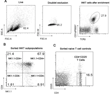
The host group has been studying the plasticity of iNKT cells when activated in the presence of TGF-β. We have shown for the first time that, like T lymphocytes, both murine (Fig. 2A) and human iNKT (Fig. 2C) lymphocytes are able to convert to Foxp3 when activated in the presence of IL-2 and TGF-β. It should be noted that these cells retain defined murine NKT-cell markers, namely the specific transcription factor PLZF (unpublished data by M. Monteiro). Moreover, in mice, expression of Foxp3 was not restricted to CD4+ but also found within the CD4- iNKT-cell subset (Fig. 2A) being Foxp3+ iNKT cells exclusively NK1.1- (Fig. 2B). This led us to hypothesize whether only NK1.1- iNKT cells can be converted towards Foxp3 expression or whether this phenotype reflects a different functional state of iNKT cells.
Fig. 2
iNKT cells can induce Foxp3 expression in vitro. (A) Murine iNKT cells wereisolated from the spleen of C57BL/6 mice, enriched for iNKT cells and FACS-sorted as CD1d/PBS57+TCRb+ cells. CD4+CD25- lymphocytes were sorted as controls. Both cell types were cultured in the presence of TGF-β and IL-2 and activated with plate bound anti-CD3 (A,
B). (A) Intracellular Foxp3 expression was analyzed 3 days later by flow cytometry. (B) Murine
Foxp3+ iNKT cells were CD4+ or CD4- and NK1.1-. (C) Human PBMCs were isolated and cultured in the presence of TGF-β and IL-2, as well as in the presence of CD28, IL-4, anti-IL-12 and anti-IFN-γ mAbs. Cultures were analyzed 5 days later. iNKT cells were identified as CD1d/PBS57+Vβ11+ cells, and Foxp3 expression assessed by intracellular FACS staining.
To address this question splenic iNKT cells from C57Bl/6 mice were enriched for iNKT cells and FACS-sorted according to the expression of NK1.1 and CD4: CD4+NK1.1-; CD4+NK1.1+; CD4-NK1.1+ CD4-NK1.1-. CD4+CD25- T cells from the negative fraction were used as controls (Fig. 3)
Fig. 3
Sorting strategy of NK1.1/CD4 iNKT-cell subpopulations. (A) iNKT cells fromC57BL/6 mice were isolated from the spleen, enriched for iNKT cells and identified after gating on the live population, followed by doublet exclusion as CD1d/PBS57+TCRb+ cells. (B) NK1.1/CD4 iNKT subpopulations were FACS-sorted (C) CD4+CD25- lymphocytes were sorted as controls.
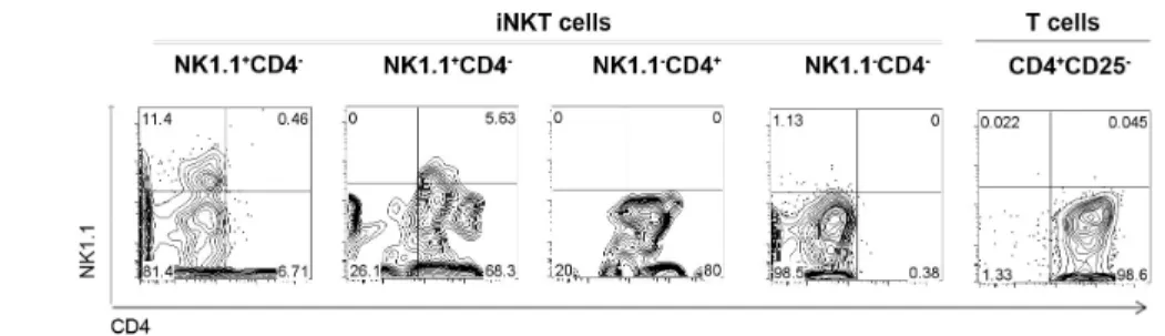
The sorted populations were stimulated for 3 days in the presence of IL-2 and TGF-β, a condition known to drive the up-regulation of Foxp3. Although Foxp3 was not detected by flow cytometry in freshly isolated splenic iNKT cells (Fig. 4 A), we showed that all subpopulations were able to induce de novo expression of Foxp3. The highest frequency of Foxp3+ cells was found in NK1.1- CD4- cell cultures (~40%), whereas the remaining subsets showed conversion rates around 20% (Fig. 4 B and C).
We also observed that, although we could not detect intracellular expression of IL-9 ex vivo in total iNKT cells isolated from naïve animals (Fig. 4A), these were able to induce IL-9 expression when activated in vitro in the presence of TGF-β, as described for conventional CD4+ T cells [6, 7] (Fig. 4D). Like iNKT lymphocytes that have up-regulated Foxp3, IL-9+ iNKT cells were all NK1.1- (Fig. 4D).
When the different NK1.1/CD4 iNKT-cell subpopulations sorted by flow cytometry, but not CD4+CD25- T-cell controls, were isolated prior to culture and stimulated in the
presence of TGF-β and IL-2, the expression of IL-9 was detected among Foxp3- cells of all subpopulations (Fig. 4E).
IL-9 expression was assessed by intracellular staining of iNKT cells (Fig. 4B and E). NK1.1+CD4- and NK1.1+CD4+ iNKT cells induced IL-9 expression in 7-15 % of the cells after 3 days of culture, twice as much as their NK1.1- counterparts. In CD4+CD25- T cells, however, IL-9 expression was only induced when exogenous IL-4 was added to cultures.
IL-4 has been described as essential for “Th9” conversion [6, 7]. Despite exogenous IL-4 was not added to the cultures, it is well established that iNKT cells respond to stimulation by secreting high amounts of IL-4 [26]. We hypothesized that iNKT-cell activation by the plate-bound anti-CD3 in our culture system was driving IL-4 secretion by iNKT cells. This cytokine, acting on iNKT cells in an autocrine way and synergizing with TGF-β, would therefore induce IL-9 expression, thus overcoming the requirement for exogenous addition of IL-4.
In fact, IL-4 was detected in culture supernatants at similar levels within all sorted iNKT-cell sorted populations, but not in CD4+ CD25- T-cell controls (Fig. 4F). This indicates that all NK1.1/CD4 iNKT-cell subpopulations were able to produce IL-4 following activation, independently of the presence of TGF-β.
In a subsequent experiment, we observed that neutralization of IL-4 in the cultures, with an anti-IL-4 antibody, resulted in a reduction of IL-9 release to the supernatant in a dose dependent manner, without affecting Foxp3 expression (Fig. 4G).
Collectively, these results show that IL-9 can be produced by all NK1.1/CD4 iNKT subpopulations, being dependent on TGF- β and IL-4.
In addition, our results indicate that cells converting to Foxp3 or IL-9 down-regulated NK1.1 and, in some cases, also CD4 expression. Importantly, cells that did not convert or that were activated in the absence of TGF- β showed the same tendency to loose NK1.1 and/or CD4 expression (Fig. 4H and data not shown).
These results demonstrate that all NK1.1/CD4 subpopulations of iNKT cells can be converted towards de novo expression of Foxp3 and IL-9 when cultured in the presence of TGF- β and IL-2, becoming NK1.1
Fig. 4
Different NK1.1/CD4 iNKT cells are susceptible to Foxp3 and “Th9” polarization in vitro in the presence of TGF-β and IL-2. (A) iNKT cells freshly isolatedfrom the spleen of C57BL/6 mice were stimulated with PMA/ionomycin for 3 hours and Foxp3 and IL9 expression was assessed by flow cytometry (isotype controls are also depicted): Foxp3 and IL9 were not detected in freshly isolated iNKT cells T cells expression. 50,000 cells of FACS-sorted NK1.1/CD4 iNKT subpopulations activated with plate-bound anti-CD3 in the presence of IL-2 and TGF-β. In some conditions, cells were deprived of TGF-β. After 3 days, cells were further stimulated for 3 hours in the presence of PMA/ionomycin and flow cytometric detection of Foxp3 and IL-9 was performed (B,C,D,G). (B) Representative dot plots are presented for the different FACS-sorted subpopulations. (C) Bar graphs depict the average frequency in culture triplicates of cells that have induced Foxp3, being the maximum achieved by NK1.1-CD4- subpopulation. (D) iNKT cells activated in presence of TGF-β can express IL-9, and IL-9+ cells are NK1.1- CD4+ or CD4-. (E) At the end point the supernatants were collected and IL-4 secretion detected by ELISA in FACS-sorted iNKT-cell subpopulations. (F) The addition of increasing doses of anti-IL4 antibody reduced IL-9 detection by CBA in the supernatants without affecting Foxp3 expression. (G) NK1.1 and/or CD4 expression of Foxp3+ (or IL9+, not shown) cells was downmodulated at the end of the cultures.
NK1.1/CD4 iNKT-cell subpopulations express IL-17 in the
presence of IL-6, IL-1
β
, anti-IFN-
γ
and TGF-
β
IL-17 production by iNKT cells was ascribed to a fixed lineage: the NK1.1- [32] or NK1.1-CD4- [31] subpopulations. Our observations that different NK1.1/CD4 splenic iNKT-cell subpopulations can convert to IL-9 or Foxp3, downmodulating NK1.1 and/or CD4, prompted us to hypothesize that different subsets of iNKT cells can be polarized towards IL-17 production, acquiring a NK1.1- phenotype as they acquire this new functional state.
Although we could not detect IL-17 expression in freshly isolated iNKT-cells from spleen of naïve mice (the existing reports agree that in naïve mice IL-17+ iNKT cells are a minute population), IL-17 became detectable after cells were cultured under the conditions described to convert conventional CD4+ T cells to Th17 [16] (Fig. 5A). Importantly, most IL-17 producers became NK1.1-, which again raised the question whether IL-17 could only be produced by NK1.1- iNKT-cells, or this reflects differentiation state of iNKT-cells.
We therefore isolated by flow cytometry the four NK1.1/CD4 iNKT cell subsets to assess their capacity to undergo Th17-like differentiation After 4 days, all NK1.1/CD4 subpopulations cultured in presence of TGF-β, IL-6, IL-1β and anti-IFN-γ induced IL-17
expression (Fig. 5B). Maximum IL-17 expression was found within NK1.1-CD4 -subpopulation, in which 23-30% of the cells upregulated IL-17. In the other subpopulations conversion rates were below 10% (Fig. 5B and C). ELISA measurements of IL-17 in culture supernatants confirmed that NK1.1- subpopulations secreted significant higher amounts of IL-17 than their counterparts and CD4+25- T cell controls (Fig. 5D). In addition, we observed that in all cultured subpopulations, the cells expressing IL-17 had downmodulated NK1.1 and/or CD4 (Fig. 5E).
In conclusion, our results show that all NK1.1/CD4 iNKT subsets are capable of induce de novo expression of IL-17, although with different efficiencies, thus disaccording with previous reports suggesting that IL-17 production by iNKT cells was restricted to NK1.1- subpopulations.
Fig. 5
Different NK1.1/CD4 iNKT cells express IL-17 in the presence of IL-1β IL-6 anti-IFN-γγγγ and TGF-β. (A) iNKT cells freshly isolated from the spleen of C57BL/6 mice werestimulated with PMA/ionomycin for 3 hours and IL-17 expression accessed by flow cytometry. IL-17 was not detected in iNKT cells ex-vivo but only when cultured in Th17 polarizing conditions. Polarized cells were NK1.1-CD4+ or NK1.1-CD4-. 50,000 cells of each FACS-sorted NK1.1/CD4 iNKT subpopulations were activated with plate-bound anti-CD3 and anti-CD28 in the presence of IL-1β, IL-6, anti-IFN-γ and TGF-β. Controls were deprived of TGF-β. After 5 days, cells were further stimulated for 3 hours in the presence of PMA/ionomycin (B,C,E). (B) IL-17 intracellular expression expression was detected by flow cytometry in different cultured subpopulations (C) Average frequency in culture triplicates of cells expressing IL-17 after culture. (D) IL-17 secretion in the culture supernatants was assessed by ELISA (non-continuous line depicts top standard curve; values represent concentrations normalized to the number of cells/well) (D). (E) NK1.1 and/or CD4 expression of Th17+ cells were downmodulated at the end of the cultures.
Co-receptor blockade with non-depleting anti-CD4 prevented
allergic sensitization in BALB/c mice
Data previously obtained by the host group indicate that a non-depleting anti-CD4 monoclonal antibody (MAb) can prevent allergic sensitization in a well established murine model of allergic airways disease (AAD), leading to allergen-specific tolerance (unpublished data by A. Agua-Doce). Animals tolerized with CD4 MAb were protected from allergic manifestations elicited by allergen exposure: they did not develop airways eosinophilia; the production of Th2 cytokines in the lung was markedly reduced, without Th1 deviation being observed; the production of allergen-specific IgE or IgG1 was abrogated; and, more importantly, no AHR was elicited in response to increased doses of inhaled methacholine (MCh) (unpublished data by A. Agua-Doce – Annexes - Supplemental Fig. 1).
As iNKT were shown to be important for induction of AAD, and as these cells can also express CD4, we investigated whether tolerance induced by CD4 blockade might also impact on the iNKT-cell compartment.
Co-receptor blockade with non-depleting anti-CD4 in AAD
reduced iNKT cell infiltrates in BAL and lung and altered the
phenotype of iNKT cells in the medLNs
Female BALB/c mice were sensitized with ovalbumin (OVA)-alum on days 1 and 14 and challenged intranasally with OVA on days 20, 21 and 22 (Fig. 6A). Experimental animals were treated with anti-CD4 at the time of sensitization. Bronchoalveolar lavage (BAL), lungs, mediastinal draining lymph nodes (medLNs), spleen and blood serum were collected 24 hours after the last OVA challenge and analyzed by flow cytometry.
iNKT cells were identified using CD1d/PBS57-labelled tetramers after CD11b, dead cells and doublet exclusion (Fig. 6B). Allergic animals exhibited an increase in the frequency and absolute number of iNKT cells in the lungs, BAL but not in medLNs (where only the number was increased) when compared to healthy controls (Fig. 6C and D). Moreover, 60-80% of the infiltrating iNKT cells in the lungs and BAL expressed the CD4 co-receptor, thus supporting the possibility of direct targeting by anti-CD4 MAb (Fig. 6E).
Mice tolerized with anti-CD4 MAb showed a significant reduction in iNKT-cell infiltrates in lungs and BAL, but not in medLNs or spleen (Fig. 5C and D), when compared to allergic animals. Although medLNs from anti-CD4 treated mice contained similar numbers of iNKT cells as AAD controls, we observed changes in the phenotype of iNKT cells: there was a marked reduction of CD103+ and CD62L+ iNKT cells from treated animals (Fig. 6E). Moreover, the antibody was able to reduce CD4+-expressing iNKT cells in lungs, BAL and medLNs without leading to direct lysis of CD4+ iNKT cells (or CD4+ T cells), as these populations in the spleen remained unchanged. This observation was not due to receptor occupancy by the MAb, as our in vitro studies showed that the antibody used in flow cytometry is able to bind CD4 on the cell surface even in the presence of the therapeutic MAb (data not shown).
Using HDM, a more physiologic allergen than OVA, to induce AAD, we also observed that not only eosinophils and lymphocyte were increased in the airways of allergic animals, but iNKT-cell infiltrates were increased as well. Notably, cell numbers were restored to basal levels after the same tolerogenic treatment with anti-CD4 MAb (Fig. 6 F).
Fig. 6 Non-depleting anti-CD4 MAb tolerization to AAD correlates with a reduction in iNKT-cell infiltrates in the airways and alteration of phenotype in the draining LNs. (A) iNKT cells were detected by FACS according to the co-expression of TCRβ
and CD1d /PBS57 tetramers, after exclusion of dead cells, doublets and CD11b expressing cells. Control staining with unloaded tetramer is also depicted. (B) Representative dot plots of iNKT-cell identification in the BAL, lungs and medLNs: untreated mice showed an increased percentage of iNKT cells, which was reduced after anti-CD4 MAb treatment (C). Absolute cell counts of infiltrating iNKT cells in medLNs, lungs, and more significantly BAL were increased in allergic when compared to healthy controls. Mice treated with anti-CD4 MAb showed a significant reduction of iNKT cells in the BAL and lungs but not in the mediastinal LNs or spleen.
(D) Mean Fluorescense intensity (MFI) of CD4 and CD103 and frequency of CD62L (Non filled
lines represent the naïve group, black filled the OVA, and grey filled the treated group. Black grey and italic numbers represented in graphs indicate the frequency of CD4+ iNKT cells in medLNs): anti-CD4 tolerization protocol reduced the expression of CD4, CD103 and CD62L. (E) In a similar protocol using HDM instead of OVA as an allergen, the anti-CD4 MAb treatment was able to reduce eosinophilic, lymphocytic and monocytic accumulation in the BAL as well as the iNKT-cell numbers.
Co-receptor blockade with non-depleting anti-CD4 prevented
EAE and led to an increase of Foxp3
+iNKT cells
Some groups have provided convincing evidence that iNKT cell content is inversely correlated with EAE manifestations, thus supporting the idea that iNKT cell play a beneficial role in this disease [54, 55].
Since we have discovered that this unconventional lymphocyte population has the ability to express Foxp3, we addressed the possibility of Foxp3 being up-regulated by iNKT cells in EAE. For that we used a relapsing-remitting EAE model in C57BL/6 mice, already established in our lab by J. Duarte, to study the effect of anti-CD4 tolerization protocol. Briefly, mice were immunized with MOG/CFA emulsion, with pertussis toxin administered on the day of immunization and two days later. In one experimental group, mice received 1 mg of anti-CD4 MAb 3 and 2 days prior to immunization (Fig. 7A). Disease manifestations were followed daily and individually mice scored according to severity of the clinical manifestations (Fig. 7B).
Mice were sacrificed when the majority was at the first peak of the disease (score 4), and before the recovery phase. Cervical lymph nodes (cervLNs), CNS and spleen were collected and analyzed. Our data show that anti-CD4-MAb administration was able to prevent MOG-induced progression of EAE, as animals treated with anti-CD4 exhibited no symptoms of the disease, in contrast to mice that did not receive the treatment (Fig. 6B). Furthermore, lymphocyte infiltrates in the CNS, characteristic of EAE pathology, were not observed in mice that had received anti-CD4 MAb (Fig. 7C), and iNKT-cell counts and frequency in the CNS, cervLNs and spleen were restored to healthy control levels (Fig. 7D and E ).
Of note, we detected, for the first time, Foxp3 expressing-iNKT cells in the cervLNs, but not in spleen, of sick and treated mice (Fig. 7F). Interestingly this subset increased in frequency and absolute numbers in the anti-CD4 treated group (Fig. 7G).
Fig. 7
Prevention of EAE following non-depleting anti-CD4 MAb treatment correlates with an increase of Foxp3+ iNKT cells. (A) Experimental model: C57BL/6mice were sensitized with MOG peptide. On one experimental group mice received 1mg antiCD4 Mab 3 and 2 days prior to immunization. (B) Average clinical scores of mice immunized with MOG in the presence or absence of antiCD4: mice that received anti-CD4 did not elicit EAE manifestations. Disease severity was scored daily on a 5 point scale: 1- tail atony; 2- hind limb weakness; 3-hind limb paralysis; 4-quadriplegia; 5- moribund (C) Anti-CD4 administration before MOG immunization was able to reduce total lymphocyte infiltrates in the CNS when compared to mice that were not tolerized (D) Immunized mice presented iNKT infiltrates in the CNS, and lower iNKT cell numbers in the spleen and cervLNs, when compared to healthy controls. Mice treated with anti-CD4 MAb prevented this effect. (E) Representative dot plots of iNKT-cell identification in the cervLNs: MOG immunized animals showed decreased percentage of iNKT cells upon treatment Foxp3 was detected by flow cytometry in the cervLNs of immunized mice, but not in naïve animals. Anti-CD4 MAb led to an increase in the percentage of Foxp3+ iNKT population (representative dot plots). (F) Total numbers of the Foxp3+ iNKT cells are increased in tolerized animals.
α
-GalCer administration prevented EAE and led to an increase
in Foxp3
+iNKT cells
It was shown that in vivo activation of iNKT cells with the specific ligand α-GalCer ameliorates EAE by preventing Th17 differentiation of auto-reactive T cells [39, 55]. To test whether iNKT activation, in this disease model, correlates to the increase in the numbers of Foxp3+ iNKT cells, we established a new experimental group where mice received α-GalCer – a specific ligand of iNKT cells – on the day of MOG immunization and 4 days later, in accordance to a protocol described elsewhere [56] (Fig. 8A). We observed that animals treated with α-GalCer did not develop clinical manifestations of the disease (Fig. 8B), nor display lymphocyte infiltrates in the CNS (Fig. 8C). Additionally, iNKT-cell frequency and total numbers were increased in the spleen and cervLNs (Fig. 8D and E), as well as the number of Foxp3+ iNKT cells detected in the cervLNs, when comparing to sick animals (Fig. 8F and G).
These data suggest that strategies effective in preventing EAE correlate with the emergence of Foxp3+ iNKT cells in CNS-draining cervLNs.
Fig. 8 Prevention of EAE by α-GalCer treatment correlates with increased Foxp3+ iNKT cells. (A) Mice were immunized with MOG as described in figure 7. One experimental group received 4 µg of α-GalCer, in the MOG/CFA emulsion, and another 4 µg 4
days later i.p. (B) Average of the clinical scores of mice immunized with MOG in the presence or absence of α-GalCer: mice in which iNKT cells were activated with α-GalCer did not elicit EAE clinical manifestations. (C) α-Galcer activation of iNKT cells along with MOG immunization was able to reduce total lymphocyte infiltrates in the CNS when compared to mice that did not receive α-GalCer treatment. (D) The numbers of iNKT cells in the cervLNs and spleen of mice receiving α-GalCer were restored to healthy control levels. (E) Representative dot plots of iNKT-cell identification in the cervLNs: the percentage of iNKT iNKT-cells was restored to healthy control levels in the group that has received α-Galcer along with immunization. (F) Foxp3 expression was detected by flow cytometry in the cervLNs: activation of iNKT cells with α-GalCer led to an increase in the percentage of Foxp3+ iNKT population (representative dot plots). (G) Total numbers of the Foxp3+ iNKT cells were also increased in the group receiving α-GalCer.